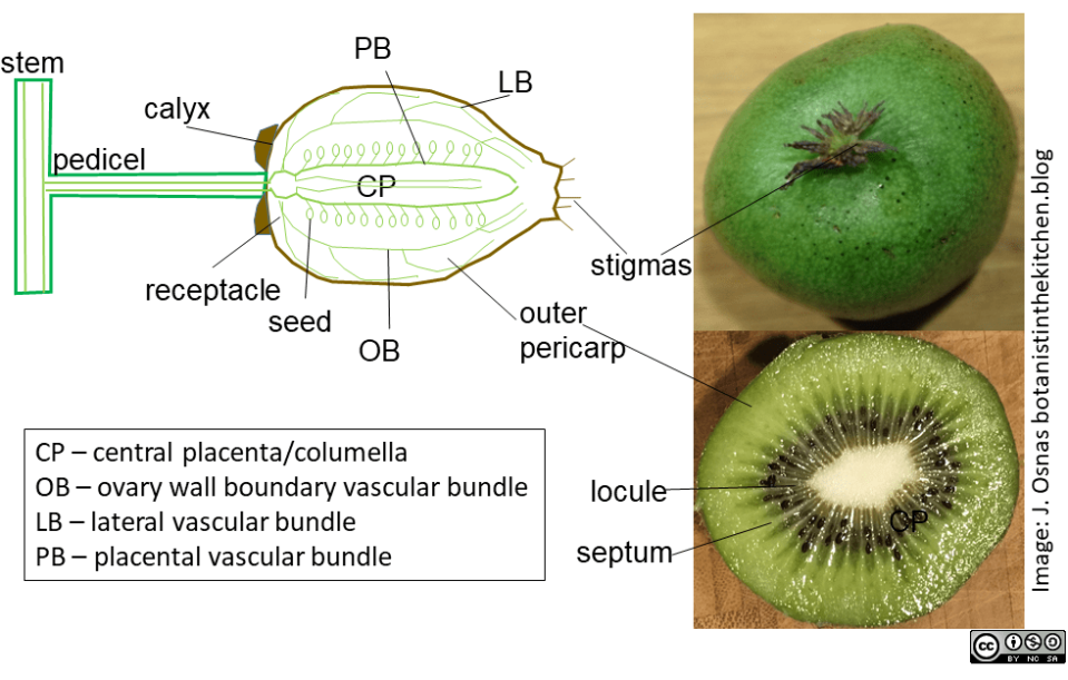
Figure 1. Position of major vascular bundles (xylem and phloem) and structures in kiwifruit. Kiwifruit is botanically classified as a berry, a fleshy fruit that develops from a single ovary. The kiwifruit ovary is composed of numerous fused carpels. Each carpel bears its own style and stigma, creating a starburst pattern in the center of the pistillate flowers (see Figure 2 below). Those stigmas can persist through fruit ripening and remain visible at an end of ripe kiwifruit (shown here on A. arguta). The same goes for the tough sepals below the petals. Those many carpels contain chambers called locules, in which seeds develop. Septa divide locules and carpels within the ovary and are visible in a kiwi cut in cross section as pale white lines (shown here for A. chinensis var. deliciosa). The carpels radiate around the large, white central (axial) placenta, which is formed of homogeneous, large parenchyma cells and vascular tissue. The major vascular bundles in the fruit are also generally visible in cross section as translucent dots. Anatomy from Clearwater et al. (2012) and Guo et al. (2013).
