After some weeks dealing with researchers of more recent times, today we are going back to ye olde naturalists from the 18th century.
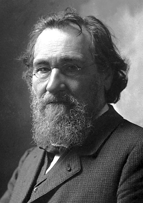
Today we commemorate the birthday of Louis-Jean-Marie Daubenton, who was born on 29 May 1716 in Montbard, Côte-d’Or, France. His father, Jean Daubenton, was a notary and wanted his son to become a priest, thus sending him to Paris to study Theology. Daubenton’s desire, however, was to study medicine. Fortunately for him (I guess), his father died in 1736, when Daubenton was 20, and so he became free to study whatever he wished.
Studying medicine at Reims, Daubenton graduated in 1741 and returned to Montbard. His intentions were to work there full time as a physician, but things changed a little bit only one year later. The naturalist Georges-Louis Leclerc de Buffon, also from Montbard, was planning to write a multi-volume work on Natural History, named Histoire naturelle, générale et particulière, and, in 1742, invited Daubenton to help him, especially with anatomical descriptions.
Due to his partnership with Buffon, Daubenton became a member of the French Academy of Sciences in 1744 as an adjunct botanist. During this time, Buffon was the curator of the Jardin du Roi (currently Jardin des plantes), the main botanical garden in France until today, and appointed Daubenton as the keeper and demonstrator of the King’s cabinet.
In 1749, the first volume of Buffon’s Histoire Naturelle was finally published, although most of Daubenton’s contributions only appeared from the fourth volume on, published in 1753. The work included detailed descriptions of 182 quadrupeds (mammals) given by Daubenton and this increased his reputation as a comparative anatomist.
Portrait of Daubenton in 1791 by Alexander Roslin.
Daubenton became one of the contributors of the famous Encyclopédie, the first encyclopedia to be written, writing several articles on natural history. He also published many articles in the memoirs of the Parisian Académie Royale des Sciences, especially on comparative anatomy, but also on agriculture and mineralogy. He became a teacher of mineralogy at the Jardin du Roi and later also taught natural history at the College of Medicine (from 1775 on) and rural economy at the Alfort School (from 1783 on).
The controversial political opinions of the writers of the Encyclopédie, who disregarded Catholicism and the Royalty, helped to prepare the scenario that led to the French Revolution. In December 1799, following the Revolution, Daubenton, who was already 83, was appointed a member of the Senate. However, during the first meeting he attended, he fell from his chair after suffering a stroke and died in Paris on 1 January 1800.
We are all familiar with animals of many species that developed interesting mechanisms to avoid being eaten. This includes, for example, animals that look like plant parts:
The famous giant leaf insect, Phyllium giganteum. Photo by Bernard Dupont.**
and animals that merge with the background:
An East African jackal, Canis mesomelas, in the Savanna. Can you spot it? Photo by Nevit Dilmen.***
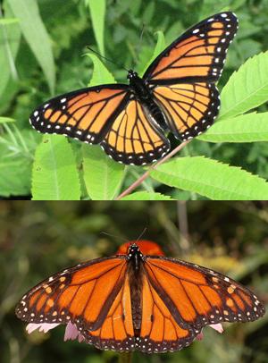
There are also animals that look like other, unpalatable or dangerous, animals, in order to push predators away:
The edible viceroy butterfly Limenitis archippus (top) mimicks the poisonous monarch butterfly Danaus plexippus (bottom). Credits to Wikimedia user DRosenbach. Photos by D. Gordon E. Robertson and Derek Ramsey.***
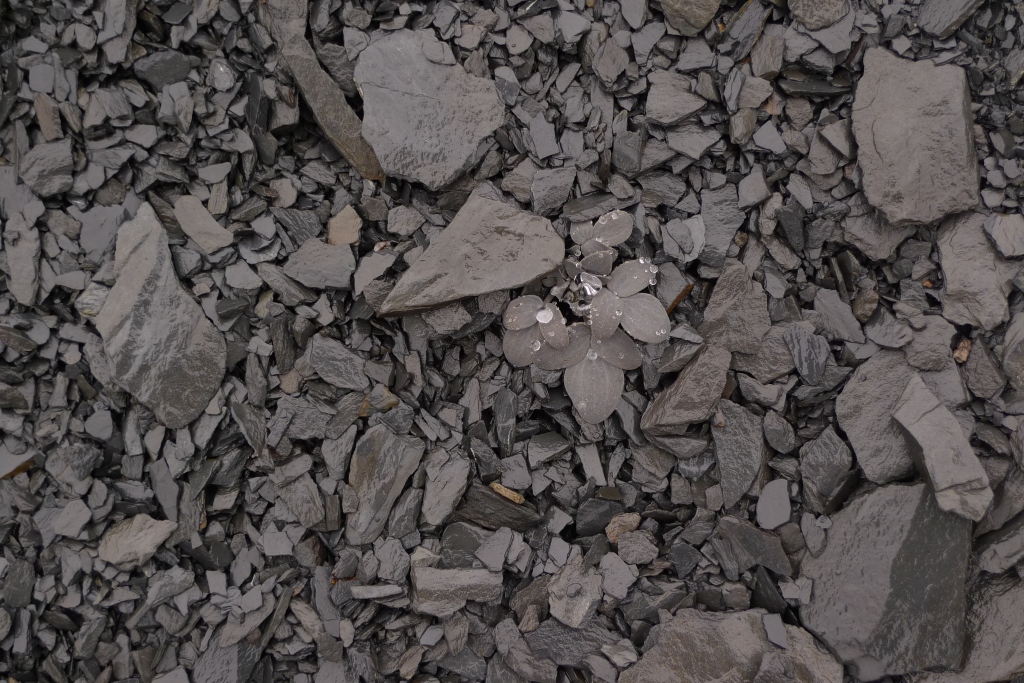
But we rarely think that plants also use this sort of mechanisms to avoid being eaten. There are, however, some recorded cases of similar behaviors in plants. One case is that of the plant Corydalis benecincta, whose leaves commonly have the brownish color of the surrounding rocks:
The leaves of Corydalis benecincta look like the rocks found in its natural habitat. Photo by Yang Niu.
Recently, a study on plants of the genus Amorphophallus found another interesting case of mimicry. This genus, which includes the famous titan arum, usually develops a single large leaf that in some species can attain the size of a small tree or shrub. Such a gigantic leaf seems to be a perfect meal for some herbivores but, to avoid them, many species of this genus developed a series of marks along the petiole of their leaf that look like lichens or cyanobacteria.
Cyanobacteria-like marks on the petiole of Amorphophallus gigas (A); Cyanobacteria-like plus lichen-like marks also on A. gigas (B); And lichen-like marks on A. hewittii (C) and A. dactylifer (D). Extracted from Claudel et al. (2019).
With this mimicry, the petioles, which are quite tender, end up looking like a hard and old trunk that does not look that interesting as a meal for most herbivores. The lichen marks are so well represented that they can even be associated with real lichen genera. For example, the marks seen on the figures B and C above look like lichens of the genus Cryptothecia.
Lichen of the species Cryptothecia striata, which seems to be mimicked by the marks in Amorphophallus gigas and A. hewittii. Photo by Jason Hollinger.*
How and why this marks evolved across Amorphophallus species is still not well understood. Despite the hypothesis that they help the plant mimic a tree trunk, some species with small leaves also have those marks, while some with large leaves do not have any marks or have them in simpler patterns. The titan arum Amorphophallus titanum is a good example of the latter:
Amorphophallus titanum is the largest species of Amorphophallus but displays a considerably simple lichen-like pattern. Photo by flickr user Björn S.**
For a long time, plants were regarded as less dynamic organisms than animals, but in recent years our knowledge about them is increasing and showing that they are actually very versatile creatures that developed similar creative and complex strategies.
Claudel C, Lev-Yadun S, Hetterscheid W, & Schultz M 2019. Mimicry of lichens and cyanobacteria on tree-sized Amorphophallus petioles results in their masquerade as inedible tree trunks. Bot J Linn Soc 190: 192–214.
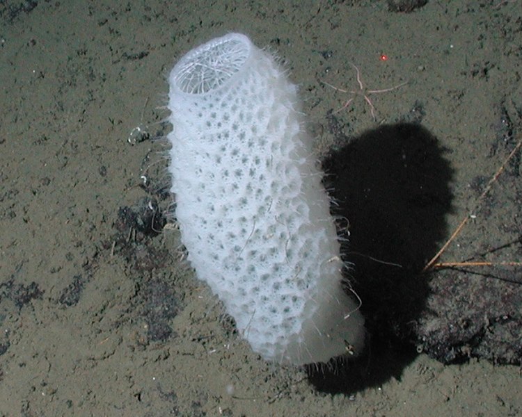
Sponges are the weirdest of all animals but also some of the most beautiful. One species of special beauty that is considerably popular is Euplectella aspergillum, popularly known as the Venus’ flower basket.
Venus’ flower basket in the Pacific Ocean.
Growing on the Ocean floor of tropical waters, the Venus’ flower basket is common around the Philippines and this may be the only place where it occurs. Other similar species are found in nearby areas such as Japan, Indonesia and Australia and are often mistaken for the Venus’ flower basket. There are, indeed, populations of this species identified in Australia and Indonesia, among other areas near the Philippines, but are considered subspecies due to subtle morphological differences and may in fact be complete separate species.
The Venus’ flower basket is a medium-sized sponge, measuring up to 1.3 m in height, althout most specimens measure between 10 and 30 cm. The body is white and has several large pores that make it look like an elongate basket, hence the common name. The osculum, the large opening at the top, is covered by a mesh of fibers that makes its interior inaccessible to large organisms.
Skeleton of the Venus’ flower basket. Credits to the Auckland Museum.*
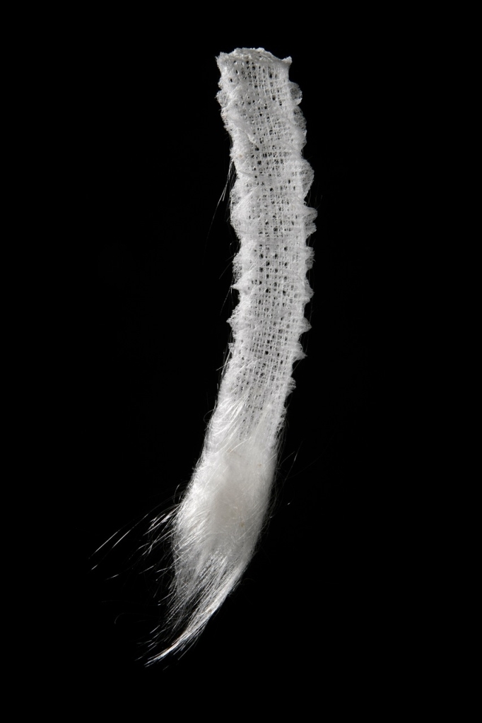
Recently, the Venus’ flower basket has called the attention of scientists because of the structural complexity of its skeleton, which is composed of silica (i.e., glass). Studies have shown that the anchor spicules, i.e., those that attach the sponge to the substrate, are similar to man-mane optical fibers regarding optical properties but are better regarding fracture resistance. Understanding the detailed pathway used by the sponge to build these spicules could lead to the development of easier ways to build optical fibers and even increase their quality.
Monn MA, Weaver JC, Zhang T, Aizenberg J, Kesari H (2015) New functional insights into the internal architecture of the laminated anchor spicules of Euplectella aspergillum. PNAS 1112(16): 4976-498. doi: 10.1073/pnas.1415502112
Shimizu K, Amano T, Bari MR, Weaver JC, Arima J, Mori N (2015) Glassin, a histidine-rich protein from the siliceous skeletal system of the marine sponge Euplectella, directs silica polycondensation. PNAS 112(37): 11449-11454. doi: 10.1073/pnas.1506968112
Today we celebrate the birthday of an important figure in the study of the fascinating copepod crustaceans.
Patricia Louise Dudley, often called Pat Dudley, was born on 22 May 1929 in Denver, Colorado, USA, the daughter of David C. Dudley, a salesman for State and School supply, and Carolyn Dudley (née Latas). Her father died in 1932, when she was only 3 years old.
During her childhood, Dudley lived with her mother and maternal grandparents in Colorado Springs. She studied in Colorado Springs High School and graduated in 1947. Soon after, she started her undergraduate studies at the University of Colorado, where she studied with the limnologist Robert William Pennak. She earned her Bachelor’s degree in 1951 and stayed in the same university for her Master’s degree, having Pennak as her advisor. Her master’s thesis was a research on the aquatic fauna of four brooks in Boulder County, Colorado.
Finishing her Master’s in 1953, she pursued a PhD Degree at the University of Washington in Seattle. Her initial intention was to continue to do limnological studies, but ended up meeting the carcinologist Paul Louis Illg, who studied copepods. At the University of Washington and its marine biological station at Friday Harbor, Dudley turned her attention to marine copepods that are associated with ascidians. This copepod group has a huge variety of forms and Dudley dedicated her doctorate to study the developmental stages of this small comensal crustaceans. She defended her thesis in 1957.
In 1959, Dudley joined the Columbia University and started to teach zoology at Barnard College. She remained there until her retirement in 1994 and dedicated her research to the study of comensal copepods, mostly those associated with ascidians, but later also those comensal on other marine invertebrates, especially polychaetes. She was one of the first researchers to use electronic microscopy for the study of copepod structures.
After her retirement, Dudley moved back to Seattle and planned to continue her research on copepods there. Unfortunately, she started to have health problems soon after, which forced her to abandon her research. She became very ill during the following years and died on 30 September 2004, aged 75.
After beetles, which make up the order Coleoptera, the second most diverse group of insects is the order Lepidoptera, which includes butterflies and moths. However, the order Hymenoptera has the potential to eventually surpass Lepidoptera and get closer to the beetles because a lot of new species are being constantly described.
The most widely known hymenopterans are bees, ants and wasps, but a huge part of their diversity is made up by the so-called sawflies. One of these species is commonly known as the turnip sawfly and scientifically named Athalia rosae.
Turnip sawfly in the Netherlands. Photo by Herman Berteler.*
The turnip sawfly is found throughout the Paleartic Ecozone, from western Europe to Japan, and his common name comes from the fact that its larvae feed on plants of the family Brassicaceae, which includes the turnip, as well as the cabbage, among others. The larvae is considerably large and resembles a caterpillar, having a dark gray, almost black dorsal color, and is lighter close to the feet. When they are about to turn into a pupa, they dig into the ground, build a cocoon and remain there until they become adults.
A larva in Denmark. Photo by Donald Hobern.**
The adults measure about 6 to 8 mm in length, the females being larger than the males. The body and the legs have a yellow to orange color, darker on the dorsal surface of the thorax, which also has two large black spots. The head and the antennae are black.
An adult in Germany. Photo by Martin Grimm.*
Hymenopterans in general are characterized by a unique sexual determination in which females are diploid, i.e., have two sets of chromosomes, and males are haploid, having only one set. Matings conducted in the laboratory with the turnip sawfly, however, were able to produce anomalous combinations, including diploid males and triploid females and males. Apparently this is possible due to sex being determined by one allele in one chromosome, so that males are always homozygous and females always heterozygous, but this must be explained in another post. The fact is that the study of this peculiar system in this species is helping to understand how sex determination evolved in hymenopterans.
In adult in Russia. Photo by Roman Providuhin.*
One last interesting thing to mention about the turnip sawfly is that it is able to bypass the defense mechanisms of the plants on which its larva feeds. Plants in the family Brassicaceae produce a group of compounds called glucosinolates that give them their characteristic pungency and bitterness, such as in mustard and horseradish. These compounds are used by the plant as a defense against pests that feed on them. However, the turnip sawfly is resistant to this compounds and is able to sequestrate them and store them in their hemolymph, i.e., their “blood” in concentrations much higher than found in the plants. When attacked by a predators, such as ants, the larva releases drops of its hemolymph in a sort of defensive bleeding and can stop the attack.
The turnip sawfly may be a nuisance for humans and their crops, but it is certainly a fascinating animal.
Müller C, Agerbirk N, Olsen CE, Boevé JL, Schaffner U, Brakefield PM (2001) Sequestration of host plant glucosinolates in the defensive hemolymph of the sawfly Athala rosae. Journal of Chemical Ecology 27(12): 2505–2516. doi: 10.1023/A:1013631616141
Müller C, Boevé JL, Brakefield PM (2002) Host plant derived feeding deterrence towards ants in the turnip sawfly Athalia rosae. In: Nielsen J.K., Kjær C., Schoonhoven L.M. (eds) Proceedings of the 11th International Symposium on Insect-Plant Relationships. Series Entomologica, vol 57. Springer, Dordrecht. doi: 10.1007/978-94-017-2776-1_18
Naito T, Suzuki H (1991) Sex determination in the sawfly, Athalia rosae ruficornis (Hymenoptera): occurrence of triploid males. Journal of Heredity 82(2): 101–104. 10.1093/oxfordjournals.jhered.a111042
Oishi K, Sawa M, Hatakeyama M, Kageyama Y (1993) Genetics and biology of the sawfly, Athalia rosae (Hymenoptera). Genetica 88(2–3): 119–127. doi: 10.1007/BF02424468
For the third week in a row, our featured scientist is a Nobel laureate!
Ilya Ilyich Mechnikov (in Russian: Илья Ильич Мечников), also known as Élie Metchnikoff, was born on 15 May 1845 in the village of Ivanovka in Ukraine. His father, Ilya Ivanovich Mechnikov was a Russian officer of the Imperial Guard and his mother, Emilia Lvovna Nevakhovich was the daughter of the writer Leo Nevakhovich.
Metchnikoff ca. 1862.
In 1856, at the age of 11, Metchnikoff entered the Kharkiv Lycée school and developed an interest in biology. Due to his mother’s influence, he was interested in science since and early age. She also convinced him to study natural sciences instead of medicine. Thus, in 1862, he tried study biology at the University of Würzburg, in Germany but, as the academic year there would only begin by the end of the year, he ended up enrolled at the Kharkiv University to study natural sciences. In 1863, he married Ludmila Feodorovitch and, in 1864, graduated at the age of 19, completing the four-year degree of natural sciences in only two. That same year, he went to Germany to study the marine fauna on the island of Heligoland in the North Sea.
After meeting the botanist Ferdinand Cohn, Metchnikoff was advised by him to work with the zoologist Rudolf Leuckart at the University of Giessen. Together with Leuckart, he studied the reproduction of nematodes and discovered intracellular digestion in flatworms. In 1866, he moved to Naples and worked on a doctoral thesis on the embryonic development of cuttlefish of the genus Sepiola. In 1867, he moved to Russia and received his doctorate degree with Alexander Kovalevsky from the University of St. Petersburg. For their work on the development of germ layers of invertebrate embryos, Metchnikoff and Kovalevsky won the Karl Ernst von Baer prize.
Due to his competence, Metchnikoff was appointed, still in 1867, professor of the new Imperial Novorossiya University (currently Odessa University). He was only 22 years old, being younger than most of his students. The next year, due to a conflict with a senior colleague, he was transferred to the University of St. Petersburg but the professional environment there was even worse. He returned to Odessa in 1870 as professor of Zoology and Comparative Anatomy.
On April 20, 1873, Metchnikoff’s wife died of tuberculosis. This event, combined with his professional problems, made him attempt suicide taking a large dose of opium. He survived and eventually recovered and, in 1875, married his student Olga Belokopytova.
The assassination of Alexander II in 1881 led to political turmoils in Russia, which made Metchnikoff resign from Odessa University in 1882. He moved to Sicily and set up a private laboratory in Messina. There, while studying starfish larvae, he noticed that, by inserting a small citrus thorn in the larvae, a group of cells started to surround the thorn. He suggested that some white cells in the blood are able to attack and kill pathogens, and the zoologist Carl Friedrich Wilhelm Claus, with whom he discussed his hypothesis, suggested the name “phagocyte” to such cells.
Élie Metchnikov ca. 1908.
Metchnikoff presented his findings on phagocytes at Odessa in 1883, but his idea was met with skepticism from many specialists, including Louis Pasteur. The idea at that time was that white blood cells carried pathogens away from the infection place and delivered them elsewhere, spreading them instead of destroying them. His main supporter was the pathologist Rudolf Virchow. Back to Odessa, Metchnikoff was appointed director of an institute created to produce Louis Pasteur’s vaccine against rabies.
In 1885, Metchnikoff’s second wife suffered from severe typhoid fever. As a result, he attempted suicide once more, this time by injecting himself with spirochaete bacteria that cause relapsing fever. He survived again, and his wife survived as well.
In 1888, Metchnikoff left Odessa again due to new difficulties and went to Paris to seek the advice of Pasteur, who gave him an appointment at the Pasteur Institute. Metchnikoff remained there for the rest of his life. In 1908, he won the Nobel prize in physiology or medicine due to his discovery of phagocytes. During his last years, he developed a theory that aging was a disease caused by toxic bacteria in the gut and that lactic acid produced by Lactobacillus could prolong life.
He died on 15 July 1916 of heart failure in Paris, aged 71.
Hemipterans constitute the fifth most numerous order of insects with about 80 thousand species currently known. Among them, one that is widely spread around the world and that you may have seen somewhere is the southern green stink bug Nezara viridula, also known as the southern green shield bug, green vegetable bug and probably other names. The name stink bug applied to these insects refers to the fact that they secrete a defensive substance with a pungent smell from their thorax when threatened.
Nezara viridula in Taiwan. Photo by Jaycee Snow.*
This species is usually green as an adult and can be recognized by a row of three small white dots at the anterior border of the scutellum, the triangular plate between the wings. There is also a small black on each of the anterior corners of the scutellum, aligned with the row of white dots. In some specimens, the anterior part of the head and the anterior border of the thorax have a different color, usually white, yellow or orange, and, sometimes, the whole body has a white border. During the winter, the green color usually fades and is replaced by a brown shade.
Variety with the whitish border in Greece. Photo by Anna Chapman.
Another, much rarer form has a completely orange or red body, which greatly contrasts with the usual green. The three small white dots, and especially the black ones, are still present though, making it easy to recognize them as belonging to the same species.
Yellow variety in Argentina. Photo by Gonzalo Roget.**
The southern green stink bug is originally from somewhere in East Africa, probably around Ethiopia, but is currently found in tropical and subtropical areas in all continents. With piercing and sucking mouthparts as all members of the order Hemiptera, it feeds on more than 30 plant families, perforating their surface, injecting digestive enzymes and later sucking the partially digested product. Some of their favorite plants include legumes such as common beans and soybeans, making them important pests of those crops. They prefer to suck on tender parts of the plant, such as growing shoots or developing fruits and, therefore, are commonly near them.
Two southern green stink bugs mating in France. One has the full green color pattern and the other has the white-bordered pattern. Photo by Bernard Dupont.***
In tropical areas, the southern green stink bug may reproduce throughout the year, but in subtropical to temperate zones their reproductive period is restricted to the warmer months of the year. In such areas, adults start to mate and search for food as soon as the temperatures rise, sometimes travelling up to 1 km per day in search for food and places to lay their eggs.
Nezara viridula in its brown winter form in France. The white structure on the head, close to the right eye, is the egg of the parasitoid fly Trichopoda pennipes. Photo by Didier Descouens.***
The mating behavior of the southern green stink bug is very complex. Males release pheromones that attract both males and females, apparently as a way to increase mating opportunities. As a result, several individuals can be found gathered and ready to copulate. The courtship behavior includes a series of stridulating sounds produced by both males and females and a specific sequence of alternating male and female songs seems to be necessary for copulation to occur.
Egg cluster laid by a female southern green stink bug in New Zealand. Photo by Robert Briggs.****
The eggs of the southern green stink bug are yellowish and the female lays them in polygonal clusters, arranged in regular rows, and glue them together. The time until hatching depends on the environment’s temperature and can take 5 days or less in the summer and up to 3 weeks in colder months.
First-instar nymphs still around the remains of the eggs in Germany. Photo by Wikimedia user Slimguy.****
The hatched first-instar nymphs are dark brown and remain close to each other near the egg shells. After molting and becoming the second instar, which is black, the nymphs start to disperse. The third and fourth instars are dark brown to black and have a series of white dots on the abdomen. It is only during the fifth and final instar of the nymph that the green color of the adult starts to appear.
Third-instar nymph in Gran Canaria. Photo by Juan Emilio.***
The southern green stink bug has several natural enemies, which include spiders and predatory ants, as well as parasitoid flies, which feed on the adults, and parasitoid wasps, which feed on the eggs. Parasitoid species are known to use pheromones released by adult stink bugs to locate them in the environment.
Fifth-instar nymph in New Zealand. Photo by Uwe Schneehagen.**
With the increasing temperatures caused by climate change, the southern green stink bug has the potential to spread to new parts of the world and may eventually become a much more serious pest.
Čokl A, Virant-Doberlet M, McDowell A (1999) Vibrational directionality in the southern green stink bug, Nezara viridula (L.), is mediated by female song. Animal Behavior 58(6): 1277–1283. doi: 10.1006/anbe.1999.1272
Čokl A, Zorović M, Žunić A, Virant-Doberlet M (2005) Tuning of host plants with vibratory songs of Nezara viridula L (Heteroptera: Pentatomidae). Journal of Experimental Biology 208: 1481–1488. doi: 10.1242/jeb.01557
Colazza S, Salerno G, Wajnberg E (1999) Volatile and Contact Chemicals Released by Nezara viridula (Heteroptera:Pentatomidae) Have a Kairomonal Effect on the Egg Parasitoid Trissolcus basalis (Hymenoptera: Scelionidae). Biological Control 16(3): 310–317. doi: 10.1006/bcon.1999.0763
Mattiacci L, Vinson SB, Williams HJ, Aldrich JR, Bin F (1993) A long-range attractant kairomone for egg parasitoid Trissolcus basalis, isolated from defensive secretion of its host, Nezara viridula. Journal of Chemical Ecology 19(6): 1167–1181. doi: 10.1007/BF00987378
Musolin DL, Numata H (2003) Photoperiodic and temperature control of diapause induction and colour change in the southern green stink bug Nezara viridula. Physiological Entomology 28(2): 65–74. doi: 10.1046/j.1365-3032.2003.00307.x
Tougou D, Musolin DL, Fujisaki K (2009) Some like it hot! Rapid climate change promotes changes in distribution ranges of Nezara viridula and Nezara antennata in Japan. Entomologia Experimentalis et Applicata 130(3): 249–258. doi: 10.1111/j.1570-7458.2008.00818.x
Today, again, just as last week, our featured scientist is a Nobel laureate.
André Michel Lwoff was born on 8 May 1902 in Ainay-le-Château, France. His mother, Marie Siminovitch, was a painter and sculptor, and his father, Solomon Lwoff, was a psychiatrist. Of Russian origin, his parents came to France to escape the oppression of the tsarist regime.
Since an early age, Lwoff showed a strong interest for experimental sciences, but his father pressed him to become a physician. After finishing secondary school at Lycée Voltaire, in Paris, Lwoff started to study medicine at the Paris Faculty of Medicine.
As Lwoff was in the neighborhood of the Muséum National d’Histoire Naturelle during this time, he took the opportunity to follow a technical course in histology at the laboratory of Edmond Perrier. His taste for natural sciences soon caught the attention of the biologist Édouard Chatton, who was just beginning his studies on protists. At the age of 19, Lwoff became Chatton’s assistant and they worked together for many years, leading to the discovery of a whole new group of ciliate protists that they called Apostomatida. Due to Chatton’s influence, Lwoff also started to work at the Pasteur Institute with the biologist Félix Mesnil.
In Mesnil’s laboratory, Lwoff studied different groups of protists and tried to establish a culture of ciliates, eventually succeeding with the species Tetrahymena pyriformis. Through his studies, he discovered the nutritional requirements of many protists and was able to arrange them in an order that expressed a progressive loss of biosynthetic functions. This idea, that evolution could lead to the loss of functions, was the main conclusion of Lwoff’s doctoral thesis. He defended this view with passion, but this was not seen with good eyes by many researchers, who saw evolution as a continuous path toward complexity.
André Lwoff around 1965.
During the following years, Lwoff, together with his future wife Marguerite (who he married in 1952), worked on this subject and managed to prove that many microorganisms are dependent of some compounds that others are able to synthesize themselves, thus proving that evolution can indeed lead to the loss of functions. Such studies later led to the development of biochemical genetics and molecular biology.
In 1938, Lwoff was appointed head of the Service de Physiologie Microbienne, which was created for him at the Pasteur Institute. In 1946, he attended the Cold Spring Harbor Symposium and discovered the work on bacteriophages, bacteria-infecting viruses, that was being conducted in the USA by a group headed by Max Delbrück. Although this group, called the phage group, had made important discoveries, Delbrück rejected any theory that was generated by someone outside of his group. He did not accept the idea that a bacterium could release bacteriophages without being previously infected by a virus. Lwoff proved that he was wrong and that a single bacterium could eventually burst into many phages that would attack other bacteria. Soon it was clear that this happened because the virus’ DNA is replicated with the bacterium’s DNA during mitosis. Lwoff gave the name “provirus” to this virus genome that is integrated in a bacterium’s genome and, in 1965, was awarded the Nobel Prize in Medicine for this discovery.
Lwoff continued to work with viruses most of his life, later changing his attention to animal viruses, and was the first to suggest a definition of virus based on their structure and not their size. He coined several new words to describe virus structures, such as virion, capsid and capsomere, and suggested the first classification of viruses.
Jacob F, Girard M (1998) André Michel Lwoff. 8 May 1902–30 September 1994. Biographical Memoirs of Fellows of the Royal Society 44: 255–263. doi: 10.1098/rsbm.1998.0017
Gastropods are the most species-rich class of animals on Earth after insects but it’s been a long time since I presented one here. So, today I’m bringing you one from the coasts of New Zealand, the ornate limpet Cellana ornata.
Two ornate limpets on the coast of Northland, New Zealand. Photo by iNaturalist user pedromalpha.*
Its cone-shaped shell, like in most limpets, has a characteristic pattern that can be used to recognize it. There is a series of elevated ridges running from the dark center toward the margins of the shell. They are usually eleven in number and have an orange tinge, sometimes very strong, almost red or brown, and sometimes very weak, almost white. The region between the ridges is darker, usually black, and has a row of white nodes running parallel to the ridges, sometimes with an additional row of smaller nodes on each side. The pattern may be obscured by other organisms growing on the shell, especially algae and barnacles.
An ornate limpet covered by barnacles. Photo by iNaturalist user pedromalpha.*
As common among limpets, the ornate limpet lives in the intertidal zone on the surface of rocks. It feeds on algae growing on the substrate, scraping them from the rock using their radula, the toothed tongue of gastropods. When the waves are striking the rock or the rock is exposed to the sun and drying, the ornate limpet lowers its shell against the substrate and remains firmly attached using its powerful foot. Only when the conditions are ideal, i.e., when the rock is wet and without strong waves, does the ornate limpet move around.
A beautiful specimen of the ornate limpet in Stewart Island. Photo by iNaturalist user naturewatchwidow.**
The ornate limpet lives about two years and its reproduction happens during summer, around February, which means that each individual reproduces a maximum of two times before dying. Environmental conditions probably affect longevity, because specimens living in less exposed rocks have a higher metabolism than those inhabiting a substrate that is constantly subject to desiccation and that forces them to remain inactive for long periods.
Compared to other closely related species, the ornate limpet has a short life and few reproductive events. Nevertheless, it is still a common species around New Zealand, having developed an increased fecundity that allows it to flourish.
Dunmore RA, Schiel DR (2000) Reproduction of the intertidal limpet Cellana ornata in southern New Zealand. New Zealand Journal of Marine and Freshwater Research 34(4): 653–660. doi: 10.1080/00288330.2000.9516966
Smith SL (1975) Physiological ecology of the limpet Cellana ornata (Dillwyn). New Zealand Journal of Marine and Freshwater Research 9(3): 395–402. doi: 10.1080/00288330.1975.9515575
Today we celebrate the birthday of a nobel laureate that is considered the father of modern Neuroscience.
Santiago Felipe Ramón y Cajal was born on 1 May 1852 in Petilla de Aragón, Spain. He was the son of Antonia Cajal and Justo Ramón Casasús, a surgeon and anatomy teacher. Because of his father profession, the family changed residence continuously and by the age of 8 he had lived in at least 6 different cities.
More than changing residence, Ramón y Cajal was constantly transferred from one school to another because of his rebellious behavior and anti-authoritarian attitude. In 1863, aged 11, he was imprisoned after destroying his neighbor’s yard gate using a homemade cannon.
Since an early age, Ramón y Cajal showed a great talent as a drawer and painter but his father did not appreciate these abilities. In 1868, aged 16, he was took by his father to graveyards to find humans remains for anatomical study. His father was hoping to make him interested in medicine, and the experience actually made him pursue studies in this field.
Ramón y Cajal attended the medical school of the University of Zaragoza, where his father was a teacher, and graduated in 1873, aged 21. Soon after, the Spanish Army opened jobs to work at the Military Health Center. Ramón y Cajal took the examination and was one of the 30 selected among more than 100 candidates.
Portrait of Santiago Ramón y Cajal in Cuba in 1874 by Izquierdo Vives.
In 1874, he took part in an expedition to Cuba, which at that time was fighting for its independence from Spain. Once reaching the Island, he was appointed to work as a doctor in the medical center of Vistahermosa in the province of Camagüey, which was in the middle of a swamp. The place was unable to attend all the sick soldiers affected by several tropical diseases and soon Ramón y Cajal contracted malaria and tuberculosis. His sickness, which started to become more serious as time passed, as well as the administrative problems that he faced, made him ask for a license to leave Cuba. His request was attended on 30 May 1875 after he was diagnosed with “severe paludal cachexia”. He returned to Europe in June and visited the Panticosa spa-town in the Pyrenees in order to heal.
Back to Spain, Ramón y Cajal received his doctorate in medicine in Madrid in 1877 and, in 1879, became director of the Zaragoza Museum. That same year, he married Silveria Fañanás García, with whom he would have 12 children (7 daughters and 5 sons). He worked until 1883 at the University of Zaragoza and from then until 1887 at the University of Valencia. At both universities, he developed microscopic studies on inflammation, cholera and the structure of epithelial cells and tissues.
In 1887, Ramón y Cajal moved to Barcelona and learned the Golgi’s method to stain neurons with a dark black color without staining surrounding cells. He improved this method and used it to turn his attention to the central nervous system and made extensive detailed drawings of neural material, including major regions of the brain. In 1892, he became professor in Madrid and, in 1899, director of the National Institute of Hygiene. In 1922, he founded the Laboratory of Biological Investigations, currently known as the Cajal Institute.
Santiago Ramón y Cajal in 1899.
Ramón y Cajal’s works were the first to reveal the true organization of the nervous system. Until then, the dominant idea was that the nervous system did not consist of individual cells as other systems, but was rather a continuous network across the body, known as the reticular theory. One of the main defenders of this theory at the time was Camillo Golgi. Ironically, Ramón y Cajal improved the staining method created by Golgi, as mentioned above, and used it to prove that Golgi was wrong, leading to the development of the current neuron theory, which states that the nervous system is formed by discrete cells just like any other system.
In 1906, Ramón y Cajal was awarded the Nobel Prize in Physiology or Medicine together with Golgi in recognition to their work on the structure of the nervous system. This was kind of awkward considering that both scientist disagreed with each other. With this award, Ramón y Cajal became the first person of Spanish origin to win a Nobel Prize.
Ramón y Cajal continued to work until his death on 17 October 1934 at the age of 82.