Adigun R, Bhimji S. Systemic Sclerosis (CREST syndrome). 2017 Jun. [QxMD MEDLINE Link]. [Full Text].
Adigun R, Goyal A, Bansal P, Hariz A. Systemic Sclerosis (CREST syndrome). StatPearls [Internet]. 2020 Jan. [QxMD MEDLINE Link]. [Full Text].
Meyer O. [From Thibierge-Weissenbach syndrome (1910) to anti-centromere antibodies (1980). Clinical and biological features of scleroderma]. Ann Med Interne (Paris). 1999 Jan. 150(1):47-52. [QxMD MEDLINE Link].
Winterbauer RH. Multiple telangiectasia, Raynaud's phenomenon, sclerodactyly, and subcutaneous calcinosis: A syndrome mimicking hereditary hemorrhagic telangiectasia. Bull Johns Hopkins Hosp. 1964 Jun. 114:361-83. [QxMD MEDLINE Link].
Frayha RA, Scarola JA, Shulman LE. Calcinosis in scleroderma: A reevaluation of the CRST syndrome, abstracted. Arthritis Rheum. 1973. 16:542.
Velayos EE, Masi AT, Stevens MB, Shulman LE. The 'CREST' syndrome. Comparison with systemic sclerosis (scleroderma). Arch Intern Med. 1979 Nov. 139(11):1240-4. [QxMD MEDLINE Link].
Rodnan GP, Jablonska S, Medsger TA. Classification and nomenclature of progressive systemic sclerosis (scleroderma). Clin Rheum Dis. 1979. 5:5-13.
Barnett AJ, Miller MH, Littlejohn GO. A survival study of patients with scleroderma diagnosed over 30 years (1953-1983): the value of a simple cutaneous classification in the early stages of the disease. J Rheumatol. 1988 Feb. 15(2):276-83. [QxMD MEDLINE Link].
Tuffanelli DL, Winkelmann RK. Diffuse systemic scleroderma. A comparison with acrosclerosis. Ann Intern Med. 1962 Aug. 57:198-203. [QxMD MEDLINE Link].
Ferri C, Valentini G, Cozzi F, Sebastiani M, Michelassi C, La Montagna G, et al. Systemic sclerosis: demographic, clinical, and serologic features and survival in 1,012 Italian patients. Medicine (Baltimore). 2002 Mar. 81(2):139-53. [QxMD MEDLINE Link].
Scussel-Lonzetti L, Joyal F, Raynauld JP, Roussin A, Rich E, Goulet JR, et al. Predicting mortality in systemic sclerosis: analysis of a cohort of 309 French Canadian patients with emphasis on features at diagnosis as predictive factors for survival. Medicine (Baltimore). 2002 Mar. 81(2):154-67. [QxMD MEDLINE Link].
LeRoy EC, Black C, Fleischmajer R, Jablonska S, Krieg T, Medsger TA Jr, et al. Scleroderma (systemic sclerosis): classification, subsets and pathogenesis. J Rheumatol. 1988 Feb. 15(2):202-5. [QxMD MEDLINE Link].
Mayes MD. Scleroderma epidemiology. Rheum Dis Clin North Am. 1996 Nov. 22(4):751-64. [QxMD MEDLINE Link].
Nadashkevich O, Davis P, Fritzler MJ. A proposal of criteria for the classification of systemic sclerosis. Med Sci Monit. 2004 Nov. 10(11):CR615-21. [QxMD MEDLINE Link].
Maricq HR, Valter I. A working classification of scleroderma spectrum disorders: a proposal and the results of testing on a sample of patients. Clin Exp Rheumatol. 2004 Jan-Feb. 22(3 Suppl 33):S5-13. [QxMD MEDLINE Link].
Wollheim FA. Classification of systemic sclerosis. Visions and reality. Rheumatology (Oxford). 2005 Oct. 44(10):1212-6. [QxMD MEDLINE Link].
Postiglione L, Montagnani S, Riccio A, Montuori N, Sciorio S, Ladogana P, et al. Enhanced expression of the receptor for granulocyte macrophage colony stimulating factor on dermal fibroblasts from scleroderma patients. J Rheumatol. 2002 Jan. 29(1):94-101. [QxMD MEDLINE Link].
Kawakami T, Soma Y, Mizoguchi M, Saito R. Immunohistochemical expression of transforming growth factor beta3 in calcinosis in a patient with systemic sclerosis and CREST syndrome. Br J Dermatol. 2000 Nov. 143(5):1098-100. [QxMD MEDLINE Link].
Postiglione L, Ladogana P, Montagnani S, di Spigna G, Castaldo C, Turano M, et al. Effect of granulocyte macrophage-colony stimulating factor on extracellular matrix deposition by dermal fibroblasts from patients with scleroderma. J Rheumatol. 2005 Apr. 32(4):656-64. [QxMD MEDLINE Link].
Kikuchi K, Kadono T, Furue M, Tamaki K. Tissue inhibitor of metalloproteinase 1 (TIMP-1) may be an autocrine growth factor in scleroderma fibroblasts. J Invest Dermatol. 1997 Mar. 108(3):281-4. [QxMD MEDLINE Link].
de Oliveira JG, Guedes AC, Lanna CC, Coelho LF, Prados RZ, Feghali C, et al. Protease nexin-1 messenger RNA levels are not affected by serum or interferon beta in cultured systemic sclerosis fibroblasts. Arch Dermatol Res. 2002 Jan. 293(11):584-9. [QxMD MEDLINE Link].
Helmbold P, Fiedler E, Fischer M, Marsch WCh. Hyperplasia of dermal microvascular pericytes in scleroderma. J Cutan Pathol. 2004 Jul. 31(6):431-40. [QxMD MEDLINE Link].
LeRoy EC. Systemic sclerosis. A vascular perspective. Rheum Dis Clin North Am. 1996 Nov. 22(4):675-94. [QxMD MEDLINE Link].
Klareskog L, Gustafsson R, Scheynius A, Hällgren R. Increased expression of platelet-derived growth factor type B receptors in the skin of patients with systemic sclerosis. Arthritis Rheum. 1990 Oct. 33(10):1534-41. [QxMD MEDLINE Link].
Nelson JL. Microchimerism and the pathogenesis of systemic sclerosis. Curr Opin Rheumatol. 1998 Nov. 10(6):564-71. [QxMD MEDLINE Link].
Gladman DD, Kung TN, Siannis F, Pellett F, Farewell VT, Lee P. HLA markers for susceptibility and expression in scleroderma. J Rheumatol. 2005 Aug. 32(8):1481-7. [QxMD MEDLINE Link].
Bovenzi M, Barbone F, Pisa FE, Betta A, Romeo L, Tonello A, et al. A case-control study of occupational exposures and systemic sclerosis. Int Arch Occup Environ Health. 2004 Jan. 77(1):10-6. [QxMD MEDLINE Link].
Maître A, Hours M, Bonneterre V, Arnaud J, Arslan MT, Carpentier P, et al. Systemic sclerosis and occupational risk factors: role of solvents and cleaning products. J Rheumatol. 2004 Dec. 31(12):2395-401. [QxMD MEDLINE Link].
Mayes MD. Endothelin and endothelin receptor antagonists in systemic rheumatic disease. Arthritis Rheum. 2003 May. 48(5):1190-9. [QxMD MEDLINE Link].
Sjogren RW. Gastrointestinal motility disorders in scleroderma. Arthritis Rheum. 1994 Sep. 37(9):1265-82. [QxMD MEDLINE Link].
Anastasopoulos G, Marinis A, Konstantinidis C, Theodosopoulos T, Fragulidis G, Vassiliou I. Adenocarcinoma of the third portion of the duodenum in a man with CREST syndrome. World J Surg Oncol. 2008 Oct 1. 6:106. [QxMD MEDLINE Link]. [Full Text].
Lauritano D, Bussolati A, Baldoni M, Leonida A. Scleroderma and CREST syndrome: a case report in dentistry. Minerva Stomatol. 2011 Sep. 60(9):443-66. [QxMD MEDLINE Link].
Mayes MD, Lacey JV Jr, Beebe-Dimmer J, Gillespie BW, Cooper B, Laing TJ, et al. Prevalence, incidence, survival, and disease characteristics of systemic sclerosis in a large US population. Arthritis Rheum. 2003 Aug. 48(8):2246-55. [QxMD MEDLINE Link].
Arnett FC, Howard RF, Tan F, Moulds JM, Bias WB, Durban E, et al. Increased prevalence of systemic sclerosis in a Native American tribe in Oklahoma. Association with an Amerindian HLA haplotype. Arthritis Rheum. 1996 Aug. 39(8):1362-70. [QxMD MEDLINE Link].
Steen VD, Powell DL, Medsger TA Jr. Clinical correlations and prognosis based on serum autoantibodies in patients with systemic sclerosis. Arthritis Rheum. 1988 Feb. 31(2):196-203. [QxMD MEDLINE Link].
Pakunpanya K, Verasertniyom O, Vanichapuntu M, Pisitkun P, Totemchokchyakarn K, Nantiruj K, et al. Incidence and clinical correlation of anticentromere antibody in Thai patients. Clin Rheumatol. 2006 May. 25(3):325-8. [QxMD MEDLINE Link].
Silman AJ, Newman J. Epidemiology of systemic sclerosis. Curr Opin Rheumatol. 1996 Nov. 8(6):585-9. [QxMD MEDLINE Link].
Alamanos Y, Tsifetaki N, Voulgari PV, Siozos C, Tsamandouraki K, Alexiou GA, et al. Epidemiology of systemic sclerosis in northwest Greece 1981 to 2002. Semin Arthritis Rheum. 2005 Apr. 34(5):714-20. [QxMD MEDLINE Link].
Allcock RJ, Forrest I, Corris PA, Crook PR, Griffiths ID. A study of the prevalence of systemic sclerosis in northeast England. Rheumatology (Oxford). 2004 May. 43(5):596-602. [QxMD MEDLINE Link].
Le Guern V, Mahr A, Mouthon L, Jeanneret D, Carzon M, Guillevin L. Prevalence of systemic sclerosis in a French multi-ethnic county. Rheumatology (Oxford). 2004 Sep. 43(9):1129-37. [QxMD MEDLINE Link].
Englert H, Small-McMahon J, Davis K, O'Connor H, Chambers P, Brooks P. Systemic sclerosis prevalence and mortality in Sydney 1974-88. Aust N Z J Med. 1999 Feb. 29(1):42-50. [QxMD MEDLINE Link].
Medsger TA. Systemic sclerosis (scleroderma): Clinical aspects. Koopman WJ, ed. Arthritis and Allied Conditions: A Textbook of Rheumatology. 13th ed. Williams and Wilkins; 1997. 1433-64.
Düzgün N. Cutaneous calcinosis in a patient with limited scleroderma: CREST Syndrome. Eur J Rheumatol. 2017 Dec. 4 (4):305-306. [QxMD MEDLINE Link].
Akesson A, Wollheim FA. Organ manifestations in 100 patients with progressive systemic sclerosis: a comparison between the CREST syndrome and diffuse scleroderma. Br J Rheumatol. 1989 Aug. 28(4):281-6. [QxMD MEDLINE Link].
Zamost BJ, Hirschberg J, Ippoliti AF, Furst DE, Clements PJ, Weinstein WM. Esophagitis in scleroderma. Prevalence and risk factors. Gastroenterology. 1987 Feb. 92(2):421-8. [QxMD MEDLINE Link].
Johnson DA, Drane WE, Curran J, Cattau EL Jr, Ciarleglio C, Khan A, et al. Pulmonary disease in progressive systemic sclerosis. A complication of gastroesophageal reflux and occult aspiration?. Arch Intern Med. 1989 Mar. 149(3):589-93. [QxMD MEDLINE Link].
Díaz-González JM, Vega-Memije ME, Mosqueda-Taylor A, García-Edgar VN, Cuevas-González JC. Dermoscopic patterns of filiform papillae of the tongue in patients with and without connective tissue autoimmune diseases. Int J Dermatol. 2018 Aug. 57 (8):938-942. [QxMD MEDLINE Link].
Lachter J, Suissa A, Schiff E, Rosner I. Anemia in CREST syndrome. Isr Med Assoc J. 2003 Jun. 5(6):449. [QxMD MEDLINE Link].
Meyer O. [Crest Syndrome]. Ann med Interne (Paris). 2002. 153(3):183-188. [QxMD MEDLINE Link].
Mavrogeni S, Bratis C, Manoussakis M. Coronary artery abnormalities in CREST syndrome revealed by cardiovascular magnetic resonance imaging. Can J Cardiol. 2011 May-Jun. 27(3):390.e5-7. [QxMD MEDLINE Link].
Kouraklis G, Glinavou A, Karatzas G. Primary biliary cirrhosis accompanied by CREST syndrome. South Med J. 2002 Sep. 95(9):1058-9. [QxMD MEDLINE Link].
Tojo J, Ohira H, Suzuki T, Takeda I, Shoji I, Kojima T, et al. Clinicolaboratory characteristics of patients with primary biliary cirrhosis associated with CREST symptoms. Hepatol Res. 2002 Mar. 22(3):187-195. [QxMD MEDLINE Link].
Hill CL, Nguyen AM, Roder D, Roberts-Thomson P. Risk of cancer in patients with scleroderma: a population based cohort study. Ann Rheum Dis. 2003 Aug. 62(8):728-31. [QxMD MEDLINE Link].
Desai K, Mian NZ. Osteonecrosis of multiple joints in a patient with limited scleroderma/CREST syndrome. J Clin Rheumatol. 2015 Apr. 21 (3):169-70. [QxMD MEDLINE Link].
van Paassen P, Damoiseaux J, Tervaert JW. Laboratory assessment in musculoskeletal disorders. Best Pract Res Clin Rheumatol. 2003 Jun. 17(3):475-94. [QxMD MEDLINE Link].
Aeschlimann A, Meyer O, Bourgeois P, Haim T, Belmatoug N, Palazzo E, et al. Anti-Scl-70 antibodies detected by immunoblotting in progressive systemic sclerosis: specificity and clinical correlations. Ann Rheum Dis. 1989 Dec. 48(12):992-7. [QxMD MEDLINE Link].
Oliva Menacho JE, Arroyo-Acevedo JL, Oliva-Candela JA, García-Hjarles MA, Domínguez-Huarcaya L. Staining of antinuclear antibodies and antibodies against removable nuclear antigens in connective tissue diseases. Allergol Immunopathol (Madr). 2020 Jan - Feb. 48 (1):18-25. [QxMD MEDLINE Link].
Gonzalez R, Storr M, Bloching H, Seige M, Ott R, Allescher HD. Autoantibody profile in progressive systemic sclerosis as markers for esophageal involvement. J Clin Gastroenterol. 2001 Feb. 32(2):123-7. [QxMD MEDLINE Link].
Bar-Sever Z, Mukamel M, Harel L, Hardoff R. Scintigraphic evaluation of calcinosis in juvenile dermatomyositis with Tc-99m MDP. Clin Nucl Med. 2000 Dec. 25(12):1013-6. [QxMD MEDLINE Link].
Strumia R. Videodermatoscopy: a useful tool for diagnosing cutaneous dystrophic calcifications. Dermatol Online J. 2005. 11(1):28. [QxMD MEDLINE Link].
Ling TC, Johnston BT. Esophageal investigations in connective tissue disease: which tests are most appropriate?. J Clin Gastroenterol. 2001 Jan. 32(1):33-6. [QxMD MEDLINE Link].
Joslin N. Early identification key to scleroderma treatment. Nurse Pract. 2004 Jul. 29(7):24-39; quiz 40-1. [QxMD MEDLINE Link].
Rus V, White B. Systemic sclerosis: multiple therapies control morbidity. J Musculoskel Med. 2002. 19:110-121.
Samuelson UK, Ahlmen EM. Development and evaluation of a patient education program for persons with systemic sclerosis (scleroderma). Arthritis Care Res. 2000 Jun. 13(3):141-8. [QxMD MEDLINE Link].
Legendre C, Allanore Y, Ferrand I, Kahan A. Evaluation of depression and anxiety in patients with systemic sclerosis. Joint Bone Spine. 2005 Oct. 72(5):408-11. [QxMD MEDLINE Link].
Matsuura E, Ohta A, Kanegae F, Haruda Y, Ushiyama O, Koarada S, et al. Frequency and analysis of factors closely associated with the development of depressive symptoms in patients with scleroderma. J Rheumatol. 2003 Aug. 30(8):1782-7. [QxMD MEDLINE Link].
Farge D, Passweg J, van Laar JM, Marjanovic Z, Besenthal C, Finke J, et al. Autologous stem cell transplantation in the treatment of systemic sclerosis: report from the EBMT/EULAR Registry. Ann Rheum Dis. 2004 Aug. 63(8):974-81. [QxMD MEDLINE Link].
Thomas-Golbanov CK, Wilke WS, Fessler BJ, Hoffman GS. Open label trial of tamoxifen in scleroderma. Clin Exp Rheumatol. 2003 Jan-Feb. 21(1):99-102. [QxMD MEDLINE Link].
Fink CW, Cook JD. Spontaneous resolution of calcinosis in childhood dermatomyositis. Arthritis Rheum. 1986. 29(suppl):S91.
Merlino G, Germano S, Carlucci S. Surgical management of digital calcinosis in CREST syndrome. Aesthetic Plast Surg. 2013 Dec. 37 (6):1214-9. [QxMD MEDLINE Link].
Hazen PG, Walker AE, Carney JF, Stewart JJ. Cutaneous calcinosis of scleroderma. Successful treatment with intralesional adrenal steroids. Arch Dermatol. 1982 May. 118(5):366-7. [QxMD MEDLINE Link].
Vayssairat M, Hidouche D, Abdoucheli-Baudot N, Gaitz JP. Clinical significance of subcutaneous calcinosis in patients with systemic sclerosis. Does diltiazem induce its regression?. Ann Rheum Dis. 1998 Apr. 57(4):252-4. [QxMD MEDLINE Link].
Berger RG, Featherstone GL, Raasch RH, McCartney WH, Hadler NM. Treatment of calcinosis universalis with low-dose warfarin. Am J Med. 1987 Jul. 83(1):72-6. [QxMD MEDLINE Link].
Cukierman T, Elinav E, Korem M, Chajek-Shaul T. Low dose warfarin treatment for calcinosis in patients with systemic sclerosis. Ann Rheum Dis. 2004 Oct. 63(10):1341-3. [QxMD MEDLINE Link].
Lassoued K, Saiag P, Anglade MC, Roujeau JC, Touraine RL. Failure of warfarin in treatment of calcinosis universalis. Am J Med. 1988 Apr. 84(4):795-6. [QxMD MEDLINE Link].
Boulman N, Slobodin G, Rozenbaum M, Rosner I. Calcinosis in rheumatic diseases. Semin Arthritis Rheum. 2005 Jun. 34(6):805-12. [QxMD MEDLINE Link].
Rabens SF, Bethune JE. Disodium etidronate therapy for dystrophic cutaneous calcification. Arch Dermatol. 1975 Mar. 111(3):357-61. [QxMD MEDLINE Link].
Metzger AL, Singer FR, Bluestone R, Pearson CM. Failure of disodium etidronate in calcinosis due to dermatomyositis and scleroderma. N Engl J Med. 1974 Dec 12. 291(24):1294-6. [QxMD MEDLINE Link].
Ambler GR, Chaitow J, Rogers M, McDonald DW, Ouvrier RA. Rapid improvement of calcinosis in juvenile dermatomyositis with alendronate therapy. J Rheumatol. 2005 Sep. 32(9):1837-9. [QxMD MEDLINE Link].
Robertson LP, Marshall RW, Hickling P. Treatment of cutaneous calcinosis in limited systemic sclerosis with minocycline. Ann Rheum Dis. 2003 Mar. 62(3):267-9. [QxMD MEDLINE Link].
Taborn J, Bole GG, Thompson GR. Colchicine suppression of local and systemic inflammation due to calcinosis universalis in chronic dermatomyositis. Ann Intern Med. 1978 Nov. 89(5 Pt 1):648-9. [QxMD MEDLINE Link].
Kalajian AH, Perryman JH, Callen JP. Intravenous immunoglobulin therapy for dystrophic calcinosis cutis: unreliable in our hands. Arch Dermatol. 2009 Mar. 145(3):334; author reply 335. [QxMD MEDLINE Link].
Goodfield MJ, Hume A, Rowell NR. The effect of simple warming procedures on finger blood flow in systemic sclerosis. Br J Dermatol. 1988 May. 118(5):661-8. [QxMD MEDLINE Link].
Freedman RR. Quantitative measurements of finger blood flow during behavioral treatments for Raynaud''s disease. Psychophysiology. 1989 Jul. 26(4):437-41. [QxMD MEDLINE Link].
Karavidas MK, Tsai PS, Yucha C, McGrady A, Lehrer PM. Thermal biofeedback for primary Raynaud's phenomenon: a review of the literature. Appl Psychophysiol Biofeedback. 2006 Sep. 31(3):203-16. [QxMD MEDLINE Link].
Meyrick Thomas RH, Rademaker M, Grimes SM, MacKay A, Kovacs IB, Cook ED, et al. Nifedipine in the treatment of Raynaud's phenomenon in patients with systemic sclerosis. Br J Dermatol. 1987 Aug. 117(2):237-41. [QxMD MEDLINE Link].
Thompson AE, Pope JE. Calcium channel blockers for primary Raynaud's phenomenon: a meta-analysis. Rheumatology (Oxford). 2005 Feb. 44(2):145-50. [QxMD MEDLINE Link].
Fischer M, Reinhold B, Falck H, Torok M, Alexander K. Topical nitroglycerin ointment in Raynaud's phenomenon. Z Kardiol. 1985 May. 74(5):298-302. [QxMD MEDLINE Link].
Anderson ME, Moore TL, Hollis S, Jayson MI, King TA, Herrick AL. Digital vascular response to topical glyceryl trinitrate, as measured by laser Doppler imaging, in primary Raynaud's phenomenon and systemic sclerosis. Rheumatology (Oxford). 2002 Mar. 41(3):324-8. [QxMD MEDLINE Link].
Teh LS, Manning J, Moore T, Tully MP, O'Reilly D, Jayson MI. Sustained-release transdermal glyceryl trinitrate patches as a treatment for primary and secondary Raynaud's phenomenon. Br J Rheumatol. 1995 Jul. 34(7):636-41. [QxMD MEDLINE Link].
Cruz JE, Ward A, Anthony S, Chang S, Bae HB, Hermes-DeSantis ER. Evidence for the Use of Epoprostenol to Treat Raynaud's Phenomenon With or Without Digital Ulcers: A Review of the Literature. Ann Pharmacother. 2016 Jul 26. [QxMD MEDLINE Link].
Torley HI, Madhok R, Capell HA, Brouwer RM, Maddison PJ, Black CM, et al. A double blind, randomised, multicentre comparison of two doses of intravenous iloprost in the treatment of Raynaud's phenomenon secondary to connective tissue diseases. Ann Rheum Dis. 1991 Nov. 50(11):800-4. [QxMD MEDLINE Link].
Milio G, Corrado E, Genova C, Amato C, Raimondi F, Almasio PL, et al. Iloprost treatment in patients with Raynaud's phenomenon secondary to systemic sclerosis and the quality of life: a new therapeutic protocol. Rheumatology (Oxford). 2006 Aug. 45(8):999-1004. [QxMD MEDLINE Link].
Kaminaga T, Takada K, Kutomi K, Imai E, Furui S. Improvement in cold-induced Raynaud's syndrome with administration of ticlopidine hydrochloride detected by Tc-99m DTPA human serum albumin scintigraphy. Clin Nucl Med. 2002 Feb. 27(2):131-2. [QxMD MEDLINE Link].
Rajagopalan S, Pfenninger D, Somers E, Kehrer C, Chakrabarti A, Mukherjee D, et al. Effects of cilostazol in patients with Raynaud's syndrome. Am J Cardiol. 2003 Dec 1. 92(11):1310-5. [QxMD MEDLINE Link].
Dean SM, Satiani B. Three cases of digital ischemia successfully treated with cilostazol. Vasc Med. 2001 Nov. 6(4):245-8. [QxMD MEDLINE Link].
Bridges AJ, Spadone DP. Tissue plasminogen activator treatment of digital thrombosis in severe Raynaud's phenomenon--a case report. Angiology. 1993 Jul. 44(7):566-9. [QxMD MEDLINE Link].
Denton CP, Howell K, Stratton RJ, Black CM. Long-term low molecular weight heparin therapy for severe Raynaud's phenomenon: a pilot study. Clin Exp Rheumatol. 2000 Jul-Aug. 18(4):499-502. [QxMD MEDLINE Link].
Jacobs MJ, Jörning PJ, Van Rhede van der Kloot EJ, Kitslaar PJ, Lemmens HA, Slaaf DW, et al. Plasmapheresis in Raynaud's phenomenon in systemic sclerosis: a microcirculatory study. Int J Microcirc Clin Exp. 1991 Feb. 10(1):1-11. [QxMD MEDLINE Link].
O'Reilly MJ, Talpos G, Roberts VC, White JM, Cotton LT. Controlled trial of plasma exchange in treatment of Raynaud's syndrome. Br Med J. 1979 Apr 28. 1(6171):1113-5. [QxMD MEDLINE Link].
Koo AP. Therapeutic apheresis in autoimmune and rheumatic diseases. J Clin Apher. 2000. 15(1-2):18-27. [QxMD MEDLINE Link].
Dziadzio M, Denton CP, Smith R, Howell K, Blann A, Bowers E, et al. Losartan therapy for Raynaud''s phenomenon and scleroderma: clinical and biochemical findings in a fifteen-week, randomized, parallel-group, controlled trial. Arthritis Rheum. 1999 Dec. 42(12):2646-55. [QxMD MEDLINE Link].
Pancera P, Sansone S, Secchi S, Covi G, Lechi A. The effects of thromboxane A2 inhibition (picotamide) and angiotensin II receptor blockade (losartan) in primary Raynaud's phenomenon. J Intern Med. 1997 Nov. 242(5):373-6. [QxMD MEDLINE Link].
Wood HM, Ernst ME. Renin-angiotensin system mediators and Raynaud's phenomenon. Ann Pharmacother. 2006 Nov. 40(11):1998-2002. [QxMD MEDLINE Link].
Baumhaekel M, Scheffler P, Boehm M. Use of tadalafil in a patient with a secondary Raynaud's phenomenon not responding to sildenafil. Microvasc Res. 2005 May. 69(3):178-9. [QxMD MEDLINE Link].
Gore J, Silver R. Oral sildenafil for the treatment of Raynaud's phenomenon and digital ulcers secondary to systemic sclerosis. Ann Rheum Dis. 2005 Sep. 64(9):1387. [QxMD MEDLINE Link].
Caglayan E, Huntgeburth M, Karasch T, Weihrauch J, Hunzelmann N, Krieg T, et al. Phosphodiesterase type 5 inhibition is a novel therapeutic option in Raynaud disease. Arch Intern Med. 2006 Jan 23. 166(2):231-3. [QxMD MEDLINE Link].
Wise RA, Wigley FM, White B, Leatherman G, Zhong J, Krasa H, et al. Efficacy and tolerability of a selective alpha(2C)-adrenergic receptor blocker in recovery from cold-induced vasospasm in scleroderma patients: a single-center, double-blind, placebo-controlled, randomized crossover study. Arthritis Rheum. 2004 Dec. 50(12):3994-4001. [QxMD MEDLINE Link].
Johnson S, Iazzetta J, Dewar C. Severe Raynaud's phenomenon with yohimbine therapy for erectile dysfunction. J Rheumatol. 2003 Nov. 30(11):2503-5. [QxMD MEDLINE Link].
Rubin LJ, Badesch DB, Barst RJ, Galie N, Black CM, Keogh A, et al. Bosentan therapy for pulmonary arterial hypertension. N Engl J Med. 2002 Mar 21. 346(12):896-903. [QxMD MEDLINE Link].
Coleiro B, Marshall SE, Denton CP, Howell K, Blann A, Welsh KI, et al. Treatment of Raynaud's phenomenon with the selective serotonin reuptake inhibitor fluoxetine. Rheumatology (Oxford). 2001 Sep. 40(9):1038-43. [QxMD MEDLINE Link].
Garcia-Porrua C, Margarinos CC, Gonzalez-Gay MA. Raynaud's phenomenon and serotonin reuptake inhibitors. J Rheumatol. 2004 Oct. 31(10):2090; author reply 2090-1. [QxMD MEDLINE Link].
Herrick AL. Treatment of Raynaud's phenomenon: new insights and developments. Curr Rheumatol Rep. 2003 Apr. 5(2):168-74. [QxMD MEDLINE Link].
Wang SJ, La JL, Chen DY, Chen YH, Hsieh TY, Lin WY. Effects of cisapride on oesophageal transit of solids in patients with progressive systemic sclerosis. Clin Rheumatol. 2002 Feb. 21(1):43-5. [QxMD MEDLINE Link].
Hulshof MM, Bouwes Bavinck JN, Bergman W, Masclee AA, Heickendorff L, Breedveld FC, et al. Double-blind, placebo-controlled study of oral calcitriol for the treatment of localized and systemic scleroderma. J Am Acad Dermatol. 2000 Dec. 43(6):1017-23. [QxMD MEDLINE Link].
Clements PJ, Furst DE, Wong WK, Mayes M, White B, Wigley F, et al. High-dose versus low-dose D-penicillamine in early diffuse systemic sclerosis: analysis of a two-year, double-blind, randomized, controlled clinical trial. Arthritis Rheum. 1999 Jun. 42(6):1194-203. [QxMD MEDLINE Link].
Alam M, Dover JS, Arndt KA. Treatment of facial telangiectasia with variable-pulse high-fluence pulsed-dye laser: comparison of efficacy with fluences immediately above and below the purpura threshold. Dermatol Surg. 2003 Jul. 29(7):681-4; discussion 685. [QxMD MEDLINE Link].
Saddic N, Miller JJ, Miller OF 3rd, Clarke JT. Surgical debridement of painful fingertip calcinosis cutis in CREST syndrome. Arch Dermatol. 2009 Feb. 145(2):212-3. [QxMD MEDLINE Link].
Melone CP Jr, McLoughlin JC, Beldner S. Surgical management of the hand in scleroderma. Curr Opin Rheumatol. 1999 Nov. 11(6):514-20. [QxMD MEDLINE Link].
Chamberlain AJ, Walker NP. Successful palliation and significant remission of cutaneous calcinosis in CREST syndrome with carbon dioxide laser. Dermatol Surg. 2003 Sep. 29(9):968-70. [QxMD MEDLINE Link].
Bottomley WW, Goodfield MJ, Sheehan-Dare RA. Digital calcification in systemic sclerosis: effective treatment with good tissue preservation using the carbon dioxide laser. Br J Dermatol. 1996 Aug. 135(2):302-4. [QxMD MEDLINE Link].
Husain MQ, Alsheikh O. Complications in Placement of a Glaucoma Drainage Device in a Patient With CREST Syndrome. J Glaucoma. 2017 Feb. 26 (2):e82-e83. [QxMD MEDLINE Link].


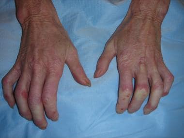
 Raynaud phenomenon showing pallor of most of the finger tips with a violaceous discoloration (hyperemia) of the thumb tip.