The science of the stomata of plants: a continuously growing list of references, abstracts and illustrations, helping researchers to data on publications.
Foliar architecture and micro-morphological characteristics of Diospyros lanceifolia Roxburgh (Ebenaceae) were studied.
Qualitative and quantitative characters of veins, vein order, areolation, epidermal cell and cell wall nature, length, breadth and number of epidermal cell; stomatal distribution, type, stomatal frequency and stomatal index were taken into consideration.
The observed characters were found useful in distinguishing the species in the genus Diospyros Linnaeus of Ebenaceae.
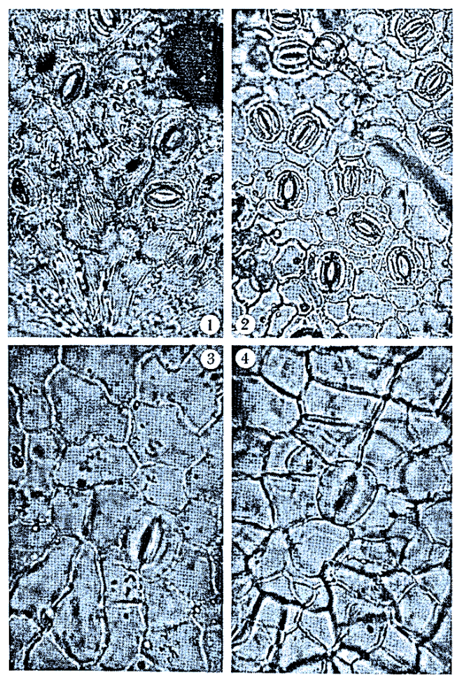
Fig. 1. D. canaliculata (adaxial): epidermal cells irregular, anticlinal cell walls slightly wavy (×400). Fig. 2. D. canaliculata (abaxial): epidermal cells irregular, anticlinal cell walls wavy (×400). Fig. 3. D. abysinica (abaxial): anticlinal cell wall straight to curved, stomata characteristically abundant (×400). Fig. 4: D. barteri (adaxial): anticlinal cell wall straight to curved (×400). Fig. 5: D. conocarpa (adaxial): anticlinal cell wall slightly wavy to wavy, epidermal cells the largest in the genus in Nigeria (×400). Fig. 6: D. conocarpa (abaxial): anticlinal cell wall slightly wavy to wavy, epidermal cells the largest in the genus in Nigeria (×400). Fig. 7. D. physocalysina (adaxial): anticlinal cell walls very wavy (×400). Fig. 8. D. tricolor (abaxial): anticlinal cell walls indistinct, being obscured by papillae (×400). Fig. 9. D. mannii (abaxial): cuticular striations obscured the anticlinal cell walls (×400). Fig. 10. D. elliotii (adaxial): the cell walls ornamented (×400). Fig. 11. D. elliotii (abaxial): the cell walls ornamented (×400). Fig. 12. D. gabunensis (adaxial): epidermal cells the smallest in the genus in Nigeria (×400)
Leaf epidermal morphology of Diospyros (Ebenaceae) in Nigeria
by Ugbogu O. A., Olowokudejo J. D., Ogundipe O. T. (2016)
Omokafe A. Ugbogu1, Jide D. Olowokudejo2 & Oluwatoyin T. Ogundipe2
1 Forestry Research Institute of Nigeria, Taxonomy Section, P.M.B. 5054, Ibadan, Oyo State, Nigeria,
2 University of Lagos, Botany Department, Lagos, Lagos State, Nigeria
The genus Diospyros L. belongs to Ebenaceae, with 30 species in Nigeria, that are either trees or shrubs, characterized by simple, estipulate, entire-margin leaves, without latex. The heartwood yields some commercial black ebony.
The observed variations in the comparative study of leaf epidermis included: cell shapes ranging from irregular, polygonal to isodiametric; anticlinal cell wall pattern which is straight, curved, slightly wavy, wavy, or very wavy; cell wall thickness between 1.30 µm to 5.50 µm; cell wall ornamentations, present or absent; cuticular striations, present or absent; coronulated papillae, present or absent; stomata types, anomocytic, polycytic, cyclocytic to staurocytic; and trichome types, glandular to non-glandular.
A study on the leaf of 31 species of Shorea Roxb. (Dipterocarpaceae) was undertaken to investigate stomatal types which could provide additional characters in the classification of the genus.
Results show that there are four types of stomata in the genus and their distribution depends on the Sections. Cyclocytic stomata are found in species belonging to Sections Pachycarpae, Mutica and Ovalis, while species in Section Anthoshorea and Brachypterae possess the staurocytic type. Species in Section Shorea and Neohopea on the other hand have the hexacytic type while the anomocytic type of stomata is found only in Section Richetioides.
This character, therefore, supports the classification of the genus by Ashton (1982) which was made using morphological characters of the flowers and fruits.
Stomatal structure, ontogeny in vegetative and floral organs of 9 genera and 12 species of Cucurbitaceae are described. The stomatal types conform to aperigenous, monoperigenous, diperigenous, hemipara‐mesoperigenous and para‐mesoperigenous types of Fryns‐Claessens & Van Cotthem (1973).
Stomatal abnormalities such as contiguous stomata, single guard cells with or without pore, one and a half stomata, degeneration of one or both the guard cells, cytoplasmic connections between guard cells of neighbouring stomata and a guard cell of a stoma and an adjacent epidermal cell, and division of guard cells are described.
Stomata index, frequency of stomata, epidermal cells, size of guard and epidermal cells and organwise distribution of stomata are given.
Stomatal studies does not support the view that the Cucurbitaceae are related to the Passifloraceae. The inclusion of 9 genera and 12 species studied in the tribe Cucumerineae is justified.
Fig. 2. Abaxial surface of Ipomoea species. A) I. triloba, B) I. eriocarpa, C) I. alba, D) I. hederifolia, E) I. muricata, F) I. intrapilosa, G) I. asarifolia, H) I. batatas, I) I. aquatica, J) I. mauritiana, K) I. carnea, L) I. involucrata, M) I. nil, N) I. heterotricha, O) I. obscura; BP = Brachyparacytic stoma, SC = Scale, L = Laticifer, ST = Stoma, GP = Granular periclinal wall, C = Crystal, S = Striae, TB = Trichome base, ET = Eglandular trichome, Scale = 0.85 μm
Taxonomic Evaluation of Fifteen Species of Ipomoea L. (Convolvulaceae) from South-Western Nigeria using Foliar Micromorphological Characters
by Folorunso A. (2013)
Abayomi Ezekiel FOLORUNSO
Obafemi Awolowo University, Department of Botany, Ile-Ife, Nigeria
Foliar micromorphological study was carried out on I. triloba, I. eriocarpa, I. alba, I. hederifolia, I. muricata, I. intrapilosa, I. asarifolia, I. batatas, I. aquatica, I. mauritiana, I. carnea, I.involucrata, I. nil, I. heterotricha and I. obscura to determine the patterns of variation in their epidermal characteristics and search for useful and stable anatomical characters for the identification of the species.
Leaf epidermal features that provided useful specific distinctions are cell shape, anticlinal wall pattern, stomata shape, stomata type, trichome, cuticular striations, crystal, granular periclinal wall, stomata size, stomata index, scale and tannin.
The characters revealed interrelationships among the Ipomoea species and also suggest their monophyly.
Stomatal apparatus
Stomatal shape is largely elliptic in most of the species but oval in I. batatas, I. carnea and circular in I. alba and I. involucrata. Brachyparacytic stomata predominantly oc- cur on both surfaces of the species except in I. hederifolia,. I. nil, I. eriocarpa, I. muricata and I. heterotricha where the stomata are paracytic.
Stomatal index
On the abaxial surface, the highest stomata index was in I. involucrata with the lowest in I. nil. Similarly, on the adaxial surface, the highest stomatal index was in I. alba with the lowest in I. triloba.
Stomatal size
I. triloba has the largest stomatal size while I. mauritiana has the smallest stomatal size on the abaxial size. On the adaxial surface, I. eriocarpa has the largest stomatal size while I. carnea and I. muricata are with the smallest stomatal size.
The present paper contributes to the anatomy of the family Convolvulaceae. In all, six genera and 10 species have been surveyed for epidermal features in detail.
They have been investigated covering stomatal type, index, frequency and number of subsidiaries, cell wall contours and cuticular striations. The leaves are amphistomatic and have commonly paracytic types, apart from rare to occasional occurrence of other stomatal types and abnormalities.
The epidermal cell walls on adaxial (a–f) and abaxial leaf surfaces (g–l) under light microscope (LM). a A. breviscapa, b A. kerrii, c A. suddeeana, d A. variabilis and f Argyreia 2, straight to slightly curved walls. e Argyreia 1, sinuate walls. g A. breviscapa and j A. variabilis, straight to slightly curved. h A. kerrii, i A. suddeeana, k Argyreia 1 and l Argyreia 2, sinuate (ep epidermal cell, st stomata, tr trichome). Scale bar 50 µm
Identification among morphologically similar Argyreia (Convolvulaceae) based on leaf anatomy and phenetic analyses
by Traiperm P., Chow J., Nopun P., Staples G., Swangpol S. C. (2017)
Paweena Traiperm,1 Janene Chow,1,2 Possathorn Nopun,1,2 G. Staples,3 and Sasivimon C. Swangpol1
1 Department of Plant Science, Faculty of Science, Mahidol University, Rama VI Road, Ratchathewi, Bangkok, 10400 Thailand
2 Department of Pharmaceutical Botany, Faculty of Pharmacy, Mahidol University, Bangkok, 10400 Thailand
3 HRA, Herbarium, Royal Botanic Gardens, Kew, Richmond, Surrey TW9 3AE UK
===
in Bot Stud. 58: 25 – doi: 10.1186/s40529-017-0178-6 –
The genus Argyreia Lour. is one of the species-rich Asian genera in the family Convolvulaceae. Several species complexes were recognized in which taxon delimitation was imprecise, especially when examining herbarium materials without fully developed open flowers. The main goal of this study is to investigate and describe leaf anatomy for some morphologically similar Argyreia using epidermal peeling, leaf and petiole transverse sections, and scanning electron microscopy. Phenetic analyses including cluster analysis and principal component analysis were used to investigate the similarity of these morpho-types.
Results
Anatomical differences observed between the morpho-types include epidermal cell walls and the trichome types on the leaf epidermis. Additional differences in the leaf and petiole transverse sections include the epidermal cell shape of the adaxial leaf blade, the leaf margins, and the petiole transverse sectional outline. The phenogram from cluster analysis using the UPGMA method represented four groups with an R value of 0.87. Moreover, the important quantitative and qualitative leaf anatomical traits of the four groups were confirmed by the principal component analysis of the first two components. The results from phenetic analyses confirmed the anatomical differentiation between the morpho-types.
Conclusions
Leaf anatomical features regarded as particularly informative for morpho-type differentiation can be used to supplement macro morphological identification.
Type of stomata and stomatal index
All of the morpho-types have amphistomatous leaves. The stomata on the adaxial leaf surface are anisocytic and paracytic in most of the morpho-types, excluding A. kerrii and A. variabilis, in which only the paracytic stomatal type was found. Conversely, a paracytic stomatal type presents on the abaxial leaf surface of every morpho-type, whereas A. breviscapa possesses both anisocytic and paracytic stomata (Table 3). On the adaxial epidermis the highest stomatal index was observed in A. breviscapa and the lowest in A. variabilis. On the other hand, the highest stomatal index on the abaxial epidermis was found in A. variabilis and the lowest in Argyreia 1 (Table 3).
Epidermis under LM. Sinuous or straight to curved anticlinal wall and stomata occurring at the adaxial surface of E. alsinoides (a), E. glomeratus (b) and E. nummularius (c). Y-shaped hair on adaxial surface of E. alsinoides from Khon Kaen (d). Y-shaped hair and capitate gland of E. glomeratus (e), capitate gland on leaf surface of E. nummularius (f). Sinuous or straight to curved anticlinal walls on abaxial epidermal layer of E. alsinoides (g), E. glomeratus (h) and E. nummularius (i). Evolvulus alsinoides (j) and E. glomeratus (k) possess the Y-shaped hairs and capitate glands, but E. nummularius (l) has no hairs. ep epidermis, tr trichome, g capitate gland
Micro-morphological study of Evolvulus spp. (Convolvulaceae): the old world medicinal plants
by Ketjarun K., Staples G. W., Swangpol S. C., Traiperm P. (2016)
Several medicinal properties have been reported for plants in the genus Evolvulus, such as a brain tonic and antifungal from Evolvulus alsinoides, and a sedative and an anthelmintic from Evolvulus nummularius. Therefore, the correct identification of the source plants is critically important. The aim of this research was to investigate the micromorphology of two Evolvulus taxa used for herbal medicines compared with one worldwide ornamental species by using peeling, paraffin embedding, acetolysis, and SEM methods in order to support species identification.
Results
Our findings indicate that all taxa share several common features, such as a single layer of epidermis on both sides of leaf surfaces, sinuous anticlinal epidermal cell walls, anomocytic, paracytic or laterocytic stomata, and capitate glandular trichomes. Y-shaped hairs were found in two species but not in E. nummularius. Similarly, isobilateral mesophyll occurs in both E. alsinoides and Evolvulus glomeratus, but a dorsiventral mesophyll is present in E. nummularius. Stems consist of a single layer of epidermis, one to four chlorenchyma layers, one to seven layers of cortical cells and a bicollateral bundle with pith in the center. The seed coat epidermal cell shapes were irregular or polygonal with raised and undulated anticlinal boundaries, and folded or flattened to concave periclinal walls. Pollens of all taxa are monads, spheroidally shaped with 28–47 µm diameter, and 15-pantocolpate apertures type with microechinate ornamentation.
Conclusions
An identification key to species is constructed based on leaf anatomy and seed coat characters. This data can be used in other subjects such as pharmaceutical botany, organic chemistry, taxonomy and horticulture, in terms of species identification.
A comprehensive study of the nodal and leaf anatomy of the Connaraceae was undertaken to provide additional evidence for evaluation in relation to the systematics of this tropical family.
The fundamental nodal pattern in both compound and simple-leaved genera is trilacunar, three-trace, whereas the multilacunar nodes in Connarus, Jollydora, and Bernardinia are derived. The prevailing petiole vasculature is a complete medullated cylinder with conditions of accessory petiole vascular tissue representing evolutionary advancement.
Characters of the lamina that show variation and are useful in generic delimitation include : mature stomatal type, trichome form, occurrence of mucilaginous epidermal cells, occurrence of epidermal papillae, presence of a hypodermis, degree of cuticularization, and the nature of high-order venation. Particularly noteworthy features are the terminal sclereids of Pseudoconnarus and the unusual closed venation pattern of Manotes.
Foliar anatomy is very consistent with the placement of the family near the Rosaceae and Leguminosae. The intrafamilial systematics of the family are discussed in light of information derived from vegetative anatomy.
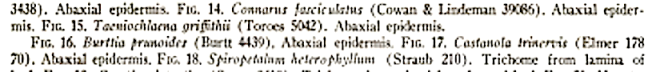
Fig. 6. Hairs, leaf epidermis, and stomata: 1. Manotes macrantha, cuticula with many more or less parallel lines, x 380; 2. Manotes expansa, stomata and glandular hair, x 3$0;3.Vismianthuspunctatus, stoma, x 950; 4. Burttia prunoides, stoma, x 950. (1. Reitsma 1728; 2. Breteler & Lemmens 8377; 3. Semsei647; 4. Peter 34193A).
The Connaraceae, a taxonomic study with special emphasis on Africa
by Jongkind C. C. H., Lemmens R. H. M. J. (1989)
Carel Christiaan Hugo Jongkind, Roeland Hendrikus Maria Julien Lemmens,
In the general part of this work the author of this thesis treats the leaves, flowers and fruits of Connaraceae, Additional chapters reflect upon the phytogeny of the genus Rourea, the phytochemistry of the family, and the relationship of Connaraceae with other families. In the taxonomic part a description of the family is presented together with keys to the genera. A revision of the genera Agelaea, Manotes, and Rourea is presented with special emphasis on Africa, including descriptions of the genera and differential keys to the species. In the revision of the genus Agelaea six species are recognized including the Asiatic species. Manotes is only found in Africa and comprises five species. The genus Rourea is revised for Africa only, twelve species are recognized for this area. The American and Asiatic species are studied but not revised. New combinations are made for American and Asiatic species that are transferred to this genus. The American species are classified into clusters. In this work Rourea also includes Bernardinia, Byrsocarpus, Jaundea, Roureopsis, Santaloidella, Santaloides, Spiropetalum, and Paxia, formely recognized as separate genera. The treatment of the African species comprises full synonymy, literature, diagnosis, a distribution map, and ecological notes. In most cases an illustration is presented as well. The author studied and collected most species of Agelaea, Manotes, and Rourea on location in the rain forest in Gabon.
4.5 Leaf epidermis and stomatal patterns by C.C.H. Jongkind
4.5.1 Introduction
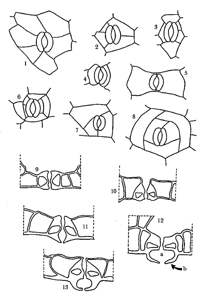
In Connaraceae the pattern of the stomata in the epidermis is a useful additional taxonomic character. It is often rather easy to recognize and it enables us, in combination with other leaf characters, to identify sterile samples down to species or at least to genus level (see tab. 23). It has been an important tool in the clustering of the South American species of Rourea (presented on pag. 368). It also reflects affinities between the taxa. Apart from the stomata the leaf epidermis ofConnaraceae provides four other characters of taxonomical value that are used in this revision.
4.5.3 Results The stomatal patterns encountered in Connaraceae are of diverse nature. The terminology by Wilkinson (1979) is used to describe the stomata. A paracytic pattern is found in Cnestis and some species of Rourea, anisocytic including helicocytic in Agelaea, Cnestidium, and some species of Rourea, anomo-cyclocytic in Burttia, Connarus, Ellipanthus, Hemandradenia, Manotes, Vismianthus, and again some species of Rourea, and bicyclic in Jollydora (fig. 6-14). Rourea is the only genus with a wide variation in stomatal patterns, therefore the results for Rourea are not given for the genus but in detail for sections or for particular species. Apart from the stomatal pattern the size of the vestibule as expressed by the size of the outer ledge of the stomata can also be an important character. This ledge is particularly large and very conspicuous in all Connarus species and in Rourea section Roureopsis. Within the Connaraceae it seems that the sequence from primitive to advanced stomata types is either from anomo-cyclocytic to bicyclic or from anomo-cyclocytic via paracytic to anisocytic and helicocytic. The presence of a papillate lower surface is not always of taxonomical significance. This character constitutes a generic difference between Connarus and Pseudoconnarus, but in Cnestis and Rourea some species are quite indifferent regarding this character. A pattern of more or less parallel lines in the lower epidermis has only been found in two species of Rourea, R. calophylla and R. camptoneura and in Manotes macrantha. The taxonomical value of the pattern of many microscopical pits in the outer cell walls of the lower epidermis is still obscure. It is only found in the leaves of Rourea prancei, at present exclusively represented by its type specimen. If only a single stomata type is reported, it means that 95-100% of the observed stomata of this taxon is of that type.


 1 Janene Chow,1,2 Possathorn Nopun,1,2 G. Staples,3 and Sasivimon C. Swangpol1


You must be logged in to post a comment.