Stomata in Verbenaceae (dicots)

Photo credit: Google

Gmelina arborea

Epidermal structure and ontogeny of stomata in some Verbenaceae

by Inamdar J. A. (1969)

Department of Botany, Sardar Patel University, Vallabh Vidyanagar, India


in Annals of Botany. 71(4) 323-370. –

Epidermal structure and ontogeny of stomata, Ani Mulyani.pdf

tfri_herb_345220000G__TBG7158
Photo credit Google: Stachytarpheta jamaicensis (L.) Vahl. – http://knowledge.taibif.tw/sites/default/files/imagecache/node-gallery-display/tfri_herb_345220000G__TBG7158.jpg

Abstract

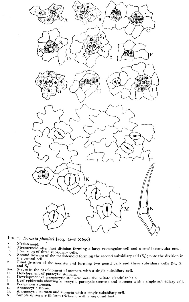
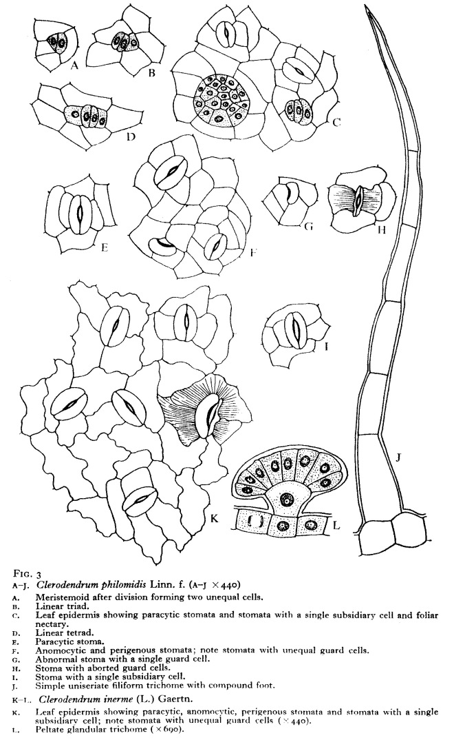
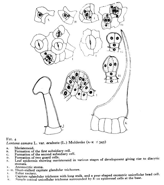
The present paper deals with epidermal structure and development of stomata in 14 species of Verbenaceae.

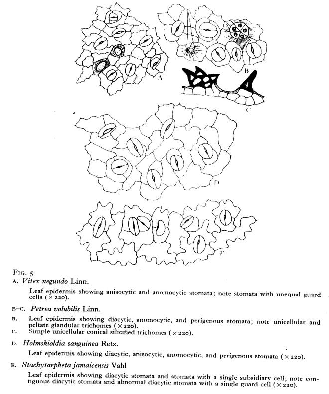
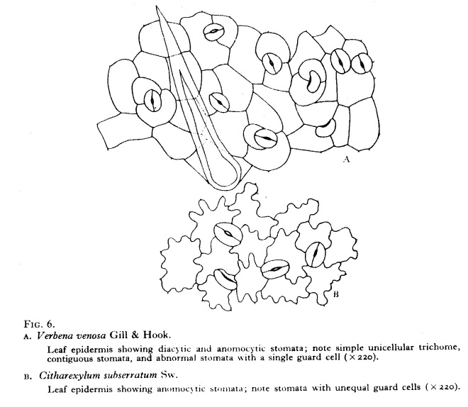
The epidermal cells are either polygonal, isodia- metric, or elongated in various directions, and irregularly arranged. The anti- clinal walls are thick, mostly sinuous, occasionally arched or straight. The surface of the cuticle shows parallel, rarely corrugated, striations. Some 12 types of eglan- dular and glandular trichomes, and foliar nectaries are noticed.

The mature stomata are diacytic, anisocytic, paracytic, with a single subsidiary cell, anomocytic and perigenous.

The development of anomocytic stomata is perigenous, while that of others is mesogenous or syndetocheilic type.

Abnormalities noticed here include contiguous stomata, stomata with a single guard cell, and aborted guard cells.


Scan

Scan 1

Scan 2

Scan 3

Scan 4

Scan 5

Scan 6


Stomata in trees of 17 dicotyledonous families


Epidermal studies in some Nigerian trees

by Nyawuame H. G. K., Adedio M. U., Gill L. S. (1993)

(With one Table) –

Feddes Repertorium Volume 104, Issue 7-8, 1993, 475–480 – DOI: 10.1002/fedr.19931040710 – 

http://onlinelibrary.wiley.com/doi/10.1002/fedr.19931040710/full

Abstract

Leaf epidermal structure of 30 trees distributed in 17 dicotyledenous families was studied.

Interspecific variation in cell wall pattern was observed in the two species of Macaranga covered in this study. The cells were straight-walled in M. barteri but sinuously-walled in M. monandra. In addition to this variation, peltate hairs were observed in M. barteri.

The two species also varied in their stomatal features. Alchornea was another species which showed interspecific variation in epidermal and stomatal features. Stellate hairs were observed in A. cordifolia but were absent in A. laxiflora.

Mature stomata were paracytic in A. cordifolia but in A. laxiflora they were anomocytic. Differences in stomatal size and frequency were also observed in the two species. Contiguous stomata were reported here for Avicennia africana, Mitragyna inermis and Pterygota macrocarpa.

Stomata in Convolvulaceae (dicots)


Cuticular studies of some species of Convolvulaceae used in traditional medicine in West Africa

by Gill L. S., Nyawaume H. O. K. (1991)

in Feddes Repertorium Volume 102, Issue 3-4, 1991, 189–198 – DOI: 10.1002/fedr.19911020308 – 

http://onlinelibrary.wiley.com/doi/10.1002/fedr.19911020308/full

Abstract

Epidermal structure and ontogeny of stomata in 30 species of the family Convolvulaceae from West Africa are described.

The epidermal cells are irregularly shaped with arched or sinuous anticlinal and periclinal walls in most of the taxa studied. The cells of Calycobolus parviflorus (Mangenot) Heine and Merremia pterygocaulos (Steud. ex Choisy) Hallier f. are striated. The trichomes are variable: peltate glandular and uniseriate eglandular types have been recorded. Septate and unbranched trichomes have been observed in Convolvulus aschersonii Engl.

The leaves are amphistomatic in the majority of the taxa presently investigated. However, hypostomatic leaves are recorded in Evolvulus alsinoides L., Ipomoea leari Paxt., I. nil (L.) Roth, I. pyrophila A. Chev., I. setosa Ker-Gawl. and Lepistemon owariense (P. Beauv.) Haller f. Anisocytic stomata in C. parviflorus are reported here for the first time.

Stomatal abnormalities viz: contiguous stomata in Convolvulus aschersonii and Ipomoea heterotricha Didr., aborted stomata in I. hederacea Jacq. and single guard-celled stomata in C. parviflorus are reported here for the first time.

Stomata in some Fabaceae from Nigeria


Observations on the cuticular features and stomatal ontogeny in the leaves of some Papilionaceae from Nigeria

by Karetala Y. Y., Hussain H. S. N., Nurani M. A. (1990)

in Feddes Repertorium Volume 101, Issue 3-4, 1990, 183–188 – DOI: 10.1002/fedr.19901010312 – 

http://onlinelibrary.wiley.com/doi/10.1002/fedr.19901010312/full

Abstract

Epidermal morphology and ontogeny of stomata in the leaves of 21 species of family Papilionaceae have been reported.

Stomata are confined on both surfaces in the species of Crotalaria, Indigofera and Mucuna and only on a lower surface in the rest of the species.

Inspite of stomatal diversity even on the same surface, the most prevalent type observed is paracytic with eumesogenous and hemimesogenous ontogeny. Anisocytic stomata with hemimesogenous ontogeny is typical of Crotalaria and anomocytic with agenous ontogeny is typical of Indigofera.

Stomata in Boraginaceae (dicots)


Epidermal morphology and ontogeny of stomata in some tropical Boraginaceae

by Nyawuame H. G. K., Gill L. S. (1990)

in Feddes Repertorium Volume 101, Issue 5-6, 1990, 289–295 – DOI: 10.1002/fedr.19901010513 – 

http://onlinelibrary.wiley.com/doi/10.1002/fedr.19901010513/full

Abstract

Epidermal morphology, the structure and development of stomata in 13 species of Boraginaceae are described.

The epidermal cells are straight-walled on the upper surface and sinuous on the lower in the majority of the investigated species.

The leaves are hypostomatic with either anomocytic or anisocytic stomata distributed on the lower surface.

The anomocytic stomata follow agenous ontogenetic pathway while the anisocytic stomata are hemimesogenously derived.

Laterally contiguous stomata in Cordia platythyrsa are recorded for the first time.

Stomata in Scrophulariaceae (dicots)


Cuticular studies and their bearing on the taxonomy of some tropical Scrophulariaceae

by Nyawuame H. G. K., Gill L. S. (1993)

(With 58 Figures and one Table)

in Feddes Repertorium Volume 104, Issue 1-2, 1993, 41–50 – DOI: 10.1002/fedr.19931040108 – 

http://onlinelibrary.wiley.com/doi/10.1002/fedr.19931040108/full

Abstract

The structure of the epidermis and the ontogeny of the stomatal complex of 26 tropical taxa of the family Scrophulariaceae are described.

The epidermal cells depict variations with the cells being straight-walled on both surfaces, straight-walled on the upper surface only or sinuously walled on both surfaces. Trichomes are also variable. The different types of trichomes observed include unicellular eglandular, peltate glandular and multicellular eglandular.

All the investigated species except Lindernia crustacea and Veronica abyssinica are hypostomatic.

During the present study three types of stomata are recorded, viz: anomocytic, anisocytic and diacytic types. Anomocytic stomata follow agenous or hemiperigenous ontogenetic development whereas the ontogeny of anisocytic stomata is either hemiperigenous or hemimesogenous.

Within limitations stomatal distribution and frequency could serve as reliable taxonomic characters in the tropical taxa of the family Scrophulariaceae.

Stomata in Apocynaceae (dicots)

Cuticular studies of some West African species of the Apocynaceae of medicinal value

by Nyawuame H. G. K., Gill L. S. (1991)

in Feddes Repertorium Volume 102, Issue 1-2, 1991, 87–104 – DOI: 10.1002/fedr.19911020110 – 

http://onlinelibrary.wiley.com/doi/10.1002/fedr.19911020110/full 

Abstract

A comparative study of the leaf epidermis of 45 West African Apocynaceous taxa using the light microscope has been undertaken.

Variations observed in anticlinal cell wall pattern, trichomes, size, distribution, frequency, morphology and ontogeny of the stomatal complex are discussed.

Aborted stomata and single guard-celles stomata are reported in Allamanda and Thevetia for the first time.

Stomata in Bicarpellatae (dicots)


Phylogenetic and Systematic Value of Stomata in Bicarpellatae (BENTHAM et HOOKER sensu stricto) Dedicated to Prof. W. R. J. VAN COTTHEM

My sincere thanks for this dedication

by Gill L. S., Nyawaume H. O. K. (1990)

L. S. GillUniversity of Benin (Benin City, Nigeria)

H. G. K. Nyawuame, University of Benin (Benin City, Nigeria)


===

in Feddes Repertorium Volume 101, Issue 9-10, 1990, 453–498 – DOI: 10.1002/fedr.19901010906 – 

http://onlinelibrary.wiley.com/doi/10.1002/fedr.19901010906/full

Abstract

Foliar epidermal morphology and ontogeny of stomata are described for 320 taxa distributed in 13 families of the Bicarpellatae.

The epidermal cells are generally straight-walled in the woody taxa but sinuous in herbs and small shrubs.

The different stomata types commonly described in the dicotyledons viz: anomocytic, anisocytic, paracytic and diacytic have been recognized during the present study.

The families Acanthaceae and Labiatae are remarkable in showing only diacytic stomata generally regarded as the most advanced. The distribution, size and frequency of stomata could be used to establish systematic divisions and to suggest phylogenetic relationships within the Bicarpellatae.

Ontogeny of stomata is uniform only in the family Oleaceae but heterogenous in all the other families. Evidence from stomatal ontogeny therefore appears to be taxonomically important only at the genus level.

Single guard-celled stomata, aborted stomata and contiguous stomata are of regular occurrence in the presently investigated families except the family Scrophulariaceae which showed no stomatal abnormalities.

Adjacent stomata with cytoplasmic connections between their guard cells are of rare occurrence and such stomata are recorded in only six taxa distributed in the families Gentianaceae, Oleaceae and Solanaceae.

Stomata in Solanaceae (dicots)


Epidermal studies of some species of family Solanaceae used in traditional medicine in West Africa

by Nyawuame H. G. K., Gill L. S. (1994)

in Feddes Repertorium Volume 105, Issue 1-2, 1994, 49–60 – DOI: 10.1002/fedr.19941050112 – 

Abstract

Leaf epidermal structure and developmental pattern of the stomatal complex of 30 species of the family Solanaceae are described.

Epidermal cells of almost all the species showed wall sinuousity. However, in Brunfelsia uniflora and some species of the genus Solanum viz. S. aethiopicum, S. dasyphyllum, S. macrocarpon and S. verbascifolium the cells were straight-walled. Several types of epidermal hairs were observed on both the upper and lower epidermis. They include bicellular eglandular, multicellular stellate and club-shaped hairs.

The leaves of most species were amphistomatic; however, a few species with hypostomatic leaves were also encountered.

Mature stomata were either anomocytic or anisocytic with hemimesogeneous developmental pattern. In Schwenkia americana, however, mature stomata were diacytic with eumesogenous developmental pathway.

Interspecific variation observed in the distribution, size and frequency of stomata may prove useful in the authentication of the foliar materials of these plants when used as crude drugs.

Stomata in Bougainvillea (dicots)


Epidermal studies in some Indian cultivars of Bougainvilleas

by Inamdar J. A., Gandadhara M., Avita Sr., Rao N. V. (1980)

in Feddes Repertorium Volume 91, Issue 4, 1980, 259–266 – DOI: 10.1002/fedr.19800910405 – 

http://onlinelibrary.wiley.com/doi/10.1002/fedr.19800910405/full

Abstract

Epidermal studies in fifteen Indian cultivars of Bougainvilleas are described.

The epidermal cells are polygonal isodiametric, or elongated with thick straight arched or slightly sinuous walls. Parallel culticular striations are radiating from guard cells.

The mature stomata are anomocytic, paracytic and with a single subsidiary cell.

The abnormal types noticed are: single guard cells with or without pores, arrested development, variously oriented contiguous stomata, cytoplasmic connections between nearby stomata and epidermal or subsidiary cells, and persistent stomatal cells.

The development of anomocytic stomata is perigenous while that of the other types is mesogenous.

Fifteen cultivars of Bougainvilleas are separated on the basis of bract colour, stomatal frequency and index per unit area.