
Therapeutic Agent-Loaded Fibrous Scaffolds for Biomedical Applications


1
Drug Manufacturing Center, Daegu-Gyeongbuk Medical Innovation Foundation (K-MEDI Hub), Daegu 41061, Republic of Korea
2
New Drug Development Center (NDDC), Daegu-Gyeongbuk Medical Innovation Foundation (K-MEDI Hub), Daegu 41061, Republic of Korea
3
Medical Device Development Center, Daegu-Gyeongbuk Medical Innovation Foundation (K-MEDI Hub), Daegu 41061, Republic of Korea
*
Author to whom correspondence should be addressed.
Pharmaceutics 2023, 15(5), 1522; https://doi.org/10.3390/pharmaceutics15051522
Submission received: 5 April 2023
/
Revised: 28 April 2023
/
Accepted: 12 May 2023
/
Published: 17 May 2023
(This article belongs to the Special Issue Fiber-Based Scaffolds as Drug Carriers: Recent Advances)
Abstract
:Tissue engineering is a sophisticated field that involves the integration of various disciplines, such as clinical medicine, material science, and life science, to repair or regenerate damaged tissues and organs. To achieve the successful regeneration of damaged or diseased tissues, it is necessary to fabricate biomimetic scaffolds that provide structural support to the surrounding cells and tissues. Fibrous scaffolds loaded with therapeutic agents have shown considerable potential in tissue engineering. In this comprehensive review, we examine various methods for fabricating bioactive molecule-loaded fibrous scaffolds, including preparation methods for fibrous scaffolds and drug-loading techniques. Additionally, we delved into the recent biomedical applications of these scaffolds, such as tissue regeneration, inhibition of tumor recurrence, and immunomodulation. The aim of this review is to discuss the latest research trends in fibrous scaffold manufacturing methods, materials, drug-loading methods with parameter information, and therapeutic applications with the goal of contributing to the development of new technologies or improvements to existing ones.
1. Introduction
Fibrous scaffolds exhibit excellent characteristics, including adjustable biodegradability, appropriate mechanical properties, and high porosity, which make them promising materials for use in regenerative medicine [1,2,3]. In addition to providing a suitable environment for cell growth [4], these scaffolds offer flexibility for drug loading [5], enabling them to be used as implantable [6] and degradable drug carriers [7] for treating diseases and repairing tissues. The local controllable release feature of these fibrous scaffolds further enhances their potential for targeted drug delivery, making them a promising option for a range of biomedical applications.
Natural materials are widely used to fabricate fibrous scaffolds for tissue engineering and regenerative medicine because of their biocompatibility and ability to mimic the extracellular matrix (ECM) of natural tissues [8,9]. Among the various natural materials, polysaccharides [10], alginate [11], and proteins such as collagen [12] have been widely explored for the fabrication of fibrous scaffolds. These natural materials are biocompatible and are less likely to cause an immune response or toxicity in the body, making them suitable for use in tissue engineering and regenerative medicine. Polysaccharides and proteins are the main components of ECM in natural tissues, and their use in scaffolds can provide a similar microenvironment to support cell attachment, proliferation, and differentiation. Moreover, the physical and chemical properties of natural materials can be tailored to fabricate a scaffold with specific properties, such as porosity, stiffness, and degradation rate, which can influence cell behavior and tissue regeneration.
Among the several methods available for fabricating nanofibrous membranes, electrospinning shows significant potential because it allows the incorporation of drugs into the membrane, which can then be released at a controlled rate over different time periods [13]. Electrospinning is a simple yet effective technique for producing fibrous materials in the micrometer to nanoscale range and can be utilized in various applications such as tissue regeneration, drug delivery, and wound dressing. Three-dimensional (3D) printing technology allows the manipulation of individual 3D shapes with high precision, including the fabrication of fiber scaffolds [7]. This approach has several advantages, such as the ability to produce complex, customized, and patient-specific designs, as well as precise control over properties such as pore size, interconnectivity, and mechanical characteristics.
Microfluidic technologies have shown promise for producing micro- and nanoscale fiber structures for a range of applications without requiring complex equipment or precise control over microflows [14].
To achieve a desired therapeutic effect, drugs or bioactive molecules can be incorporated into fibrous scaffolds via a process known as drug loading [15]. When selecting a fiber matrix for drug loading, it is crucial to consider the interactions between the drug and the fiber scaffolds. Covalent bonds, hydrogen bonds, and electrostatic interactions can be used to link the drug to the fibers.
In this review, natural fiber materials, methods for fabricating fibrous scaffolds, loading methods, and state-of-the-art applications of fabricated fibrous scaffolds are discussed.
2. Natural Materials for Fibrous Scaffolds
Tissue-engineering scaffolds are designed to temporarily replace the native ECM, thus providing mechanical support for cells and regulating cell behavior. The ECM is primarily composed of collagen, elastin, glycoproteins, and polysaccharides; therefore, natural polymers, such as proteins and polysaccharides, have been extensively studied for scaffold production using various methods.
2.1. Polysaccharides
Natural polysaccharides such as arginate, chitosan, and hyaluronic acid are highly valued in biomedical science owing to their outstanding properties [9]. (Figure 1) Compared to other polymers, they are easily customizable, biocompatible, bioactive, homogeneous, and possess bioadhesive properties. Moreover, they can replicate the ECM microenvironment, making them superior to other materials. Polysaccharides are natural polymers that have been extensively studied for the production of fibrous scaffolds in tissue engineering. One of their advantages is that they are naturally occurring molecules in the human body, making them well-tolerated by host tissues and reducing the likelihood of an immune response or adverse reactions. In addition, polysaccharides are often biodegradable, indicating that they can be safely broken down by enzymes in the body. The abundance and diversity of polysaccharides, which are available from various sources such as plants, animals, and microorganisms, allow the production of scaffolds with unique mechanical, chemical, and biological properties [16]. All these factors make polysaccharides promising materials for scaffold production in tissue engineering.
2.1.1. Alginate
Alginate is a natural polymer derived from brown seaweed that is widely used in tissue engineering because of its unique properties. Alginate is composed of two types of monomers, guluronic acid and mannuronic acid, arranged in different ratios, depending on the seaweed source [22] (Figure 1a). One advantage of alginate as a natural material for fibrous scaffolds is its biocompatibility and non-toxicity, as it is naturally derived and does not cause an immune response [23]. Alginate hydrogels can also be easily modified with various chemical groups or bioactive molecules to improve their properties and mimic the ECM. Alginate-based biomaterials can be further modified by combining them with other biomaterials, immobilizing specific ligands (such as peptides and sugar molecules), and subjecting them to crosslinking treatments. These modifications allow for the fine-tuning of the structural and functional properties of alginate, including its biodegradability, mechanical strength, gelation behavior, and cell interactions [24]. Alginate-based biomaterials can potentially function as drug delivery vehicles [25] and cell carriers [26] in tissue-engineering applications.
2.1.2. Chitosan
Chitosan, a natural polymer derived from chitin (Figure 1b), is often used to fabricate fibrous scaffolds for tissue-engineering applications. Its unique properties such as biocompatibility, biodegradability, and antimicrobial activity make it a promising candidate for tissue engineering applications [27]. Chitosan can be processed into fibrous scaffolds using various techniques such as electrospinning [28] and gas foaming [29], which results in a 3D structure that mimics the ECM tissues. Moreover, chitosan can form blends with other polymers to form scaffolds with the desired properties. The cationic nature of chitosan enables it to form polyelectrolyte complexes with anionic glycosaminoglycans, which have been shown to regulate the activity of growth factors and cytokines, making them suitable for bone and cartilage regeneration [30]. Additionally, the degradation rate of chitosan can be adjusted by modifying its degree of deacetylation, enabling it to satisfy the regenerative requirements of different tissues [31]. The functional groups on chitosan also facilitate conjugation with biologically active molecules, such as adhesive proteins and peptides, to support cell growth, differentiation, and proliferation in various fields, including dentistry, ophthalmology, veterinary science, biomedicine, the drink industry, and biotechnology [32].
2.1.3. Hyaluronic Acid
Hyaluronic acid (HA) is a polysaccharide naturally found in the body (Figure 1c), particularly in the ECM of connective tissues, including the skin, cartilage, and connective tissue. It exhibits good biocompatibility and minimizes the immune response when implanted in the body. HA is degraded by hyaluronidase, which cleaves the glycosidic bonds between sugar molecules [33]. Hyaluronidase enzymes are present in various tissues of the body, including the liver, kidney, spleen, and lymph nodes, and degraded fragments can be metabolized and eliminated from the body through the lymphatic system.
Low-molecular-weight HA has been found to promote the proliferation and migration of vascular endothelial cells. Moreover, it has been reported that low-molecular-weight HA can stimulate the expression of signaling molecules such as ezrin, which is a crucial protein for cellular adhesion [33]. Furthermore, HA exhibits antioxidant properties, and it is hypothesized that high-molecular-weight HA may protect against the effects of reactive oxygen species. HA possesses antioxidative properties such as the ability to reduce ultraviolet (UV) light-induced apoptosis and acid-induced DNA damage [34]. These properties make HA an attractive candidate for use in various fibrous scaffolds, including wound healing, cartilage repair, and tissue regeneration.
2.2. Proteins
Proteins are naturally biodegradable materials that have garnered significant attention in the field of tissue engineering for scaffold production. Their remarkable biocompatibility and biodegradability, coupled with their degradation products and amino acids, make them prime candidates for nutrient resorption. Moreover, some proteins induce minimal tissue inflammatory responses [35].
Among proteins, fibrous proteins such as collagen are of particular interest for scaffold production because of their unique properties. Collagen is a natural component of the ECM in many tissues, including skin, bone, and cartilage [12]. This makes it highly biocompatible and facilitates cellular attachment and proliferation, making it an ideal material for tissue-engineering applications. These fibrous proteins exhibit highly repetitive amino acid sequences that primarily serve mechanical and architectural functions. Repetitive primary sequences give rise to relatively uniform secondary structures through self-assembly, resulting in the formation of fibrous structural materials. Such structures offer several benefits, including high mechanical strength, stability, and enhanced cellular attachment and proliferation, all of which are critical for tissue regeneration and repair [36].
2.2.1. Collagen
Collagen, an abundant protein in the human body (Figure 2), is a highly investigated biomolecule of the ECM because of its wide distribution in connective tissues and superior mechanical properties, which make it an ideal material for load-bearing tissues such as bones and tendons [37]. Collagen is a useful material for applications in drug delivery systems and tissue engineering, owing to its tensile strength, biomimetic nature, low antigenicity, and safety profile [38]. Collagen is biocompatible and is degraded by human collagenases. Moreover, cell–collagen interactions are naturally facilitated as specific peptide sequences within collagen molecules are recognized by cell receptors [39]. Therefore, collagen can support the growth and differentiation of various cell types, making it a versatile material for various tissue-engineering applications. In addition, the mechanical strength and degradation properties of collagen-based scaffolds can be controlled using various chemical, physical, and enzymatic treatments.
Furthermore, collagen can be easily extracted from natural sources such as bovine hide, pig skin, and human placenta, making it a readily available and cost-effective material for scaffold production [40]. In addition, marine organisms are being increasingly investigated as a potential source of collagen due to a lower risk of transmissible diseases [41]. Unfortunately, the fishing industry discards a significant amount of marine biomass, up to 85%, leading to environmental concerns [42]. However, extracting collagen from these wastes can offer both environmental and economic benefits. Marine collagens have unique properties, such as biocompatibility, reduced zoonotic and immunological risks, and fewer religious restrictions. Finally, collagen can be processed into various forms such as gels, fibers, and films, allowing for the fabrication of scaffolds with different properties and structures to suit specific tissue-engineering needs.

2.2.2. Silk Fibroin
Silk fibroin (SF) is a natural protein-based polymer that has attracted significant attention as a material for fibrous scaffolds in tissue engineering and regenerative medicine. Silkworms are commonly used in silk research and the most common silk originates from Bombyx mori, a mulberry-feeding silkworm [48]. Silkworm cocoons consist of SF, a load-bearing protein, and sericin, a gumming agent. Sericin is often removed from SF to ensure biocompatibility in tissue-engineering applications. The sodium carbonate degumming is the most commonly used method for sericin removal [49]. However, after degumming, the average diameter of silk fibers is reduced to 10–25 µm. Researchers have designed chimeric recombinant spider silk proteins that can produce large quantities of artificial spider silk in bacterial shake-flask culture.
SF is composed of heavy (H) and light (L) chains linked by disulfide bonds along with a glycoprotein called P25 [6] (Figure 3). The H-chain, L-chain, and P25 are three polypeptides that form the cocoon of Bombyx mori at a molar ratio of 6:6:1 [50]. The H-chain is rich in glycine and alanine with a repeating Gly-X dipeptide motif, including two hexapeptides with specific peptide sequences. SF has two crystalline structures, silk I and silk II, with silk II being the most stable owing to its strong hydrogen bonding [51]. SF also exists in an amorphous state with α-helices, turns, and random-coil structures. The unstable crystal structure of SF is silk III, which exists at the air–water interface of the regenerated SF solutions.
SF fibers from the Bombyx mori cocoons have exceptional mechanical properties, including large break strain, ultimate strength, and toughness, surpassing those of natural and synthetic fibers [52]. SF has been used as a scaffold material in load-bearing tissue-engineering applications, particularly in musculoskeletal tissue engineering [53]. However, SF scaffolds fabricated from regenerated SF (RSF) solutions are typically brittle and weak compared with raw SF fibers because of the lack of hierarchical and secondary structures [54]. Various strategies such as crosslinking, porogens [55], and 3D bioprinting [56] have been used to improve the mechanical properties of RSF-produced silk scaffolds, resulting in scaffolds with mechanical properties similar to those of the native tissue being repaired.
SF is a biologically inert and biocompatible natural polymer approved by the U.S. Food and Drug Administration (FDA) for use as a biomaterial in 1993 [57]. SF has been shown to have blood compatibility and has been used successfully in cell culture to guide bone regeneration [58]. In vitro and in vivo studies have demonstrated that there is no significant immune reaction or inflammatory response to SF films or fibers [59], and SF has been shown to be a suitable alternative to collagen in some applications.

2.2.3. Elastin
Elastin is a fibrous protein that is predominantly found in the elastic tissues of the body, such as the skin, lung tissue, and blood vessels. Elastogenesis, which primarily occurs during the late fetal and early neonatal periods, is regulated by various cell types, including smooth muscle cells, fibroblasts, endothelial cells, chondroblasts, and mesothelial cells [65]. It is highly elastic and can stretch and recoil without deformation. Elastin-based scaffolds have been used in tissue-engineering applications such as vascular grafts and skin substitutes.
Tropoelastin is a water-soluble protein that serves as the precursor to the crosslinked elastin. It is composed of alternating hydrophobic and hydrophilic domains and has a molecular weight of approximately 72 kDa (Figure 4). After secretion into the ECM, tropoelastin interacts with microfibrillar scaffolds to facilitate elastic fiber formation [66]. Crosslinking is accomplished through an entropically driven process of coacervation, which causes tropoelastin molecules to aggregate with increasing temperature. This process is closely regulated to match the physiological conditions. Finally, copper-dependent lysyl oxidase is responsible for the crosslinking of tropoelastin [67]. Elastin can withstand billions of extension and recoil cycles without failure [68].
Various techniques have been explored to produce elastin-based molecules owing to the significance of elastin in tissues. These approaches include the extraction and purification of elastin from animal sources, expression of tropoelastin using recombinant methods, and manipulation of elastin-like peptides through engineering.
Elastin can be obtained from animal sources through the partial hydrolysis of the peptide chain using agents such as hot alkali, guanidine, oxalic acid, and potassium hydroxide [68]. Various methods yield different versions of soluble elastin with similar frameworks, including the removal of other macromolecules and fragmentation of elastin by cleaving peptide bonds [69]. The fragmented peptides possessed properties similar to those of tropoelastin, including the capacity to coacervate. However, the final product obtained from animal sources is a heterogeneous mixture of partially crosslinked elastin peptides with compromised biological properties because of the degradation and high levels of contaminants, depending on the protocol used [70].
Researchers have explored various methods for producing tropoelastin [71]. One approach involves obtaining tropoelastin from animal sources at the late fetal or neonatal stage before it is crosslinked into elastin by feeding newborn animals a copper-deficient diet, which is a critical cofactor for lysyl oxidase activity [72]. However, the extracted tropoelastin is more susceptible to degradation by proteases and has an overall positive charge owing to the presence of uncrosslinked lysine residues.
Recombinant human tropoelastin using Escherichia coli (E. coli) has been used as a model to study the behavior of human elastin and fabricated into tropoelastin-based biomaterials for tissue-engineering applications [73]. However, the extensive purification of proteins from bacterial systems is required. Elastin-like peptides (ELPs) have been studied as alternatives [74]. ELPs are synthesized by modifying the amino acid sequence, side chains, and synthesis, and can be used to control the chemical and biological properties of the material. ELPs have been shown to elicit minimal inflammatory responses when implanted in vivo and the addition of a cell-binding sequence to the ELP chain promotes cell adhesion, proliferation, and migration [75].
Figure 4.
Structural illustration of elastin. (Reprinted with permission from ref. [76]. Copyright 2019 Springer Nature).
Figure 4.
Structural illustration of elastin. (Reprinted with permission from ref. [76]. Copyright 2019 Springer Nature).

3. Fabrication Methods
Various methods, including electrospinning, 3D printing, and microfluidics, have been employed to fabricate fibrous scaffolds. Electrospinning is a versatile and widely used technique for producing fine fibers from polymer solutions or melts. Three-dimensional printing allows for the precise control over of scaffold geometry and ability to incorporate multiple materials and functional gradients. Three-dimensional printing can also be used to produce scaffolds with high mechanical strength and structural integrity. Microfluidics uses microscale channels and chambers to control the flow of fluids and deposition of fibers. It offers the advantages of high-throughput fabrication and the ability to create complex scaffold architectures. This section aims to provide a summary of the methods for the fabrication of fibrous scaffolds with a detailed classification and principles of the mentioned technologies, along with recent research examples.
3.1. Electrospinning
Over the past few decades, electrospinning has become a crucial method for fabricating 3D fibrous scaffolds with fiber diameters ranging from nanometers to micrometers. This technique is the most extensively studied and widely used method for scaffold fabrication. Electrospinning utilizes a high electric voltage to synthesize nanofibers, and a typical electrospinning setup consists of a syringe pump, power supply, metallic needle, and metallic collector [13]. The electric field causes the polymeric solution to flow in the direction of the electric field and generates ultrafine fibers from the tip of the metallic needle, which are collected on the collector drum. The diameters of the resulting fibers range from nanometers to micrometers and are called electrospun micro- and nanofibers. To achieve uniformly sized and beadless fibers with well-developed inter- and intra-porous morphology of the scaffold, certain factors need to be optimized, including the applied voltage, solution flow rate, viscosity of the solution, concentration of the solution, distance between the needle and collector, and humidity of the electrospinning chamber. Fiber mats manufactured using this method possess large surface areas and controllable pores, making them potentially useful for diagnosing and treating cancer cells [77]. One limitation of electrospinning is that non-conductive polymers cannot be electrospun; however, this limitation can be overcome by adding appropriate conductive additives to the polymer solution. Various techniques have been developed to produce complex nanofibers using electrospinning methods [13,28,78] (Figure 5).
3.1.1. Bicomponent Spinning
Bicomponent spinning is used to fabricate scaffolds with two polymers to provide a particular combination of mechanical, chemical, or biological properties that are desired for the scaffold. The bicomponent spinning method uses a spinning nozzle with two or more separate openings to extrude two different polymer solutions simultaneously. The two solutions are arranged in a sheath–core configuration, with one polymer forming the core and the other forming the sheath. As the fibers are extruded, the two polymers combine to form a single composite fiber with a well-defined sheath–core structure.
Wang et al. developed bicomponent electrospun scaffolds for bone tissue regeneration by incorporating calcium phosphate (Ca-P) and bone morphogenetic protein 2 (BMP2) in separate fibers [79]. Bicomponent scaffolds were fabricated using a dual-source dual-power electrospinning technique, resulting in evenly distributed fibers. The bicomponent scaffolds exhibited enhanced cell proliferation, alkaline phosphatase activity, cell mineralization, and the gene expression of osteogenic markers compared to the monocomponent scaffolds because of the synergistic effect of BMP2 and Ca-P nanoparticles.
Liu et al. investigated the formation and properties of fibrous bicomponent scaffolds for the controlled dual delivery of glial cell line-derived neurotrophic factor (GDNF) and nerve growth factor (NGF) for peripheral nerve tissue regeneration [80]. Bicomponent scaffolds with various ratios of GDNF/lactic-co-glycolic acid (PLGA) and NGF/poly-d,l-lactide (PDLLA) fibers were fabricated using bicomponent electrospinning techniques. The release profiles of GDNF and NGF were controlled, and a concurrent and sustained release from the bicomponent scaffolds was achieved. The released GDNF and NGF exhibited a synergistic effect in promoting neural differentiation. Bicomponent scaffolds are promising for use in tissue engineering for peripheral nerve regeneration.
3.1.2. Coaxial Electrospinning
The coaxial electrospinning process allows the synthesis of diverse nanofibers with multiple types of polymer solutions as well as non-polymeric materials without filament-forming properties, such as ceramics and semiconducting materials. Coaxial electrospinning uses a setup similar to that of standard electrospinning but with an ejection device that consists of concentric needles. As the solutions are ejected from these needles, core–sheath fibers are formed [81]. Coaxial electrospinning has been modified to fabricate more layers [82] and multichannel structures, expanding its applications to various fields, such as biomedicine, electrochemistry, and environmental chemistry. However, controlling the interfacial behavior between adjacent polymeric liquids is complex because it involves the control of multiple parameters. Identifying the specific process parameters is important for realizing useful one-dimensional nanostructures for various applications.
Coaxial electrospinning involves the use of two or more solutions, with one solution encapsulated within the other, resulting in a core–shell structure. In contrast, bicomponent electrospinning involves spinning two or more solutions side-by-side, resulting in side-by-side structures.
Reise et al. fabricated poly(L-lactide-co-D,L-lactide) fiber mats with different distributions of metronidazole using coaxial and conventional electrospinning techniques [83]. The coaxially fabricated fibers exhibited improved and sustained drug release compared with conventional fibers and had a higher antibacterial effect and better biocompatibility with human gingival fibroblasts. This study suggests that coaxially fabricated fibers have potential as drug delivery systems for the treatment of local periodontitis.
3.1.3. Multijet Electrospinning
Multijet electrospinning is a technique that involves the use of multiple needles or nozzles to generate multiple jets of polymer solution or melt [83]. Multijet electrospinning allows the deposition of multiple fibers of different materials and sizes on the same substrate, thus enabling the fabrication of multilayered structures or composite materials. This technique offers increased production rates, the ability to control fiber morphology and diameter, and the production of fibers with various chemical and physical properties [84,85].
We et al. examined the impact of various solution parameters such as the dielectric constant, conductivity, polarity, and surface tension on the formation of multiple jets during electrospinning [84]. The results showed that solutions with higher dielectric constants and larger surface tensions were more likely to generate two to six jets with shorter stable jet lengths under low voltages. This study provides insight into the jet evolution process and its relationship with the mass production of nanofibers during electrospinning.
3.1.4. Emulsion Electrospinning
Emulsion electrospinning is a novel and simple method for fabricating core–shell nanofibers. This technique can utilize either a water-in-oil (W/O) or oil-in-water (O/W) emulsion to produce core–shell fibers that directly encapsulate hydrophilic or hydrophobic substances, respectively. This method eliminates the need for organic solvents, which are heavily regulated in food applications. Emulsion electrospun fibers can encapsulate a variety of food ingredients such as vitamins, carotenoids, polyphenols, enzymes, peptides, oils, flavors, and probiotics, including hydrophilic and hydrophobic compounds [86].
Norouzi et al. fabricated sodium alginate/poly(ε-caprolactone) (PCL) core–shell nanofibers using water-in-oil emulsion electrospinning for potential use as drug vehicles and tissue-engineering scaffolds [87].
Emulsion electrospinning can be used to fabricate biofunctional tissue-engineered scaffolds. Xiaoqiang et al. used emulsion electrospinning to develop biodegradable fibrous mats with encapsulated human NGF [88]. The NGF protein was effectively encapsulated and released in a sustained manner. The bioactivity of NGF released from the fibrous mats was confirmed by testing the neurite outgrowth of rat pheochromocytoma cells (PC12).
The main features of all the electrospinning methods are listed in Table 1.
3.2. Three-Dimensional Printing
Manipulation of individual 3D shapes with high precision can be achieved using 3D printing technology. Three-dimensional printing-based fiber scaffold fabrication has several advantages, including the ability to create complex, customized, and patient-specific designs, as well as precise control over pore size, interconnectivity, and mechanical properties. Three-dimensional printing can be categorized based on the specific techniques employed in the process. These techniques include fused deposition modeling (FDM), stereolithography (SLA), selective laser sintering (SLS), and selective laser melting (SLM) (Figure 6).
3.2.1. Fused Deposition Modeling
FDM is a commonly used additive technique in the manufacturing industry. It involves the use of molten thermoplastic polymers such as acrylonitrile butadiene styrene and polylactic acid, which are heated above their glass transition temperatures and sequentially sprayed layer-by-layer to build the desired 3D model. The speed of plotting, rate of filament given, and thickness of the layer are the key factors affecting the FDM performance. Researchers have explored the potential of FDM for drug delivery and drug-eluting implantation devices. For instance, Stewart et al. developed an implantable system that employed 3D printing technology to deliver diverse samples [89], and Giri et al. proposed a novel approach for generating gastro-retentive floating tablets with zero-order drug release patterns [90]. These studies highlight the potential of FDM as an efficient and cost-effective method for developing controlled-release dosage forms with improved release rates.
3.2.2. Stereolithography
The SLA technique utilizes a specialized photopolymer that undergoes a chemical reaction when exposed to UV/IR light, resulting in alterations in its physicochemical properties. Despite its advantages, such as excellent surface texture and the fastest and finest resolution 3D printers, this technique has limitations such as brittleness, low impact strength, and limited lifespan owing to the degradation of physical characteristics with time. Xu et al. utilized SLA 3D printing to manufacture novel indwelling bladder devices using an elastic polymer for the extended and localized delivery of lidocaine hydrochloride [91]. In vitro studies demonstrated acceptable blood compatibility, good resistance to compressive and stretching forces, and a complete release of lidocaine within 4 days for hollow devices and sustained release for up to 14 days for solid devices.
However, the authors reported unforeseen drug–polymer reactions in SLA 3D printing in another article [92]. This underscores the importance of screening for photoreactive monomers to ensure the compatibility of drug-loaded oral dosage forms produced through SLA.
3.2.3. Selective Laser Sintering
In SLS, a laser is used to melt and harden polymer mixtures for fabricating 3D objects. SLS printers use a CO2 laser for high-resolution models at room temperature. However, the mutagenic properties of the polymer limit their use. By adjusting the SLS process parameters, users can have excellent control over the microstructures of the fabricated scaffolds [93]. The parameters used in SLS, such as temperature and laser momentum, have a significant impact on the performance and stability of the final product.
Du et al. fabricated a bioinspired osteochondral scaffold using SLS with PCL and hydroxyapatite/PCL microspheres [94]. These scaffolds demonstrated biocompatibility and supported cell adhesion and proliferation in vitro. The scaffolds were implanted into rabbit models to evaluate their repair effects on osteochondral defects. The multilayer scaffolds induced articular cartilage formation, accelerated subchondral bone regeneration, and integrated well with native tissues. This study suggests that SLS can be used to fabricate well-designed multilayer scaffolds for osteochondral repair.
Thakkar et al. investigated the effect of processing parameters and composition on drug degradation, crystallinity, and quality of SLS-based 3D-printed dosage forms using a light-sensitive drug, nifedipine [95]. The results showed that formulation and processing parameters have a critical impact on stability and performance. Candurin and laser speed had a strong negative correlation with drug degradation, hardness, and weight, whereas surface temperature had a strong positive correlation with drug degradation, amorphous conversion, and hardness.
3.2.4. Selective Laser Melting
SLM is a type of powder bed fusion technique that involves using a bed of powder granules with a specified density. Heat from a thermal source is used to fuse the particles and regulate the fusion process. Laser light is projected onto the powder bed, generating heat that melts the construction material. The molten material solidifies as it cools to form the desired object. A portion of the bed remains in the unmelted form to support the object. The unused powder bed is removed after the object is fully formed.
Shuai et al. used SLM to fabricate an iron–manganese (Fe-Mn) biodegradable scaffold [96]. The scaffold exhibited suitable mechanical properties including yield strength and good cytocompatibility. This study concluded that the SLM-processed Fe-Mn scaffold is a promising material for bone repair.
Table 2 outlines the advantages and disadvantages of each method discussed above. Despite these limitations, 3D printing is promising for the fabrication of fibrous scaffolds. In addition to the abovementioned 3D printing methods (FDM, SLA, SLS, and SLM), other methods such as EHD printing [97] and inkjet printing [98] have been utilized in 3D-printing applications.
3.3. Microfluidics
Microfluidic technologies have recently demonstrated considerable potential for producing microscale and nanoscale fiber structures for various applications without the need for complicated devices and with excellent manipulation of microflows [14]. In this section, the principles of microfluidic systems are discussed, followed by an explanation of the microfluidic strategies employed for fiber production from the perspective of their platforms and geometries.
3.3.1. Principle of the Microfluidic Synthesis of Fiber
Microfluidic systems typically operate under laminar flow conditions (i.e., at low Reynolds numbers), providing superior control over the synthesis process. Therefore, the structure and functionality of the produced fibers can be finely tuned by manipulating the flows in microchannels, which are affected by surface tension and fluid viscosity and are mixed only through diffusion at the interface between flows under laminar flow conditions [99]. An example of the glass capillary used for the synthesis of the fiber is shown in Figure 7.
During a typical microfluidic fiber fabrication process, sample fluids containing polymer precursors and potentially non-polymerizable sheath solutions are introduced into separate input ports of the microchannels. Under this condition, core–sheath flow is generated. In this fluidic channel, the outer liquid plays a crucial role in carrying the inner liquid, shaping the geometry of the inner fluid, preventing contact with the channel walls, and avoiding channel clogging after fiber formation [101]. By controlling the flow rates of various fluids and the dimensions, shapes, and arrangements of the microchannels, the size and shape of the final fibers can be precisely modulated. Another important part of the fabrication of fibers is the fluid solidification process, which must be sufficiently fast to control and restore the desired fiber shapes [102].
Another factor to consider is the behavior of non-Newtonian fluids [103]. At the microscale, the fluid flow can be influenced by a variety of factors that are not significant at larger scales. These factors include the confinement of the fluid to small channels, presence of solid walls that can affect the flow, and influence of surface tension and viscous dissipation. Due to these factors, microfluidic fluids can exhibit non-Newtonian behavior, implying that their viscosity is not constant but instead depends on the shear rate or applied stress.
One of the most common types of non-Newtonian behaviors observed in microfluidic fluids is shear thinning, in which the viscosity of the fluid decreases as the shear rate increases [104]. This behavior is typically attributed to the deformation of the fluid microstructure under shear, such as the alignment of particles or stretching of polymer chains. Another type of non-Newtonian behavior observed in microfluidic fluids is viscoelasticity [105], in which the fluid exhibits both viscous and elastic properties. Viscoelasticity arises from the ability of the fluid to store and release energy when deformed, such as when a polymer chain stretches and then recoils. This behavior can lead to complex flow phenomena such as flow instability and vortex formation in microfluidic channels. Microfluidic fluids that exhibit viscoelastic behavior include polymer, protein, and surfactant solutions. These factors must be considered when designing microchannels for the fabrication of fibrous scaffolds.
3.3.2. Microfluidic Devices
Microfluidic devices developed for fiber production typically utilize microchannels made from cylindrical or rectangular shapes using materials such as polydimethylsiloxane (PDMS) [106], cyclic olefin copolymers [107], poly(methyl methacrylate) (PMMA) [108], and glass.
Soft lithography using elastomeric materials such as PDMS or computer numerical control/milling machines with materials such as PMMA, fluoropolymers such as polytetrafluoroethylene (PTFE), or metals can be used to fabricate two-dimensional microfluidic chips. These chips have numerous benefits such as high reproducibility, compatibility with bio/chemical components, low production costs, microchannels with precise designs, and rapid prototyping. While the conventional soft lithography technique mostly results in microchannels with rectangular cross-sections, some researchers have proposed alternative methods to generate channels with different cross-sectional shapes. In contrast, the fabrication process for microfluidic devices using a master mold fabricated via soft lithography is relatively affordable. However, this method requires an initial investment in cleanroom facilities.
Wang et al. developed a microfluidic technology to generate islet-laden fibers in a single step to protect them from immune responses [109]. Islet-laden fibers with mesenchymal stromal cells (MSCs) transplanted into diabetic rats restored normoglycemia for an extended period and improved the glucose-stimulated response of the islets. Fibers including islets and MSCs provide a versatile platform for improving cell preservation and function after in vivo transplantation.
Nguyen et al. presented a novel technique for producing hollow fibers using a triple-flow microfluidic device made of PDMS [110]. Alginate, induced with CaCl2, was used to form hollow fibers in a coaxial flow system. Two PDMS replicas with a semicylindrical microchannel were combined to create a circular cross-section and produce mild and continuous coaxial flows for fiber fabrication without the need for complex glass microcapillaries. The resulting hollow fibers were flexible, mechanically strong, permeable, and biocompatible, making them suitable scaffolds for the attachment and proliferation of human umbilical vein endothelial cells (HUVECs) to mimic blood vessels.
Regarding glass materials, researchers have utilized the advantages of glass to fabricate microfluidic devices with diverse structures by using a pipette puller and cutting to different orifice diameters. The benefits of glass materials include hydrophilicity, circular cross-sections to form a stable coaxial flow, and easy surfaces. Although glass capillary-based systems are relatively inexpensive and rapid, they require labor-intensive and time-consuming procedures, specialized tools, such as microforges or microcapillary pullers, and skilled personnel.
Wang et al. developed a microfluidic 3D printing technique using a glass capillary [111] for the fabrication of fibrous scaffolds incorporating black phosphorus and photothermally responsive channels, which could enhance vascularization and bone regeneration. Scaffolds are promising candidates for tissue and vascular ingrowth in various tissue-engineering applications.
Microfluidic systems offer a wide range of benefits including energy efficiency, cost-effectiveness, precise controllability, and consistent production. These advantages enable the production of micro/nanoscale fibers with diverse structures and functionalities [112] as well as allow spatiotemporal control over the safe loading of multiple components owing to system flexibility.
3.4. Solution Blowing
Solution blowing utilizes compressed air as the driving force for fiber formation [113,114,115] (Figure 8). The most commonly used model is a concentric nozzle system, in which a polymer solution is pumped into the inner nozzle and a high-pressure gas is delivered through the outer nozzle. The resulting polymer solution leaves the nozzle as multiple strands and is stretched by the high-speed gas. High shear forces at the gas–solution interface cause solvent evaporation and the attenuation of the blown jet.
Li et al. developed a needleless blow-spinning system that utilizes a roll-to-roll nylon thread to deliver a spinning solution, combined with a vertically blowing airflow to draw high-quality nanofibers with a high throughput [114]. The system produced a wide range of nanofibers, including polymers, carbon, ceramics, and composites, with adjustable diameters at ultrahigh rates. The system can be upgraded from a single thread to multiple parallel threads and meshes, allowing the production of nanofibers on the kilogram scale without compromising their quality.
3.5. Centrifugal Spinning
Centrifugal spinning offers advantages over electrospinning in terms of production rate, cost, and safety [116,117,118]. This process involves feeding a polymer solution or melting it in a rotating chamber with multiple orifices. As the chamber rotates, the centrifugal force pushes the solution/melt toward the orifices. When the force exceeds the surface tension and viscosity, the polymer jet exits the orifices and is elongated as the solvent evaporates. The resulting nanofibers are collected in the collector plane. Centrifugal spinning is a promising technique for large-scale nanofiber production.
Mary et al. developed fibrous structures using the centrifugal spinning technique for tissue-engineered implantable materials and wound-dressing applications [116] because the centrifugal method is relatively simple and cost-effective for producing nanofibers. It can also be easily scaled up for large-scale production, rendering it suitable for industrial applications. In vitro cell line studies showed that the nanofiber mat was highly biocompatible and supported cell proliferation.
Martin-Alonso et al. developed multifunctional fibers by incorporating functionalized silver nanoparticles into poly acetic acid solutions by centrifugal force spinning [117]. The authors tested the suitability of the centrifugal force spinning technique for producing shape-memory fiber mats and found that the nanoparticle concentration of 2 wt% resulted in a good thermally activated shape-memory effect with high fixity and recovery ratios.
The parameters that should be considered for each fabrication method are listed in Table 3.
4. Drug-Loading Methods
Drug loading involves the incorporation of drugs or other bioactive molecules into a scaffold to achieve desired therapeutic effects. When selecting a fiber matrix for drug loading to achieve a specific drug release profile, it is essential to consider the interactions between the drug and the fiber scaffolds. In addition to hydrogen bonds and electrostatic interactions, covalent bonds can also be used to link the drug with fibers. The solubility of the drug in the polymer solution is another crucial factor to determine the optimal loading technique. However, the release process depends heavily on the drug-loading technique.
The composition, molecular weight, hydrophilicity, and degradation rate of the fiber polymer matrix affect the drug release behavior. Furthermore, release behavior is influenced by the relative molecular mass, crystallinity, solubility, and other properties of the drug. In this section, various strategies for loading drugs onto fibers are discussed, and a brief introduction to the methods for drug loading is illustrated in Figure 9.
4.1. Blending
The technique involving the blending of drugs with fibers and polymeric solutions remains crucial for drug loading and integration with fibers because it is the simplest and most cost-effective method, requiring only mixing and electrospinning [122,123]. The drug product is dispersed or dissolved in a polymer solution to produce encapsulated medicines by using a fabrication process for fibrous scaffolds. Specifically, drugs or functional nanoparticles with organic solvent resistance and chemical stability can be combined with polymers to form homogeneous solutions of fibrous scaffolds. The resulting microfibers or nanofibers contain one or more drugs and can be fabricated [124].
This method provides several advantages such as high drug loading, uniform distribution of drug molecules, and increased initial release. However, there are some limitations to this approach, such as the potential loss of bioactivity of the bioactive agents in the presence of solvents or electric fields [125] and the aggregation of drug, leading to an initial burst release that results in toxicity and reduces the effective lifespan of the medical device.
Electrospun halloysite clay nanotubes were investigated as drug containers with poly(caprolactone)/gelatin microfibers [126]. The nanotubes were aligned within the electrospun fibers, resulting in biocompatible halloysite-doped membranes. These membranes also demonstrated a sustained release of metronidazole for 20 days, indicating the potential use of halloysite clay nanotubes incorporated into electrospun membranes as drug containers for clinical applications.
Recently, a curcumin-loaded covalent organic framework (CUR@COF) was developed by condensation and the Schiff base reaction to establish pH-triggered drug release for wound dressings by incorporation into PCL nanofibrous membranes (CUR@COF/PCL NFMs) [127]. A pH-responsive CUR release profile was observed for the CUR@COF-based membrane, which had a high CUR-loading capacity. It was demonstrated that the CUR@COF/PCL NFM platform could accelerate wound healing and skin regeneration by controlling the expression of tumor necrosis factor-α (TNF-α) or vascular endothelial growth factor (VEGF) in an in vivo skin defect model, providing a novel application for COF-based drug/nanocomposites in wound healing.
4.2. Chemical Immobilization
Chemical treatment of the fiber surface for immobilization is an alternative strategy [128]. This enables the modification of the fiber adhesion properties, which can be further modified by the incorporation of pertinent functional groups, such as amines, carboxyls, hydroxyls, and thiols. Chemical conjugation techniques are better suited than physical adsorption onto the surfactant surface for accurately determining the quantity of drugs incorporated into the nanofiber mesh.
Electrospun nanocellulose and elastin hybrid scaffolds comprising gelatin/PDLLA, gelatin/PDLLA/nanocellulose, and gelatin/PDLLA/cellulose nanocrystal/elastin were fabricated [123]. The scaffolds exhibited a porous structure that persisted even after crosslinking. The scaffolds containing nanocellulose and elastin exhibited higher elongation at rupture than those containing pure gelatin/PDLLA, indicating that the composition could be modulated to tune the toughness of the scaffolds. The scaffolds were non-cytotoxic to fibroblasts in the extraction media and supported the attachment and proliferation of fibroblasts on their surfaces.
The surface of nanofibrillated bacterial cellulose (NFBC) was hydrophobized by surface modification with methyltrimethoxysilane (MTMS) in water [129]. Different NFBC: MTMS ratios were used to prepare cellulose samples with varying degrees of molar substitution and their structural characteristics and molecular dynamics were studied. This study suggests that surface-modified NFBC can be used as a filler in fiber-reinforced nanocomposite resins.
4.3. Physical Adsorption
Physical adsorption based on various forces, such as electrostatic, hydrophobic, hydrogen, and van der Waals forces and fiber–surface interactions, can be another strategy for drug loading onto fibrous scaffolds. Surface-modified pre-fabricated fibrous meshes possess a remarkably high surface-area-to-volume ratio, enabling drug-friendly physical immobilization on the surface and resulting in higher drug loading per unit mass compared with other devices [130]. However, non-specific adsorption may lead to the formation of weak bonds, causing large bursts of drug release. The rapid release of drugs from the nanofiber surface facilitates a facile management of the dosages of certain therapeutic agents, which should be considered for specialized applications.
Hydrophobic PCL nanofibers were modified with an amphiphilic triblock copolymer (PCL-b-PEG-b-PCL) to reinforce the physical hydrogel formed by the micellar crosslinking of the same copolymer [131]. The copolymer played a dual role in dispersing and stabilizing the nanofibers and in providing a framework for the hydrogel matrix. The addition of modified PCL nanofibers significantly enhanced the mechanical strength of the hydrogel, and the gel modulus could be tuned by varying the concentrations of the copolymer and nanofibers. The nanofiber-reinforced hydrogel has considerable potential as a new type of tissue sealant owing to its injectable and self-healing properties.
The anticancer drug camptothecin was physisorbed onto the surface of cellulose nanofibers [132] and the controlled release of camptothecin was studied. The results showed that enzyme-mediated cellulose nanofibers could be optimal carriers for controlled drug release formulations without any chemical excipients.
4.4. Emulsion Electrospinning
Bioactive factors such as growth factors, proteins, and nucleic acids must enter the cells to be effective; therefore, it is important to avoid contact with organic solvents and deliver them without inactivation [133]. Emulsion electrospinning can be used to partition bioactive substances into the aqueous phase, which enhances their activity. It also allows the simultaneous loading of multiple drugs and mitigates the explosive release of drugs.
Li et al. developed a local drug delivery system using an emulsion electrospun polymer patch with a core–sheath structure containing hydrophobic 10-hydroxycamptothecin (HCPT) and hydrophilic tea polyphenols (TPs) [134]. HCPT was used to suppress the growth of liver cancer cells, whereas TP was used to reduce the levels of free oxygen radicals and prevent the spread of cancer cells. The core–sheath structure allowed for the sustained and sequential release of the drugs, with HCPT being released first, followed by TP. This study demonstrated the potential benefits of using nanofiber membranes for the localized treatment of both early and advanced liver cancer.
However, this system has poor solution stability and low drug-loading efficiency. Microsol electrospinning can be used to achieve the efficient loading and slow release of hydrophilic drugs or easily deactivated biomolecules. Xia et al. successfully incorporated gemcitabine (GEM) into electrospun fibers using microsol electrospinning technology, resulting in a stable core–shell fibrous structure [135]. The release rate of GEM was modulated by adjusting the thickness of the hyaluronan-sol inner fiber and the quantity of the loaded GEM, leading to a sustained release for up to three weeks. Overall, this study presents a promising core–shell electrospun fiber approach for the controlled and localized delivery of GEM, which can enhance the treatment of residual tumors and prevent pancreatic tumor recurrence.
4.5. Coaxial Electrospinning
The coaxial electrospinning method can be used to encapsulate unstable bioactive molecules in electrospun fibers [81,136]. Compared to other electrospinning methods, coaxial electrospinning minimizes the interactions between organic polymer solutions and biomolecules, thus maintaining their biological activity. Coaxial electrospinning also allows for the simultaneous loading of hydrophilic and hydrophobic drugs, avoiding the biological toxicity caused by the late crosslinking of hydrophilic polymers. By loading different drugs or bioactive molecules into the core and sheath layers, coaxial electrospinning can fabricate a core–sheath structure that slows the release rate of the drug in the core layer [137].
Darbasizadeh et al. prepared polyethylene oxide/polycaprolactone core/sheath nanofibers via coaxial electrospinning and loaded them with doxorubicin hydrochloride (DOX) [138]. Sustained drug release and cytotoxicity against MCF-7 breast cancer cells were evaluated, and the nanofibers showed controlled release of the drug for up to 28 days, with no toxic effects on the cells. The DOX-loaded nanofibers exhibited high activity against MCF-7 breast cancer cells.
Rui et al. fabricated ultrathin C-SiC/ZrSixOyCz core–shell fibers using coaxial electrospinning and high-temperature pyrolysis with polycarbosilane as the core precursor and zirconium butoxide as the shell precursor [139]. The fibers exhibited excellent thermal stability and antioxidation properties owing to the protective effect of the shell layer and the self-healing mechanism. In addition, the fibers maintained good flexibility and deformability, indicating their potential as excellent thermal insulation materials for high-temperature applications.
4.6. Electrospray Technology
Electrospray technology can be used to integrate nanoparticles loaded with bioactive molecules or drugs onto or into fibers, allowing the deposition of particles to protect their activity and prolong the drug release. By combining electrospray and electrospinning technologies, spatiotemporally controlled drug release can be achieved, enabling the design of on-demand drug delivery systems that are responsive to external stimuli.
Xue et al. proposed a new approach to improve the mechanical properties of a fibrous electrospun nanofiber membrane by sandwiching it between two layers of electrospray-deposited microparticles [78]. The tensile strengths and elastic moduli of the composite membrane were significantly higher than those of the fibrous membrane. This study suggests that the layer-by-layer deposition technique using electrospraying and electrospinning has considerable potential in microfiltration applications.
Boda et al. introduced a new method for fabricating nanofiber microspheres using a combination of electrospinning and electrospraying techniques with potential applications in cell therapy [140]. This process involves electrospraying aqueous dispersions of electrospun nanofiber segments. The size and morphology of the microspheres can be controlled by varying the voltage applied during the electrospray process. This study demonstrated that nanofiber microspheres are better at carrying stem cells than solid microspheres and can form microtissue-like structures in situ when injected into microchannel devices.
Xue et al. used electrospray to form density gradients of biomacromolecular particles on nanofibers [141]. This method has been successfully applied to various types of biomacromolecules, including collagen, fibronectin, and laminin. For example, a gradient of collagen particles can promote the migration of bone marrow stem cells or NIH-3T3 fibroblasts in the direction of increasing particle density. This study suggests that scaffolds consisting of uniaxially aligned nanofibers and biomacromolecular particles in a density gradient can be used in a range of biological studies and biomedical applications.
In Table 4, the parameters and considerations for each drug-loading method are listed.
5. Biomedical Applications of the Therapeutic Agent-Loaded Fibrous Scaffolds
5.1. Scaffolds for Cardiovascular Regeneration and Vascularization
The cardiovascular system comprises the heart, blood vessels, and blood, which work together to circulate essential nutrients throughout the body and remove waste products. The heart is responsible for pumping blood throughout the body, while the blood vessels transport blood vessels transport blood, oxygen, nutrients, and other signaling molecules, including hormones. The cardiovascular system is involved in a wide range of functions, including the regulation of body temperature and maintenance of homeostasis. Therefore, it is necessary to regenerate the cardiovascular system properly when damaged. Injury to cardiac vascular tissue is generally accompanied by an insufficient blood supply to important tissues, causing health problems that could lead to death.
An example of a use for fibrous scaffolds for cardiovascular regeneration is in heart failure. Together with surgery, one strategy to cure infarcted hearts is the implantation of scaffolds for heart regeneration. Although the administration of cells that regenerate tissue could also be used for regeneration, the poor survival of the administered cells makes it difficult to induce the expected therapeutic effect [142]. Therefore, there is a persistent need to develop scaffolds to repair heart failure [143].
Richard et al. recently investigated therapeutic agent-coated PCL-based nanofiber-woven scaffolds for heart regeneration [144] (Figure 10). The woven scaffold, manufactured by electrospinning with a collecting device to yarn the fibers followed by weaving [145], exhibited suitable mechanical strength and could serve as an appropriate structure for cell adhesion. In addition, α-mangostin, a dip-coated woven scaffold, exhibited antioxidant scavenging activity that could promote cardiomyocyte growth and enhance heart repair [145,146,147,148]. In addition, the antioxidant activity on the surface could serve as a cardioprotective activity, resulting in the protection of myocardial tissues from oxidative stress. This synergistic effect between the fibrous scaffold structure and the therapeutic agent improved vascular generation and represented considerable potential for cardiac tissue regeneration.
In addition to implanting regenerating scaffolds, developing regeneration strategies to prevent unwanted platelet aggregation and thrombosis is crucial [149]. Thrombosis is blood clot formation that occurs in response to vascular injury, thereby preventing excessive bleeding. However, inappropriate thrombosis blocks blood vessels such as arteries that reach important organs, thereby inhibiting blood supply to the organs and causing serious health problems. Therefore, the development and functionalization of scaffolds that inhibit thrombosis and platelet adhesion to the scaffold surface should be considered [149,150].
With regard to this point, M. Guo et al. recently developed antithrombosis agent-loaded fibrous scaffolds for vascular tissue engineering [151] (Figure 11). Heparin, an antithrombosis agent used for clinical anticoagulant and antiplatelet activity in medical devices [152,153,154,155], was encapsulated in PCL fibers using the drug-mix electrospinning method. In addition, the fibers crystallize to construct a shish-kebab structure [156,157], which is expected to increase vascular endothelial cell adhesion and promote cell growth. The combination of heparin and shish-kebab structures can induce a synergistic effect in the generation of vascular tissue.
In the case of cardiac tissue regeneration, including the previously introduced research cases, most of the cardiac regeneration strategies are accompanied by new vascularization or angiogenesis [10,158,159,160]. Vascularization and angiogenesis often play a crucial role for cardiac regeneration as well as other tissue regeneration [161,162]. Vascularization is particularly important for larger tissues or organs to provide all the necessary nutrients and oxygen through blood vessels. Without the formation of proper blood vessels around the tissue, the engineered tissue cannot survive and maintain itself properly because of an inadequate supply of nutrients or oxygen. Therefore, to facilitate tissue regeneration, vascularization strategies are frequently combined with the construction of scaffolds that support the target tissue. In the following subsections, which discuss scaffolds for regenerating other tissues, such as nerve tissue, bone, or wounds, many developed scaffolds accompanying the induction of vascularization or angiogenesis for tissue regeneration are introduced.
5.2. Regenerating Agent-Loaded Fibrous Scaffolds for Nerve Regeneration
The nervous system consists of the central nervous system (CNS) and peripheral nervous system (PNS) and is the most important part of the human body that controls human actions and sensory information by communicating with different parts of the body using electrical signals or hormones [163]. Therefore, nervous system injuries can be a serious problem. For example, damage to the nerve tissue in the spinal cord can result in paralysis or loss of sensation in the limbs, whereas damage to the nerve tissue in the brain can result in cognitive or motor deficits [164].
It is necessary to regenerate damaged nerve tissue. However, nerve tissue has a limited capacity for self-repair [165]. Neurons have a limited ability to divide and regenerate. Therefore, developing an optimal environment for growth factor delivery and modulation is crucial for successful regeneration. Therefore, various types of therapeutic agent-delivering scaffolds have been developed.
Zhang et al. developed microRNAs (miRs) to deliver fibrous scaffolds for CNS and PNS regeneration [166]. MiRs are regulatory RNAs that modulate protein expression by RNA interference and can be used for neuroregeneration by manipulating local cell populations [167,168]. To prepare these fibrous scaffolds, they first prepared the polymeric fibrous scaffold made of PCL using the electrospinning method. The miRs were incorporated into the fiber after coating with polydopamine. The developed fibers can provide orientation and promote the growth of neurites by delivering miRs. When the developed fiber scaffold oriented in the collagen matrix was implanted in addition to transection in a spinal cord injury (SCI) model, nerve growth was observed in 2 weeks. In addition, the direction of neurofilament growth was parallel to the spinal cord by providing guidance in the direction owing to the aligned fibers.
In addition to nerve tissue treatment via gene delivery, small drug-loaded fibrous scaffolds can regenerate the nervous system. For example, deferoxamine (DFO)-loaded nerve scaffolds have the potential for peripheral nerve regeneration through the synergistic combination of drugs and scaffold orientation [169]. DFO, an iron chelator that stabilizes HIF1-α which enhances angiogenesis and suppresses radical oxygen species (ROS) such as hydroxyl radical, could be used for neuron regeneration by promoting angiogenesis with anti-inflammatory activity [170,171]. In addition, the scaffold construct of DFO-loaded fibers oriented in cylindrical PTFE rods served as nerve guidance conduits that guide axonal regrowth to induce nerve regeneration [169,172]. The combination of the orientation structure of the nanofiber and the DFO-releasing property could effectively enhance cell migration, vascularization, and accelerate axon growth (Figure 12). In this study, the orientation of the nanofiber could reduce the reduction of the classical M1 activation of the macrophages, whereas the oriented fiber structure could express proinflammatory factor TNF-α 0.7-fold less than the randomly structured fiber construct [169]. In terms of the oriented structure, the release of DFO activated tube formations in HUVECs resulted in rapid angiogenesis to promote nerve regeneration.
In addition to directly affecting the neurological system, immune regulation can regenerate the nerve system [173]. The classical M1 activation of macrophages could serve as a killing function for pathogens such as bacteria or tumor cells, accompanied by inflammation and downregulation of the proliferation of the surrounding tissue [174,175]. However, alternative M2 activation of macrophages could reduce the inflammation and induce the proliferation of the surrounding tissue [174,175]. Therefore, regulating the state of macrophages between M1 and M2 is a key factor for tissue regeneration, and this strategy could be used for nerve regeneration [176].
Consequently, Xi et al. prepared a macrophage-regulating fibrous scaffold for the regeneration of the nervous system [176] (Figure 13). The plasmid DNA (pDNA) encoding the DNA sequence to activate the macrophages toward the M2 phase was loaded into an aldehyde-containing cationic liposome via electrostatic interactions. Simultaneously, an amine-functionalized fibrous scaffold was manufactured using microsol electrospinning. The pDNA-loaded liposome was then loaded onto the surface of the scaffold by a condensation reaction between the amine of the scaffold and the aldehyde of the liposome. This pDNA-containing scaffold could regulate the M2-type polarization of macrophages and promote the neural differentiation of mesenchymal stem cells. In addition, when this scaffold was implanted into an SCI model, a significant reduction in the inflammatory response accompanied by a positive biological function of the nervous system regeneration, such as angiogenesis, which supplies nutrients and highly differentiated neurons, was observed within 8 weeks.
5.3. Bone Regeneration
Bone, a mineralized connective tissue, performs important functions such as mechanical support and shielding of important tissues in the body [177,178]. In addition to these functions, the bone marrow forms new blood cells, such as red blood cells, white blood cells, and platelets, to maintain the cardiovascular system. Therefore, the administration of proper regeneration strategies is necessary when bone defects occur.
Scaffold implantation into bone tissue is a common strategy used in bone tissue engineering and regeneration. The structure of the scaffold can be designed to mimic the natural structure of bone tissue, providing mechanical support, promoting osteoblast cell attachment and proliferation, and enhancing bone regeneration [179,180,181]. In addition to structural support, scaffolds can be loaded with various therapeutic agents to promote osteoblast differentiation, new vascular formation, or immune modulation, resulting in increased bone tissue regeneration. Therefore, in recent years, many strategies have been investigated for developing scaffolds loaded with therapeutic molecules for bone regeneration.
Geng et al. described the development of 3D scaffolds to enhance bone tissue growth by increasing vascularization (Figure 14). For the construction of the scaffold, poly(glycerol-cosebacic acid-co-L-lactic acid-co-polyethylene glycol) was mixed with salt particles to prepare a 3D-printing bioink. The bioink was printed using a 3D printer, and salt particles were removed after crosslinking. In the 3D-printed scaffold structure, macroporous gelatin nanofiber was accompanied by using the thermally induced phase separation (TIPS) technique [182]. After that, DFO, which induces angiogenesis, was loaded into a scaffold for bone regeneration. The developed scaffold exhibited a synergistic effect between the therapeutic activity of the drug and the skeletal support of the scaffold for bone regeneration. DFO increased the proliferation and migration of venous endothelial cells, thereby inducing vascularization surrounding the implanted tissue. Together with vascularization, the structural support of the scaffold could simultaneously promote osteogenesis and angiogenesis, thereby facilitating successful regeneration of the bone tissue.
In addition to scaffold manufacturing using 3D printing, electrospinning has been used for the development of bone regeneration scaffolds. Cui et al. recently developed DFO- and dexamethasone (DEX)-loaded fibrous scaffolds by coaxial electrospinning [15] (Figure 15). Coaxial electrospinning formed core–sheath layered fibers from different materials to sequentially release dual drugs [4,183]. DFO, an angiogenic agent, was rapidly released from the electrospun fibrous scaffold, which promoted vascularization surrounding the bone tissue to supply nutrients and oxygen. DEX, which modulates osteogenic differentiation and osteogenesis [184,185], was released sustainably during bone regeneration to differentiate bone mesenchymal stem cells into osteoblasts for a long time. This study could provide a potential strategy for the dual modulation of angiogenesis and osteogenesis to promote vascularized bone regeneration.
A promising approach to bone tissue engineering involves the use of small molecules to load drugs onto scaffolds and gene delivery systems to enhance the regeneration process. This has been demonstrated in recent studies and has the potential for effective and efficient bone repair treatments. Fang et al. developed plasmid DNA (pDNA)-loaded core–sheath-structured fiber scaffolds for bone defect repair [186] (Figure 16). The pDNAs corresponding to these sequences express two important growth factors, VEGF and bone morphogenetic protein 2 (BMP2). VEGF can induce vascularization around the tissue. BMP2 promotes bone tissue growth by stimulating osteogenic differentiation [187]. Together, these effects can lead to more effective and efficient bone regeneration, making this approach a promising avenue for future tissue-engineering research. For successful delivery of pDNA into the surrounding tissue, pDNAs were loaded onto the non-viral vector, and the vectors were then incorporated onto the electrospun PCL fiber sheath and alginate hydrogel core. This core–sheath structure of the scaffold could load two different therapeutic agents and release them sequentially, promoting osteogenesis and inducing successful vascularized bone regeneration.
In addition to applying vascularization and osteogenic differentiation to bone regeneration, osteoimmunomodulation of the surrounding bone tissue has the potential to promote bone regeneration. The activation of macrophages toward the inflammatory M1 phase is necessary during the initial stage of repair [188]. However, prolonged M1 phase activation can induce chronic inflammation, thereby damaging tissues. Therefore, the modulation of macrophages toward the anti-inflammatory M2 phenotype at the proper stage could be a promising strategy for successful bone regeneration [189]. Based on this strategy, He et al. developed an immunomodulatory nanomedicine-loaded fibrous membrane for enhanced bone regeneration [190] (Figure 17). The anti-inflammatory agents tannic acid (TA) and indomethacin (IND) [191,192] were assembled into a nanomedicine and released from a PCL-based nanofibrous scaffold after implantation. The release of TA and IND could modulate the macrophage phase toward the M2 phenotype and suppress inflammation, thereby exhibiting enhanced and effective bone regeneration.
5.4. Scaffolds for Wound Healing
A wound is an injury or break in the skin or underlying tissue caused by an external force, such as a cut, scrape, or burn [193]. Wounds can be acute or chronic and range in severity from minor cuts to deep lacerations, surgical incisions, or severe burns. In general, matrix components and other biological factors are activated to heal wounds [193,194]. However, pathological states such as severe burns or bacterial infections can induce a deficiency in wound healing [195]. In particular, severe burns or injuries cause defects in the skin layer, resulting in the infiltration of the pathogen deep inside the skin. Bacterial infections cause severe inflammation, delay tissue regeneration, and increase the risk of scarring.
Severe burns or bacterial infections can inhibit wound healing, and a patient’s chronic disease such as diabetes may also interfere with wound healing. Hyperglycemia resulting from diabetes can induce atherosclerosis, preventing the circulation of nutrients and impairing healing [196]. In addition, diabetes can weaken the immune system and cause a dysfunction of the immune response [197], resulting in increased bacterial infection and chronic inflammation, which inhibit wound healing. Therefore, scaffolds have been developed to promote wound healing.
Kanhassamy et al. reported that fibrous scaffolds containing antibacterial agents have the potential to promote effective wound healing by inhibiting additional bacterial infection [198] (Figure 18). Fibrous scaffolds produced by electrospinning that emulate the ECM can offer structural and biological support to expedite wound healing [199]. Carnosine peptides conjugated with vitamin K3 (VKC) exhibit antibacterial properties against both Gram-negative (e.g., E. coli and Pseudomonas aeruginosa) and Gram-positive (e.g., Staphylococcus aureus) bacteria. This agent promotes wound healing by inhibiting bacterial infections. Additionally, the VKC peptide can impart a hydrophilic surface to the scaffold, which can enhance cell attachment and differentiation, resulting in the faster vascularization of the neighboring tissue and wound-healing process [200,201]. To this end, the authors developed an electrospun fibrous scaffold consisting of SF and the VKC peptide. When this scaffold was implanted into a STZ-induced diabetic mouse model [202], it prevented bacterial infection and accelerated wound closure. This approach holds the potential for the development of advanced scaffolds for patients with diabetic wounds.
Similar to other biomedical applications, such as nerve and bone tissue regeneration, the modulation of the immune response could help drive the wound-healing process. When a wound occurs, inflammation is induced to protect the body from infection by microorganisms [203]. However, prolonged or excess inflammation can dysregulate matrix deposition and wound remodeling, resulting in hypertrophic scar formation, impaired wound regeneration, and chronic skin wounds [204,205,206]. Therefore, immune modulation is a promising strategy for successful wound healing. Among immune-regulating factors, interleukin-10 (IL-10) plays an essential role in decreasing the expression of inflammatory cytokines, regulating the ECM through fibroblast modulation, and promoting proper wound regeneration with normal physiological integrity [207,208,209]. Thus, IL-10 is a promising therapeutic agent for wound regeneration.
Due to the promising activity of IL-10, Chen et al. developed IL-10-incorporated electrospun fibrous scaffolds for complete wound regeneration [210] (Figure 19). The scaffolds were prepared by a microsol electrospinning method using recombinant IL-10 containing HA to encapsulate IL-10 in the core of the fiber. Along with electrospinning fabrication, additional IL-10 was immobilized on the fibers by plasma treatment [211], resulting in an initial burst release of IL-10 when the scaffolds were implanted in the wound. This core–shell structure releases IL-10 from the scaffold during the initial and subsequent phases of wound repair, resulting in scarless skin regeneration [210].
In addition to the research mentioned earlier, studies are being conducted on the utilization of scaffolds containing signaling molecules to facilitate tissue regeneration within the body. This approach has considerable potential for enhancing the efficacy of tissue regeneration and has generated considerable interest. For example, nitric oxide is a gaseous signaling molecule that promotes angiogenesis and antimicrobial activity depending on its concentration [212,213]. Therefore, it is expected that a nitric oxide-generating scaffold can be used for wound healing by inhibiting microbial infection.
Li et al. developed antimicrobial fibrous dressings for wound healing by using nitric oxide [214] (Figure 20). The wound-healing scaffold was prepared as follows. First, fibers containing PCL and gelatin were prepared via electrospinning. The fibers were then crosslinked in a genipin-containing solution. Genipin can interact with primary amines in gelatin, resulting in the formation of genipin-crosslinked gelatin scaffolds [215,216]. After the preparation of the scaffold, nitric oxide was charged into the scaffold to functionalize the N-diazenium diolates, which are nitric oxide-releasing moieties that have the potential for antibacterial activity and wound healing [217,218,219]. The resultant dressing scaffolds exhibited effective antimicrobial activities against E. coli and Staphylococcus aureus (S. aureus) with minimal cytotoxic effects against keratinocytes and fibroblasts. Although the wound-healing and antibacterial properties of the dressing scaffolds could be elucidated more clearly in vivo, these experimental results are expected to be applicable for wound dressings in the future.
5.5. Anticancer Drug-Loaded Fibrous Scaffolds for Inhibiting the Recurrence and Metastasis of Cancer
In the previous sections, we discussed various types of scaffolds for regenerating target tissues. Although the development of scaffolds is primarily focused on tissue regeneration, they can also be employed as adjunctive therapies to treat diseases. In this case, scaffolds can be implanted around the diseased tissue to enhance the efficacy of the treatment and inhibit disease recurrence.
Most therapeutic scaffolds have been developed for cancer treatments. Cancer, the second leading cause of death in the United States, is a major health problem worldwide [220]. Several methods such as surgery, chemotherapy, and radiotherapy have been used to treat cancer [221]. Among the treatment methods, surgery is the primary treatment for as much removal of the solidified tumor tissue as possible [222]. However, tumor metastasis and cancer recurrence remain serious problems in the successful treatment of cancer [223]. Therefore, the implantation of a fibrous scaffold that releases anticancer agents with surgery is a promising adjunctive strategy to treat cancer successfully [224].
Fang et al. developed a multiple-layered scaffold, including a combretastin A4 (CA4)-loaded 3D-printed scaffold and DOX-loaded electrospun fiber, for inhibiting tumor recurrence by the sequential release of the drugs [225] (Figure 21). The outer layer, the 3D-printed scaffold, induces hemolysis to inhibit bleeding after tumor tissue surgery. In addition, the release of CA4 from the scaffold blocks blood flow into the center of the tumor tissue, resulting in the inhibition of tumor growth. The DOX-loaded fiber, the inner layer of the scaffold, releases DOX to inhibit peripheral and metastatic tumor cells to inhibit cancer recurrence. The synergistic effect of bleeding, blocking blood flow, and the effect of the anticancer drugs exhibited an enhanced antitumor effect compared with other control groups that lack scaffolds or drugs.
In addition to the anticancer effect, the inhibition of bacterial infection is required for implanted scaffolds. Pathogen infection can have harmful effects on patients, resulting in decreased survival rates and increased healthcare cost [226]. Moreover, certain bacterial species reduce the anticancer effects of drugs [227]. For example, Gammaproteobacteria, present in pancreatic cancer tissue, can metabolize and inactivate GEM which inhibits tumor growth, resulting in a reduced efficacy of the anticancer effect [227]. Therefore, the incorporation of antitumor and antibacterial effects is a promising strategy to inhibit the tumor growth effectively.
Zhang et al. developed a fiber scaffold that included an anticancer agent and an antibacterial agent for the prevention of cancer recurrence [228] (Figure 22). To fabricate this scaffold, an anticancer agent GEM-loaded fibrous membrane was first prepared by electrospinning by blending GEM with poly-L-lactic acid (PLLA). Subsequently, the GEM-loaded membranes (GEM@PLLA) were immersed in a TA solution for TA coating on the membrane. Finally, silver nanoparticles (AgNPs) were incorporated onto the TA-coated GEM@PLLA by a TA-mediated dipping process. AgNPs are antibacterial agents that combat both Gram-positive and Gram-negative bacteria [229,230]. The AgNP-incorporated fibrous membranes (GEM@PLLA-TA-Ag) exhibited high antirecurrence effects on the cancer when implanted after tumor resection. In addition, the inhibition of tumor growth was considerably greater than that in the GEM-treated group in vivo.
5.6. Additional Therapeutic Applications of the Fibrous Scaffolds
In the previous sections, we have discussed the development of numerous fibrous scaffolds for various biomedical applications such as cardiovascular regeneration, nerve regeneration, bone regeneration, and wound healing, in addition to the use of fibrous scaffolds to inhibit disease recurrence. In addition, fibrous scaffolds can also be used for the regeneration of other tissues.
Another biomedical application is periodontal regeneration [231]. Periodontal tissue (periodontium), a specialized oral tissue that plays a crucial role in providing support to the teeth, is composed of a sophisticated structure comprising hard tissues, such as bone, and soft tissues, such as gingiva [232]. Therefore, the strategies for regenerating periodontal tissue resemble those for regenerating bone [231,232]. Osteogenesis and vascularization have been used to regenerate the alveolar bone that supports the teeth [231,233]. In addition to bone regeneration, the wound-healing process is also applicable for regenerating the soft tissue that covers the periodontium [2,233,234].
Another possible application of fibrous scaffolds is in tendon regeneration [1,235,236,237]. The tendon, a connective tissue between the muscle and bone, plays a crucial role in the musculoskeletal system [235]. Globally, over 15 million people suffer from tendon injuries annually, and numerous therapeutic strategies have been suggested for tendon repair [235,236]. However, successful tendon repair is yet to be achieved [1,235]. Therefore, the development of scaffolds for tendon regeneration has been researched [1,237,238]. In a recent study, fibrous scaffolds loaded with angiogenic agents were shown to increase vascular formation near the implanted tendon site, promoting tendon regeneration [237].
In addition to the aforementioned cases, extensive studies have been conducted using fibrous scaffolds loaded with therapeutic agents for a wide range of tissue regeneration and disease treatment applications. To provide a comprehensive overview, we present our findings in Table 5.
6. Future Perspective and Limitations
The growing demand for the regeneration of injured or diseased tissues has led to the emergence of tissue engineering as a promising field. Tissue engineering uses scaffolds to provide structural and biological support for tissue regeneration [8]. The mechanical support of the scaffold facilitates cell attachment and proliferation, resulting in tissue regeneration with a proper orientation [258,259]. The scaffold can also be utilized as a reservoir for loading and releasing bioactive agents, such as drugs, genes, proteins, and peptides, leading to efficient and effective tissue regeneration. Therefore, the development of therapeutic agent-loaded scaffolds is a promising strategy for achieving successful tissue regeneration.
Despite their potential, fibrous scaffolds loaded with therapeutic agents have not yet been approved for primary use. To fully realize their clinical application, various factors, such as the potential toxicity of solvents, crosslinkers, or materials must be thoroughly assessed [211,260]. Regulatory guidelines should also be considered to develop a clinically usable scaffold in the future [211,261]. Although pre-clinical studies have been conducted to clarify the therapeutic effects of fibrous scaffolds, the significant differences between humans and animals tested in these studies could lead to unexpected results during clinical trials [261,262]. One example is the notable variations in the immune systems of mice and humans. This difference implies that the immune responses observed during pre-clinical tests in mice may not correspond to those observed in humans [261,263]. Therefore, it is necessary to prepare humanized animal models to evaluate the therapeutic effects and biocompatibility of scaffolds for further clinical trials [264,265]. Along with the regulatory aspects and proper strategies to reveal pre-clinical data, mass production with the reproducibility of the scaffold must also be considered for clinical applications and market release [211,262]. One example of a commercialized fibrous scaffold is Biogide, a collagen membrane used for guided tissue regeneration in dentistry. One limitation of commercialized fibrous scaffold is the lack of optimal mechanical properties and biocompatibility for some specific applications. Additionally, the cost of production and regulatory approval can be significant barriers to commercialization. Furthermore, the degradation rate and stability of the scaffold material may not be suitable for long-term applications. These factors can limit the widespread use of commercialized fibrous scaffolds in regenerative medicine.
Despite existing challenges, therapeutic agent-loaded fibrous scaffolds hold immense promise for future clinical applications. The future direction of regenerative fibrous scaffolds may involve addressing the limitations of current scaffolds and exploring new approaches to scaffold design and fabrication. This may include developing novel biomaterials with improved mechanical properties, biocompatibility, and degradation rates, as well as incorporating advanced technologies such as 3D printing and microfluidics to achieve more precise control over scaffold structure and function. In addition, by addressing various factors such as regulatory guidelines, mass production, reproducibility, and animal studies that accurately mimic human conditions, the development of advanced scaffolds with superior tissue regeneration capabilities is expected to be successful.
7. Conclusions
In this review, we present an overview of the use of fibrous scaffolds loaded with therapeutic agents in tissue engineering. Various methods can be used to prepare fibrous scaffolds, including electrospinning methods and 3D printing. Electrospinning is the most commonly used method for preparing fibrous scaffolds because it allows for the facile processing of a wide range of materials into fibrous scaffolds. Three-dimensional printing technology has recently been used to fabricate fibrous scaffolds to create customized structures that closely match the target tissue. These scaffolds are loaded with a range of therapeutic agents to enhance their regenerative effects upon implantation. Various methods have been used to load therapeutic agents into scaffolds, such as pre-mixing before scaffold fabrication, physical adsorption or surface coating, and immobilization after scaffold fabrication.
Furthermore, this review focuses on the recent therapeutic applications of therapeutic agent-loaded fibrous scaffolds. These scaffolds have been utilized for various tissue regeneration applications such as cardiovascular tissue, nerves, bones, and wound healing. Strategies such as vascularization, growth factor stimulation, and immune modulation have been implemented to achieve effective tissue regeneration. Additionally, these scaffolds have been used to treat diseases such as tumors and microbial infections. Scaffolds can help prevent tumor recurrence and inhibit bacterial infections to enhance therapeutic efficacy and effectively treat tissues. The results of these studies demonstrate the diverse potential of therapeutic agent-loaded fibrous scaffolds and provide valuable insights into the development of advanced scaffolds that can be used for the treatment of other diseases or tissue regeneration in the future.
Author Contributions
Conceptualization, D.K.C. and J.-W.P.; writing—original draft preparation, D.P., S.J.L., D.K.C. and J.-W.P.; writing—review and editing, D.P., S.J.L., D.K.C. and J.-W.P.; funding acquisition, S.J.L. and J.-W.P.; visualization, D.P. and S.J.L.; supervision, J.-W.P. All authors have read and agreed to the published version of the manuscript.
Funding
This work was supported by National Research Foundation of Korea (NRF) grants funded by the Korean government (MSIT) (No. 2021R1A2C201339612 and No. 2021R1F1A105197112). This research was supported by a research grant from the Creative KMEDI Hub for 2022 [Method Development of new drugs for IND filing/B-E-N-22-02] [Development of a portable colorimetric LAMP device for quantitative detection of target nucleic acids./B-C-N-22-09].
Institutional Review Board Statement
Not applicable.
Informed Consent Statement
Not applicable.
Data Availability Statement
Not applicable.
Conflicts of Interest
The authors declare no conflict of interest.
References
- Wang, Y.; Jin, S.; Luo, D.; He, D.; Shi, C.; Zhu, L.; Guan, B.; Li, Z.; Zhang, T.; Zhou, Y.; et al. Functional Regeneration and Repair of Tendons Using Biomimetic Scaffolds Loaded with Recombinant Periostin. Nat. Commun. 2021, 12, 1293. [Google Scholar] [CrossRef]
- Varghese, J.; Rajagopal, A.; Shanmugasundaram, S. Role of Biomaterials Used for Periodontal Tissue Regeneration—A Concise Evidence-Based Review. Polymers 2022, 14, 3038. [Google Scholar] [CrossRef]
- Pattnaik, A.; Sanket, A.S.; Pradhan, S.; Sahoo, R.; Das, S.; Pany, S.; Douglas, T.E.L.; Dandela, R.; Liu, Q.; Rajadas, J.; et al. Designing of Gradient Scaffolds and Their Applications in Tissue Regeneration. Biomaterials 2023, 296, 122078. [Google Scholar] [CrossRef] [PubMed]
- Park, J.K.; Pham-Nguyen, O.V.; Yoo, H.S. Coaxial Electrospun Nanofibers with Different Shell Contents to Control Cell Adhesion and Viability. ACS Omega 2020, 5, 28178–28185. [Google Scholar] [CrossRef] [PubMed]
- Zeinali, R.; Del Valle, L.J.; Torras, J.; Puiggalí, J. Recent Progress on Biodegradable Tissue Engineering Scaffolds Prepared by Thermally-Induced Phase Separation (Tips). Int. J. Mol. Sci. 2021, 22, 3504. [Google Scholar] [CrossRef] [PubMed]
- Qi, Y.; Wang, H.; Wei, K.; Yang, Y.; Zheng, R.Y.; Kim, I.S.; Zhang, K.Q. A Review of Structure Construction of Silk Fibroin Biomaterials from Single Structures to Multi-Level Structures. Int. J. Mol. Sci. 2017, 18, 237. [Google Scholar] [CrossRef]
- Wang, Z.; Wang, Y.; Yan, J.; Zhang, K.; Lin, F.; Xiang, L.; Deng, L.; Guan, Z.; Cui, W.; Zhang, H. Pharmaceutical Electrospinning and 3D Printing Scaffold Design for Bone Regeneration. Adv. Drug Deliv. Rev. 2021, 174, 504–534. [Google Scholar] [CrossRef]
- Krishani, M.; Shin, W.Y.; Suhaimi, H.; Sambudi, N.S. Development of Scaffolds from Bio-Based Natural Materials for Tissue Regeneration Applications: A Review. Gels 2023, 9, 100. [Google Scholar] [CrossRef]
- Aravamudhan, A.; Ramos, D.M.; Nada, A.A.; Kumbar, S.G. Natural Polymers: Polysaccharides and Their Derivatives for Biomedical Applications; Elsevier Inc.: Amsterdam, The Netherlands, 2014; ISBN 9780123969835. [Google Scholar]
- Gonçalves, R.C.; Banfi, A.; Oliveira, M.B.; Mano, J.F. Strategies for Re-Vascularization and Promotion of Angiogenesis in Trauma and Disease. Biomaterials 2021, 269, 120628. [Google Scholar] [CrossRef]
- Zhang, Y.; Wang, C.; Jiang, W.; Zuo, W.; Han, G. Influence of Stage Cooling Method on Pore Architecture of Biomimetic Alginate Scaffolds. Sci. Rep. 2017, 7, 16150. [Google Scholar] [CrossRef]
- Ma, C.; Wang, H.; Chi, Y.; Wang, Y.; Jiang, L.; Xu, N.; Wu, Q.; Feng, Q.; Sun, X. Preparation of Oriented Collagen Fiber Scaffolds and Its Application in Bone Tissue Engineering. Appl. Mater. Today 2021, 22, 100902. [Google Scholar] [CrossRef]
- Rodríguez-Tobías, H.; Morales, G.; Grande, D. Comprehensive Review on Electrospinning Techniques as Versatile Approaches toward Antimicrobial Biopolymeric Composite Fibers. Mater. Sci. Eng. C 2019, 101, 306–322. [Google Scholar] [CrossRef] [PubMed]
- Choi, N.W.; Cabodi, M.; Held, B.; Gleghorn, J.P.; Bonassar, L.J.; Stroock, A.D. Microfluidic Scaffolds for Tissue Engineering. Nat. Mater. 2007, 6, 908–915. [Google Scholar] [CrossRef] [PubMed]
- Cui, J.; Yu, X.; Yu, B.; Yang, X.; Fu, Z.; Wan, J.; Zhu, M.; Wang, X.; Lin, K. Coaxially Fabricated Dual-Drug Loading Electrospinning Fibrous Mat with Programmed Releasing Behavior to Boost Vascularized Bone Regeneration. Adv. Healthc. Mater. 2022, 11, 2200571. [Google Scholar] [CrossRef]
- Gopinath, V.; Saravanan, S.; Al-Maleki, A.R.; Ramesh, M.; Vadivelu, J. A Review of Natural Polysaccharides for Drug Delivery Applications: Special Focus on Cellulose, Starch and Glycogen. Biomed. Pharmacother. 2018, 107, 96–108. [Google Scholar] [CrossRef] [PubMed]
- Pawar, S.N.; Edgar, K.J. Alginate Derivatization: A Review of Chemistry, Properties and Applications. Biomaterials 2012, 33, 3279–3305. [Google Scholar] [CrossRef]
- Urbanova, M.; Pavelkova, M.; Czernek, J.; Kubova, K.; Vyslouzil, J.; Pechova, A.; Molinkova, D.; Vyslouzil, J.; Vetchy, D.; Brus, J. Interaction Pathways and Structure-Chemical Transformations of Alginate Gels in Physiological Environments. Biomacromolecules 2019, 20, 4158–4170. [Google Scholar] [CrossRef]
- Novoa-Carballal, R.; Fernandez-Megia, E.; Riguera, R. Dynamics of Chitosan by 1H NMR Relaxation. Biomacromolecules 2010, 11, 2079–2086. [Google Scholar] [CrossRef]
- Levengood, S.K.L.; Zhang, M. Chitosan-Based Scaffolds for Bone Tissue Engineering. J. Mater. Chem. B 2014, 2, 3161–3184. [Google Scholar] [CrossRef]
- Gupta, R.C.; Lall, R.; Srivastava, A.; Sinha, A. Hyaluronic Acid: Molecular Mechanisms and Therapeutic Trajectory. Front. Vet. Sci. 2019, 6, 192. [Google Scholar] [CrossRef]
- Rosellini, E.; Cascone, M.G. Microfluidic Fabrication of Natural Polymer-Based Scaffolds for Tissue Engineering Applications: A Review. Biomimetics 2023, 8, 74. [Google Scholar] [CrossRef] [PubMed]
- Abasalizadeh, F.; Moghaddam, S.V.; Alizadeh, E.; Akbari, E.; Kashani, E.; Fazljou, S.M.B.; Torbati, M.; Akbarzadeh, A. Alginate-Based Hydrogels as Drug Delivery Vehicles in Cancer Treatment and Their Applications in Wound Dressing and 3D Bioprinting. J. Biol. Eng. 2020, 14, 8, Erratum in J. Biol. Eng. 2020, 14, 1–22. [Google Scholar] [CrossRef] [PubMed]
- Sun, J.; Tan, H. Alginate-Based Biomaterials for Regenerative Medicine Applications. Materials 2013, 6, 1285–1309. [Google Scholar] [CrossRef] [PubMed]
- Tønnesen, H.H.; Karlsen, J. Alginate in Drug Delivery Systems. Drug Dev. Ind. Pharm. 2002, 28, 621–630. [Google Scholar] [CrossRef] [PubMed]
- Novikova, L.N.; Mosahebi, A.; Wiberg, M.; Terenghi, G.; Kellerth, J.-O.; Novikov, L.N. Alginate Hydrogel and Matrigel as Potential Cell Carriers for Neurotransplantation. J. Biomed. Mater. Res. Part A 2006, 77, 242–252. [Google Scholar] [CrossRef] [PubMed]
- Islam, M.M.; Shahruzzaman, M.; Biswas, S.; Nurus Sakib, M.; Rashid, T.U. Chitosan Based Bioactive Materials in Tissue Engineering Applications—A Review. Bioact. Mater. 2020, 5, 164–183. [Google Scholar] [CrossRef] [PubMed]
- Qasim, S.B.; Zafar, M.S.; Najeeb, S.; Khurshid, Z.; Shah, A.H.; Husain, S.; Rehman, I.U. Electrospinning of Chitosan-Based Solutions for Tissue Engineering and Regenerative Medicine. Int. J. Mol. Sci. 2018, 19, 407. [Google Scholar] [CrossRef]
- Ji, C.; Annabi, N.; Khademhosseini, A.; Dehghani, F. Fabrication of Porous Chitosan Scaffolds for Soft Tissue Engineering Using Dense Gas CO2. Acta Biomater. 2011, 7, 1653–1664. [Google Scholar] [CrossRef]
- Lim, S.G.; Seo, S.E.; Park, S.J.; Kim, J.; Kim, Y.; Kim, K.H.; An, J.E.; Kwon, O.S. Real-Time Monitoring of Serotonin with Highly Selective Aptamer-Functionalized Conducting Polymer Nanohybrids. Nano Converg. 2022, 9, 31. [Google Scholar] [CrossRef]
- Pérez-Álvarez, L.; Ruiz-Rubio, L.; Vilas-Vilela, J.L. Determining the Deacetylation Degree of Chitosan: Opportunities To Learn Instrumental Techniques. J. Chem. Educ. 2018, 95, 1022–1028. [Google Scholar] [CrossRef]
- Al-Rooqi, M.M.; Hassan, M.M.; Moussa, Z.; Obaid, R.J.; Suman, N.H.; Wagner, M.H.; Natto, S.S.A.; Ahmed, S.A. Advancement of Chitin and Chitosan as Promising Biomaterials. J. Saudi Chem. Soc. 2022, 26, 101561. [Google Scholar] [CrossRef]
- Snetkov, P.; Zakharova, K.; Morozkina, S.; Olekhnovich, R.; Uspenskaya, M. Hyaluronic Acid: The Influence of Molecular Weight on Structural, Physical, Physico-Chemical, and Degradable Properties of Biopolymer. Polymers 2020, 12, 1800. [Google Scholar] [CrossRef]
- Moseley, R.; Leaver, M.; Walker, M.; Waddington, R.J.; Parsons, D.; Chen, W.Y.J.; Embery, G. Comparison of the Antioxidant Properties of HYAFF®-11p75, AQUACEL® and Hyaluronan towards Reactive Oxygen Species In Vitro. Biomaterials 2002, 23, 2255–2264. [Google Scholar] [CrossRef] [PubMed]
- Thurber, A.E.; Omenetto, F.G.; Kaplan, D.L. In Vivo Bioresponses to Silk Proteins. Biomaterials 2015, 71, 145–157. [Google Scholar] [CrossRef]
- Wang, X.; Kim, H.J.; Wong, C.; Vepari, C.; Matsumoto, A.; Kaplan, D.L. Fibrous Proteins and Tissue Engineering. Mater. Today 2006, 9, 44–53. [Google Scholar] [CrossRef]
- Parenteau-Bareil, R.; Gauvin, R.; Berthod, F. Collagen-Based Biomaterials for Tissue Engineering Applications. Materials 2010, 3, 1863–1887. [Google Scholar] [CrossRef]
- Arun, A.; Malrautu, P.; Laha, A.; Luo, H.; Ramakrishna, S. Collagen Nanoparticles in Drug Delivery Systems and Tissue Engineering. Appl. Sci. 2021, 11, 11369. [Google Scholar] [CrossRef]
- Elango, J.; Hou, C.; Bao, B.; Wang, S.; Maté Sánchez de Val, J.E.; Wenhui, W. The Molecular Interaction of Collagen with Cell Receptors for Biological Function. Polymers 2022, 14, 876. [Google Scholar] [CrossRef]
- Schmidt, M.M.; Dornelles, R.C.P.; Mello, R.O.; Kubota, E.H.; Mazutti, M.A.; Kempka, A.P.; Demiate, I.M. Collagen Extraction Process. Int. Food Res. J. 2016, 23, 913–922. [Google Scholar]
- Coppola, D.; Oliviero, M.; Vitale, G.A.; Lauritano, C.; D’Ambra, I.; Iannace, S.; de Pascale, D. Marine Collagen from Alternative and Sustainable Sources: Extraction, Processing and Applications. Mar. Drugs 2020, 18, 214. [Google Scholar] [CrossRef]
- Liu, S.; Lau, C.-S.; Liang, K.; Wen, F.; Teoh, S.H. Marine Collagen Scaffolds in Tissue Engineering. Curr. Opin. Biotechnol. 2022, 74, 92–103. [Google Scholar] [CrossRef] [PubMed]
- Fan, L.; Ren, Y.; Emmert, S.; Vučković, I.; Stojanovic, S.; Najman, S.; Schnettler, R.; Barbeck, M.; Schenke-Layland, K.; Xiong, X. The Use of Collagen-Based Materials in Bone Tissue Engineering. Int. J. Mol. Sci. 2023, 24, 3744. [Google Scholar] [CrossRef] [PubMed]
- Copes, F.; Pien, N.; Van Vlierberghe, S.; Boccafoschi, F.; Mantovani, D. Collagen-Based Tissue Engineering Strategies for Vascular Medicine. Front. Bioeng. Biotechnol. 2019, 7, 166. [Google Scholar] [CrossRef] [PubMed]
- Rezvani Ghomi, E.; Nourbakhsh, N.; Akbari Kenari, M.; Zare, M.; Ramakrishna, S. Collagen-Based Biomaterials for Biomedical Applications. J. Biomed. Mater. Res.-Part B Appl. Biomater. 2021, 109, 1986–1999. [Google Scholar] [CrossRef] [PubMed]
- Buehler, M.J. Nature Designs Tough Collagen: Explaining the Nanostructure of Collagen Fibrils. Proc. Natl. Acad. Sci. USA 2006, 103, 12285–12290. [Google Scholar] [CrossRef]
- Ferreira, A.M.; Gentile, P.; Chiono, V.; Ciardelli, G. Collagen for Bone Tissue Regeneration. Acta Biomater. 2012, 8, 3191–3200. [Google Scholar] [CrossRef]
- Fang, G.; Sapru, S.; Behera, S.; Yao, J.; Shao, Z.; Kundu, S.C.; Chen, X. Exploration of the Tight Structural-Mechanical Relationship in Mulberry and Non-Mulberry Silkworm Silks. J. Mater. Chem. B 2016, 4, 4337–4347. [Google Scholar] [CrossRef]
- Dou, H.; Zuo, B. Effect of Sodium Carbonate Concentrations on the Degumming and Regeneration Process of Silk Fibroin. J. Text. Inst. 2015, 106, 311–319. [Google Scholar] [CrossRef]
- Inoue, S.; Tanaka, K.; Arisaka, F.; Kimura, S.; Ohtomo, K.; Mizuno, S. Silk Fibroin of Bombyx mori Is Secreted, Assembling a High Molecular Mass Elementary Unit Consisting of H-Chain, L-Chain, and P25, with a 6:6:1 Molar Ratio. J. Biol. Chem. 2000, 275, 40517–40528. [Google Scholar] [CrossRef]
- Cebe, P.; Partlow, B.P.; Kaplan, D.L.; Wurm, A.; Zhuravlev, E.; Schick, C. Silk I and Silk II Studied by Fast Scanning Calorimetry. Acta Biomater. 2017, 55, 323–332. [Google Scholar] [CrossRef]
- Koh, L.D.; Cheng, Y.; Teng, C.P.; Khin, Y.W.; Loh, X.J.; Tee, S.Y.; Low, M.; Ye, E.; Yu, H.D.; Zhang, Y.W.; et al. Structures, Mechanical Properties and Applications of Silk Fibroin Materials. Prog. Polym. Sci. 2015, 46, 86–110. [Google Scholar] [CrossRef]
- Ma, D.; Wang, Y.; Dai, W. Silk Fibroin-Based Biomaterials for Musculoskeletal Tissue Engineering. Mater. Sci. Eng. C 2018, 89, 456–469. [Google Scholar] [CrossRef] [PubMed]
- Altman, G.H.; Horan, R.L.; Lu, H.H.; Moreau, J.; Martin, I.; Richmond, J.C.; Kaplan, D.L. Silk Matrix for Tissue Engineered Anterior Cruciate Ligaments. Biomaterials 2002, 23, 4131–4141. [Google Scholar] [CrossRef] [PubMed]
- Li, M.; Tao, W.; Lu, S.; Zhao, C. Porous 3-D Scaffolds from Regenerated Antheraea Pernyi Silk Fibroin. Polym. Adv. Technol. 2008, 19, 207–212. [Google Scholar] [CrossRef]
- Gregory, D.A.; Zhang, Y.; Smith, P.J.; Zhao, X.; Ebbens, S.J. Reactive Inkjet Printing of Biocompatible Enzyme Powered Silk Micro-Rockets. Small 2016, 12, 4048–4055. [Google Scholar] [CrossRef]
- Melke, J.; Midha, S.; Ghosh, S.; Ito, K.; Hofmann, S. Silk Fibroin as Biomaterial for Bone Tissue Engineering. Acta Biomater. 2016, 31, 1–16. [Google Scholar] [CrossRef]
- Kim, J.-Y.; Yang, B.-E.; Ahn, J.-H.; Park, S.O.; Shim, H.-W. Comparable Efficacy of Silk Fibroin with the Collagen Membranes for Guided Bone Regeneration in Rat Calvarial Defects. J. Adv. Prosthodont. 2014, 6, 539–546. [Google Scholar] [CrossRef]
- Meinel, L.; Hofmann, S.; Karageorgiou, V.; Kirker-Head, C.; McCool, J.; Gronowicz, G.; Zichner, L.; Langer, R.; Vunjak-Novakovic, G.; Kaplan, D.L. The Inflammatory Responses to Silk Films in Vitro and in Vivo. Biomaterials 2005, 26, 147–155. [Google Scholar] [CrossRef]
- Rastogi, S.; Kandasubramanian, B. Progressive Trends in Heavy Metal Ions and Dyes Adsorption Using Silk Fibroin Composites. Environ. Sci. Pollut. Res. 2020, 27, 210–237. [Google Scholar] [CrossRef]
- Reizabal, A.; Costa, C.M.; Pérez-Álvarez, L.; Vilas-Vilela, J.L.; Lanceros-Méndez, S. Silk Fibroin as Sustainable Advanced Material: Material Properties and Characteristics, Processing, and Applications. Adv. Funct. Mater. 2023, 33, 2210764. [Google Scholar] [CrossRef]
- Murphy, A.R.; Kaplan, D.L. Biomedical Applications of Chemically-Modified Silk Fibroin. J. Mater. Chem. 2009, 19, 6443–6450. [Google Scholar] [CrossRef] [PubMed]
- Tomeh, M.A.; Hadianamrei, R.; Zhao, X. Silk Fibroin as a Functional Biomaterial for Drug and Gene Delivery. Pharmaceutics 2019, 11, 494. [Google Scholar] [CrossRef]
- Wongpinyochit, T.; Johnston, B.F.; Seib, F.P. Degradation Behavior of Silk Nanoparticles-Enzyme Responsiveness. ACS Biomater. Sci. Eng. 2018, 4, 942–951. [Google Scholar] [CrossRef] [PubMed]
- Debelle, L.; Tamburro, A.M. Elastin: Molecular Description and Function. Int. J. Biochem. Cell Biol. 1999, 31, 261–272. [Google Scholar] [CrossRef] [PubMed]
- Rock, M.J.; Cain, S.A.; Freeman, L.J.; Morgan, A.; Mellody, K.; Marson, A.; Shuttleworth, C.A.; Weiss, A.S.; Kielty, C.M. Molecular Basis of Elastic Fiber Formation: Critical interactions and a tropoelastin-fibrillin-1 cross-link. J. Biol. Chem. 2004, 279, 23748–23758. [Google Scholar] [CrossRef] [PubMed]
- Kagan, H.M.; Cai, P. Isolation of Active Site Peptides of Lysyl Oxidase. Methods Enzymol. 1995, 258, 122–132. [Google Scholar] [CrossRef]
- Keeley, F.W.; Bellingham, C.M.; Woodhouse, K.A. Elastin as a Self-Organizing Biomaterial: Use of Recombinantly Expressed Human Elastin Polypeptides as a Model for Investigations of Structure and Self-Assembly of Elastin. Philos. Trans. R. Soc. B Biol. Sci. 2002, 357, 185–189. [Google Scholar] [CrossRef]
- Daamen, W.F.; Hafmans, T.; Veerkamp, J.H.; Van Kuppevelt, T.H. Comparison of Five Procedures for the Purification of Insoluble Elastin. Biomaterials 2001, 22, 1997–2005. [Google Scholar] [CrossRef]
- Mecham, R.P. Methods in Elastic Tissue Biology: Elastin Isolation and Purification. Methods 2008, 45, 32–41. [Google Scholar] [CrossRef]
- Sato, F.; Wachi, H.; Starcher, B.C.; Murata, H.; Amano, S.; Tajima, S.; Seyama, Y. The Characteristics of Elastic Fiber Assembled with Recombinant Tropoelastin Isoform. Clin. Biochem. 2006, 39, 746–753. [Google Scholar] [CrossRef]
- Wise, S.G.; Weiss, A.S. Tropoelastin. Int. J. Biochem. Cell Biol. 2009, 41, 494–497. [Google Scholar] [CrossRef] [PubMed]
- Indik, Z.; Abrams, W.R.; Kucich, U.; Gibson, C.W.; Mecham, R.P.; Rosenbloom, J. Production of Recombinant Human Tropoelastin: Characterization and Demonstration of Immunologic and Chemotactic Activity. Arch. Biochem. Biophys. 1990, 280, 80–86. [Google Scholar] [CrossRef] [PubMed]
- Despanie, J.; Dhandhukia, J.P.; Hamm-Alvarez, S.F.; MacKay, J.A. Elastin-like Polypeptides: Therapeutic Applications for an Emerging Class of Nanomedicines. J. Control. Release 2016, 240, 93–108. [Google Scholar] [CrossRef] [PubMed]
- Ravi, S.; Haller, C.A.; Sallach, R.E.; Chaikof, E.L. Cell Behavior on a CCN1 Functionalized Elastin-Mimetic Protein Polymer. Biomaterials 2012, 33, 2431–2438. [Google Scholar] [CrossRef]
- Jenkins, T.L.; Little, D. Synthetic Scaffolds for Musculoskeletal Tissue Engineering: Cellular Responses to Fiber Parameters. NPJ Regen. Med. 2019, 4, 15. [Google Scholar] [CrossRef]
- Rabionet, M.; Yeste, M.; Puig, T.; Ciurana, J. Electrospinning PCL Scaffolds Manufacture for Three-Dimensional Breast Cancer Cell Culture. Polymers 2017, 9, 328. [Google Scholar] [CrossRef]
- Xue, Y.; Zheng, Z.; Shen, S.; Liu, G.; Xu, R.X. Combinatory Electrospray and Electrospinning to Produce Multi-Layered Membrane with Enhanced Mechanical Property. Environ. Eng. Res. 2022, 28, 220023. [Google Scholar] [CrossRef]
- Wang, C.; Lu, W.W.; Wang, M. Bicomponent Fibrous Scaffolds Made through Dual-Source Dual-Power Electrospinning: Dual Delivery of RhBMP-2 and Ca-P Nanoparticles and Enhanced Biological Performances. J. Biomed. Mater. Res. Part A 2017, 105, 2199–2209. [Google Scholar] [CrossRef]
- Liu, C.; Li, X.; Zhao, Q.; Xie, Y.; Yao, X.; Wang, M.; Cao, F. Nanofibrous Bicomponent Scaffolds for the Dual Delivery of NGF and GDNF: Controlled Release of Growth Factors and Their Biological Effects. J. Mater. Sci. Mater. Med. 2021, 32, 9. [Google Scholar] [CrossRef]
- Yoon, J.; Yang, H.-S.; Lee, B.-S.; Yu, W.-R. Recent Progress in Coaxial Electrospinning: New Parameters, Various Structures, and Wide Applications. Adv. Mater. 2018, 30, 1704765. [Google Scholar] [CrossRef]
- Lee, B.-S.; Yang, H.-S.; Yu, W.-R. Fabrication of Double-Tubular Carbon Nanofibers Using Quadruple Coaxial Electrospinning. Nanotechnology 2014, 25, 465602. [Google Scholar] [CrossRef] [PubMed]
- Reise, M.; Kranz, S.; Guellmar, A.; Wyrwa, R.; Rosenbaum, T.; Weisser, J.; Jurke, A.; Schnabelrauch, M.; Heyder, M.; Watts, D.C.; et al. Coaxial Electrospun Nanofibers as Drug Delivery System for Local Treatment of Periodontitis. Dent. Mater. 2023, 39, 132–139. [Google Scholar] [CrossRef]
- Wu, Y.-K.; Wang, L.; Fan, J.; Shou, W.; Zhou, B.-M.; Liu, Y. Multi-Jet Electrospinning with Auxiliary Electrode: The Influence of Solution Properties. Polymers 2018, 10, 572. [Google Scholar] [CrossRef] [PubMed]
- Varesano, A.; Carletto, R.A.; Mazzuchetti, G. Experimental Investigations on the Multi-Jet Electrospinning Process. J. Mater. Process. Technol. 2009, 209, 5178–5185. [Google Scholar] [CrossRef]
- Zhang, C.; Feng, F.; Zhang, H. Emulsion Electrospinning: Fundamentals, Food Applications and Prospects. Trends Food Sci. Technol. 2018, 80, 175–186. [Google Scholar] [CrossRef]
- Norouzi, M.-R.; Ghasemi-Mobarakeh, L.; Itel, F.; Schoeller, J.; Fashandi, H.; Borzi, A.; Neels, A.; Fortunato, G.; Rossi, R.M. Emulsion Electrospinning of Sodium Alginate/Poly(ε-Caprolactone) Core/Shell Nanofibers for Biomedical Applications. Nanoscale Adv. 2022, 4, 2929–2941. [Google Scholar] [CrossRef]
- Li, X.; Su, Y.; Liu, S.; Tan, L.; Mo, X.; Ramakrishna, S. Encapsulation of Proteins in Poly(l-Lactide-Co-Caprolactone) Fibers by Emulsion Electrospinning. Colloids Surf. B Biointerfaces 2010, 75, 418–424. [Google Scholar] [CrossRef]
- Stewart, S.A.; Domínguez-Robles, J.; McIlorum, V.J.; Mancuso, E.; Lamprou, D.A.; Donnelly, R.F.; Larrañeta, E. Development of a Biodegradable Subcutaneous Implant for Prolonged Drug Delivery Using 3D Printing. Pharmaceutics 2020, 12, 105. [Google Scholar] [CrossRef]
- Giri, B.R.; Song, E.S.; Kwon, J.; Lee, J.-H.; Park, J.-B.; Kim, D.W. Fabrication of Intragastric Floating, Controlled Release 3D Printed Theophylline Tablets Using Hot-Melt Extrusion and Fused Deposition Modeling. Pharmaceutics 2020, 12, 77. [Google Scholar] [CrossRef]
- Xu, X.; Goyanes, A.; Trenfield, S.J.; Diaz-Gomez, L.; Alvarez-Lorenzo, C.; Gaisford, S.; Basit, A.W. Stereolithography (SLA) 3D Printing of a Bladder Device for Intravesical Drug Delivery. Mater. Sci. Eng. C 2021, 120, 111773. [Google Scholar] [CrossRef]
- Xu, X.; Robles-Martinez, P.; Madla, C.M.; Joubert, F.; Goyanes, A.; Basit, A.W.; Gaisford, S. Stereolithography (SLA) 3D Printing of an Antihypertensive Polyprintlet: Case Study of an Unexpected Photopolymer-Drug Reaction. Addit. Manuf. 2020, 33, 101071. [Google Scholar] [CrossRef]
- Tan, K.H.; Chua, C.K.; Leong, K.F.; Cheah, C.M.; Cheang, P.; Abu Bakar, M.S.; Cha, S.W. Scaffold Development Using Selective Laser Sintering of Polyetheretherketone-Hydroxyapatite Biocomposite Blends. Biomaterials 2003, 24, 3115–3123. [Google Scholar] [CrossRef] [PubMed]
- Du, Y.; Liu, H.; Yang, Q.; Wang, S.; Wang, J.; Ma, J.; Noh, I.; Mikos, A.G.; Zhang, S. Selective Laser Sintering Scaffold with Hierarchical Architecture and Gradient Composition for Osteochondral Repair in Rabbits. Biomaterials 2017, 137, 37–48. [Google Scholar] [CrossRef]
- Thakkar, R.; Davis, D.A.J.; Williams, R.O.I.I.I.; Maniruzzaman, M. Selective Laser Sintering of a Photosensitive Drug: Impact of Processing and Formulation Parameters on Degradation, Solid State, and Quality of 3D-Printed Dosage Forms. Mol. Pharm. 2021, 18, 3894–3908. [Google Scholar] [CrossRef]
- Shuai, C.; Yang, W.; Yang, Y.; Pan, H.; He, C.; Qi, F.; Xie, D.; Liang, H. Selective Laser Melted Fe-Mn Bone Scaffold: Microstructure, Corrosion Behavior and Cell Response. Mater. Res. Express 2020, 7, 15404. [Google Scholar] [CrossRef]
- Su, L.; Jing, L.; Zeng, X.; Chen, T.; Liu, H.; Kong, Y.; Wang, X.; Yang, X.; Fu, C.; Sun, J.; et al. 3D-Printed Prolamin Scaffolds for Cell-Based Meat Culture. Adv. Mater. 2023, 35, 2207397. [Google Scholar] [CrossRef]
- Uehlin, A.F.; Vines, J.B.; Feldman, D.S.; Dean, D.R.; Thomas, V. Inkjet Printing of Nanohydroxyapatite Gradients on Fibrous Scaffold for Bone–Ligament Enthesis. JOM 2022, 74, 3336–3348. [Google Scholar] [CrossRef]
- Kenis, P.J.A.; Ismagilov, R.F.; Whitesides, G.M. Microfabrication inside Capillaries Using Multiphase Laminar Flow Patterning. Science 1999, 285, 83–85. [Google Scholar] [CrossRef]
- Honaker, L.W.; Vats, S.; Anyfantakis, M.; Lagerwall, J.P.F. Elastic Sheath-Liquid Crystal Core Fibres Achieved by Microfluidic Wet Spinning. J. Mater. Chem. C 2019, 7, 11588–11596. [Google Scholar] [CrossRef]
- Haynl, C.; Hofmann, E.; Pawar, K.; Förster, S.; Scheibel, T. Microfluidics-Produced Collagen Fibers Show Extraordinary Mechanical Properties. Nano Lett. 2016, 16, 5917–5922. [Google Scholar] [CrossRef]
- Daniele, M.A.; Boyd, D.A.; Adams, A.A.; Ligler, F.S. Microfluidic Strategies for Design and Assembly of Microfibers and Nanofibers with Tissue Engineering and Regenerative Medicine Applications. Adv. Healthc. Mater. 2015, 4, 11–28. [Google Scholar] [CrossRef] [PubMed]
- Lu, X.; Liu, C.; Hu, G.; Xuan, X. Particle Manipulations in Non-Newtonian Microfluidics: A Review. J. Colloid Interface Sci. 2017, 500, 182–201. [Google Scholar] [CrossRef] [PubMed]
- Burrell, G.L.; Dunlop, N.F.; Separovic, F. Non-Newtonian Viscous Shear Thinning in Ionic Liquids. Soft Matter 2010, 6, 2080–2086. [Google Scholar] [CrossRef]
- Zhou, J.; Papautsky, I. Viscoelastic Microfluidics: Progress and Challanges. Microsyst. Nanoeng. 2020, 6, 113. [Google Scholar] [CrossRef]
- Dahlberg, T.; Stangner, T.; Zhang, H.; Wiklund, K.; Lundberg, P.; Edman, L.; Andersson, M. 3D Printed Water-Soluble Scaffolds for Rapid Production of PDMS Micro-Fluidic Flow Chambers. Sci. Rep. 2018, 8, 3372. [Google Scholar] [CrossRef] [PubMed]
- Agha, A.; Waheed, W.; Alamoodi, N.; Mathew, B.; Alnaimat, F.; Abu-Nada, E.; Abderrahmane, A.; Alazzam, A. A Review of Cyclic Olefin Copolymer Applications in Microfluidics and Microdevices. Macromol. Mater. Eng. 2022, 307, 2200053. [Google Scholar] [CrossRef]
- Chen, Y.; Zhang, L.; Chen, G. Fabrication, Modification, and Application of Poly(Methyl Methacrylate) Microfluidic Chips. Electrophoresis 2008, 29, 1801–1814. [Google Scholar] [CrossRef]
- Navaei-Nigjeh, M.; Mirzababaei, S.; Ghiass, M.A.; Roshanbinfar, K.; Gholami, M.; Abdollahi, M. Microfluidically Fabricated Fibers Containing Pancreatic Islets and Mesenchymal Stromal Cells Improve Longevity and Sustained Normoglycemia in Diabetic Rats. Biofabrication 2023, 15, 15013. [Google Scholar] [CrossRef]
- Nguyen, T.P.T.; Tran, B.M.; Lee, N.Y. Microfluidic Approach for the Fabrication of Cell-Laden Hollow Fibers for Endothelial Barrier Research. J. Mater. Chem. B 2018, 6, 6057–6066. [Google Scholar] [CrossRef]
- Wang, X.; Yu, Y.; Yang, C.; Shao, C.; Shi, K.; Shang, L.; Ye, F.; Zhao, Y. Microfluidic 3D Printing Responsive Scaffolds with Biomimetic Enrichment Channels for Bone Regeneration. Adv. Funct. Mater. 2021, 31, 2105190. [Google Scholar] [CrossRef]
- Du, X.Y.; Li, Q.; Wu, G.; Chen, S. Multifunctional Micro/Nanoscale Fibers Based on Microfluidic Spinning Technology. Adv. Mater. 2019, 31, e1903733. [Google Scholar] [CrossRef] [PubMed]
- Polat, Y.; Pampal, E.S.; Stojanovska, E.; Simsek, R.; Hassanin, A.; Kilic, A.; Demir, A.; Yilmaz, S. Solution Blowing of Thermoplastic Polyurethane Nanofibers: A Facile Method to Produce Flexible Porous Materials. J. Appl. Polym. Sci. 2016, 133, 1–9. [Google Scholar] [CrossRef]
- Li, Z.; Cui, Z.; Zhao, L.; Hussain, N.; Zhao, Y.; Yang, C.; Jiang, X.; Li, L.; Song, J.; Zhang, B.; et al. High-Throughput Production of Kilogram-Scale Nanofibers by Kármán Vortex Solution Blow Spinning. Sci. Adv. 2022, 8, eabn3690. [Google Scholar] [CrossRef]
- Jia, C.; Li, L.; Song, J.; Li, Z.; Wu, H. Mass Production of Ultrafine Fibers by a Versatile Solution Blow Spinning Method. Accounts Mater. Res. 2021, 2, 432–446. [Google Scholar] [CrossRef]
- Mary, S.A.; Ariram, N.; Gopinath, A.; Chinnaiyan, S.K.; Raja, I.S.; Sahu, B.; Giri Dev, V.R.; Han, D.-W.; Madhan, B. Investigation on Centrifugally Spun Fibrous PCL/3-Methyl Mannoside Mats for Wound Healing Application. Polymers 2023, 15, 1293. [Google Scholar] [CrossRef]
- Martín-Alonso, M.D.; Salaris, V.; Leonés, A.; Hevilla, V.; Muñoz-Bonilla, A.; Echeverría, C.; Fernández-García, M.; Peponi, L.; López, D. Centrifugal Force-Spinning to Obtain Multifunctional Fibers of PLA Reinforced with Functionalized Silver Nanoparticles. Polymers 2023, 15, 1240. [Google Scholar] [CrossRef]
- Zhang, X.; Lu, Y. Centrifugal Spinning: An Alternative Approach to Fabricate Nanofibers at High Speed and Low Cost. Polym. Rev. 2014, 54, 677–701. [Google Scholar] [CrossRef]
- Amariei, N.; Manea, L.R.; Bertea, A.P.; Bertea, A.; Popa, A. The Influence of Polymer Solution on the Properties of Electrospun 3D Nanostructures. IOP Conf. Ser. Mater. Sci. Eng. 2017, 209, 012092. [Google Scholar] [CrossRef]
- Bakar, S.S.S.; Fong, K.C.; Eleyas, A.; Nazeri, M.F.M. Effect of Voltage and Flow Rate Electrospinning Parameters on Polyacrylonitrile Electrospun Fibers. IOP Conf. Ser. Mater. Sci. Eng. 2018, 318, 012076. [Google Scholar] [CrossRef]
- Zhong, H.; Huang, J.; Wu, J.; Du, J. Electrospinning Nanofibers to 1D, 2D, and 3D Scaffolds and Their Biomedical Applications. Nano Res. 2022, 15, 787–804. [Google Scholar] [CrossRef]
- Hu, X.; Liu, S.; Zhou, G.; Huang, Y.; Xie, Z.; Jing, X. Electrospinning of Polymeric Nanofibers for Drug Delivery Applications. J. Control. Release 2014, 185, 12–21. [Google Scholar] [CrossRef] [PubMed]
- Balaji, A.; Vellayappan, M.V.; John, A.A.; Subramanian, A.P.; Jaganathan, S.K.; Supriyanto, E.; Razak, S.I.A. An Insight on Electrospun-Nanofibers-Inspired Modern Drug Delivery System in the Treatment of Deadly Cancers. RSC Adv. 2015, 5, 57984–58004. [Google Scholar] [CrossRef]
- Schaub, N.J.; Corey, J.M. A Method to Rapidly Analyze the Simultaneous Release of Multiple Pharmaceuticals from Electrospun Fibers. Int. J. Pharm. 2020, 574, 118871. [Google Scholar] [CrossRef] [PubMed]
- Nikmaram, N.; Roohinejad, S.; Hashemi, S.; Koubaa, M.; Barba, F.J.; Abbaspourrad, A.; Greiner, R. Emulsion-Based Systems for Fabrication of Electrospun Nanofibers: Food, Pharmaceutical and Biomedical Applications. RSC Adv. 2017, 7, 28951–28964. [Google Scholar] [CrossRef]
- Xue, J.; Niu, Y.; Gong, M.; Shi, R.; Chen, D.; Zhang, L.; Lvov, Y. Electrospun Microfiber Membranes Embedded with Drug-Loaded Clay Nanotubes for Sustained Antimicrobial Protection. ACS Nano 2015, 9, 1600–1612. [Google Scholar] [CrossRef]
- Zou, Y.; Wang, P.; Zhang, A.; Qin, Z.; Li, Y.; Xianyu, Y.; Zhang, H. Covalent Organic Framework-Incorporated Nanofibrous Membrane as an Intelligent Platform for Wound Dressing. ACS Appl. Mater. Interfaces 2022, 14, 8680–8692. [Google Scholar] [CrossRef]
- Lategan, M.; Kumar, P.; Choonara, Y.E. Functionalizing Nanofibrous Platforms for Neural Tissue Engineering Applications. Drug Discov. Today 2022, 27, 1381–1403. [Google Scholar] [CrossRef]
- Kono, H.; Uno, T.; Tsujisaki, H.; Anai, H.; Kishimoto, R.; Matsushima, T.; Tajima, K. Nanofibrillated Bacterial Cellulose Surface Modified with Methyltrimethoxysilane for Fiber-Reinforced Composites. ACS Appl. Nano Mater. 2020, 3, 8232–8241. [Google Scholar] [CrossRef]
- Reddy, V.S.; Tian, Y.; Zhang, C.; Ye, Z.; Roy, K.; Chinnappan, A.; Ramakrishna, S.; Liu, W.; Ghosh, R. A Review on Electrospun Nanofibers Based Advanced Applications: From Health Care to Energy Devices. Polymers 2021, 13, 3746. [Google Scholar] [CrossRef]
- Zhong, H.; Li, Z.; Zhao, T.; Chen, Y. Surface Modification of Nanofibers by Physical Adsorption of Fiber-Homologous Amphiphilic Copolymers and Nanofiber-Reinforced Hydrogels with Excellent Tissue Adhesion. ACS Biomater. Sci. Eng. 2021, 7, 4828–4837. [Google Scholar] [CrossRef]
- Kumari, P.; Meena, A. Application of Enzyme-Mediated Cellulose Nanofibers from Lemongrass Waste for the Controlled Release of Anticancer Drugs. Environ. Sci. Pollut. Res. 2021, 28, 46343–46355. [Google Scholar] [CrossRef] [PubMed]
- Agarwal, S.; Greiner, A. On the way to clean and safe electrospinning-green electrospinning: Emulsion and suspension electrospinning. Polym. Adv. Technol. 2011, 22, 372–378. [Google Scholar] [CrossRef]
- Li, J.; Xu, W.; Li, D.; Liu, T.; Zhang, Y.S.; Ding, J.; Chen, X. Locally Deployable Nanofiber Patch for Sequential Drug Delivery in Treatment of Primary and Advanced Orthotopic Hepatomas. ACS Nano 2018, 12, 6685–6699. [Google Scholar] [CrossRef]
- Xia, G.; Zhang, H.; Cheng, R.; Wang, H.; Song, Z.; Deng, L.; Huang, X.; Santos, H.A.; Cui, W. Localized Controlled Delivery of Gemcitabine via Microsol Electrospun Fibers to Prevent Pancreatic Cancer Recurrence. Adv. Healthc. Mater. 2018, 7, e1800593. [Google Scholar] [CrossRef]
- Liao, I.C.; Chew, S.Y.; Leong, K.W. Aligned Core-Shell Nanofibers Delivering Bioactive Proteins. Nanomedicine 2006, 1, 465–471. [Google Scholar] [CrossRef]
- Lan, X.; Wang, H.; Bai, J.; Miao, X.; Lin, Q.; Zheng, J.; Ding, S.; Li, X.; Tang, Y. Multidrug-Loaded Electrospun Micro/Nanofibrous Membranes: Fabrication Strategies, Release Behaviors and Applications in Regenerative Medicine. J. Control. Release 2021, 330, 1264–1287. [Google Scholar] [CrossRef]
- Darbasizadeh, B.; Mortazavi, S.A.; Kobarfard, F.; Jaafari, M.R.; Hashemi, A.; Farhadnejad, H.; Feyzi-barnaji, B. Electrospun Doxorubicin-Loaded PEO/PCL Core/Sheath Nanofibers for Chemopreventive Action against Breast Cancer Cells. J. Drug Deliv. Sci. Technol. 2021, 64, 102576. [Google Scholar] [CrossRef]
- Rui, J.; Li, J.; Li, Y.; Liu, H.; Zhang, Y.; Huang, J.; Ma, P.; Chen, Z. Preparation of C–SiC/ZrSixOyCz Core-Shell Fibers by Coaxial Electrospinning and the High-Temperature Performance. Ceram. Int. 2022, 48, 2625–2631. [Google Scholar] [CrossRef]
- Boda, S.K.; Chen, S.; Chu, K.; Kim, H.J.; Xie, J. Electrospraying Electrospun Nanofiber Segments into Injectable Microspheres for Potential Cell Delivery. ACS Appl. Mater. Interfaces 2018, 10, 25069–25079. [Google Scholar] [CrossRef] [PubMed]
- Xue, J.; Wu, T.; Qiu, J.; Rutledge, S.; Tanes, M.L.; Xia, Y. Promoting Cell Migration and Neurite Extension along Uniaxially Aligned Nanofibers with Biomacromolecular Particles in a Density Gradient. Adv. Funct. Mater. 2020, 30, 2002031. [Google Scholar] [CrossRef] [PubMed]
- Kwon, S.G.; Kwon, Y.W.; Lee, T.W.; Park, G.T.; Kim, J.H. Recent Advances in Stem Cell Therapeutics and Tissue Engineering Strategies. Biomater. Res. 2018, 22, 36. [Google Scholar] [CrossRef] [PubMed]
- Schmitt, P.R.; Dwyer, K.D.; Coulombe, K.L.K. Current Applications of Polycaprolactone as a Scaffold Material for Heart Regeneration. ACS Appl. Bio Mater. 2022, 5, 2461–2480. [Google Scholar] [CrossRef] [PubMed]
- Sunil Richard, A.; Verma, R.S. Antioxidant α-Mangostin Coated Woven Polycaprolactone Nanofibrous Yarn Scaffold for Cardiac Tissue Repair. ACS Appl. Nano Mater. 2022, 5, 5075–5086. [Google Scholar] [CrossRef]
- Wu, S.; Dong, T.; Li, Y.; Sun, M.; Qi, Y.; Liu, J.; Kuss, M.A.; Chen, S.; Duan, B. State-of-the-Art Review of Advanced Electrospun Nanofiber Yarn-Based Textiles for Biomedical Applications. Appl. Mater. Today 2022, 27, 101473. [Google Scholar] [CrossRef]
- Farías, J.G.; Molina, V.M.; Carrasco, R.A.; Zepeda, A.B.; Figueroa, E.; Letelier, P.; Castillo, R.L. Antioxidant Therapeutic Strategies for Cardiovascular Conditions Associated with Oxidative Stress. Nutrients 2017, 9, 966. [Google Scholar] [CrossRef]
- Herrera-Aco, D.R.; Medina-Campos, O.N.; Pedraza-Chaverri, J.; Sciutto-Conde, E.; Rosas-Salgado, G.; Fragoso-González, G. Alpha-Mangostin: Anti-Inflammatory and Antioxidant Effects on Established Collagen-Induced Arthritis in DBA/1J Mice. Food Chem. Toxicol. 2019, 124, 300–315. [Google Scholar] [CrossRef]
- Tao, G.; Kahr, P.C.; Morikawa, Y.; Zhang, M.; Rahmani, M.; Heallen, T.R.; Li, L.; Sun, Z.; Olson, E.N.; Amendt, B.A.; et al. Pitx2 Promotes Heart Repair by Activating the Antioxidant Response after Cardiac Injury. Nature 2016, 534, 119–123. [Google Scholar] [CrossRef]
- Labarrere, C.A.; Dabiri, A.E.; Kassab, G.S. Thrombogenic and Inflammatory Reactions to Biomaterials in Medical Devices. Front. Bioeng. Biotechnol. 2020, 8, 123. [Google Scholar] [CrossRef]
- Boccafoschi, F.; Mosca, C.; Cannas, M. Cardiovascular Biomaterials: When the Inflammatory Response Helps to Efficiently Restore Tissue Functionality? J. Tissue Eng. Regen. Med. 2014, 8, 253–267. [Google Scholar] [CrossRef]
- Guo, M.; Wang, X.; Liu, Y.; Yu, H.; Dong, J.; Cui, Z.; Bai, Z.; Li, K.; Li, Q. Hierarchical Shish–Kebab Structures Functionalizing Nanofibers for Controlled Drug Release and Improved Antithrombogenicity. Biomacromolecules 2022, 23, 1337–1349. [Google Scholar] [CrossRef]
- Hirsh, J.; Fuster, V. Guide to Anticoagulant Therapy. Part 1: Heparin. Circulation 1994, 89, 1449–1468. [Google Scholar] [CrossRef] [PubMed]
- Biran, R.; Pond, D. Heparin Coatings for Improving Blood Compatibility of Medical Devices. Adv. Drug Deliv. Rev. 2017, 112, 12–23. [Google Scholar] [CrossRef] [PubMed]
- Onishi, A.; St Ange, K.; Dordick, J.S.; Linhardt, R.J. Heparin and Anticoagulation. Front. Biosci.-Landmark 2016, 21, 1372–1392. [Google Scholar] [CrossRef]
- Chen, J.; Verni, C.C.; Jouppila, A.; Lassila, R.; Diamond, S.L. Dual Antiplatelet and Anticoagulant (APAC) Heparin Proteoglycan Mimetic with Shear-Dependent Effects on Platelet-Collagen Binding and Thrombin Generation. Thromb. Res. 2018, 169, 143–151. [Google Scholar] [CrossRef]
- Jing, X.; Mi, H.-Y.; Wang, X.-C.; Peng, X.-F.; Turng, L.-S. Shish-Kebab-Structured Poly(ε-Caprolactone) Nanofibers Hierarchically Decorated with Chitosan–Poly(ε-Caprolactone) Copolymers for Bone Tissue Engineering. ACS Appl. Mater. Interfaces 2015, 7, 6955–6965. [Google Scholar] [CrossRef]
- Guo, X.; Wang, X.; Li, X.; Jiang, Y.C.; Han, S.; Ma, L.; Guo, H.; Wang, Z.; Li, Q. Endothelial Cell Migration on Poly(ϵ-Caprolactone) Nanofibers Coated with a Nanohybrid Shish-Kebab Structure Mimicking Collagen Fibrils. Biomacromolecules 2020, 21, 1202–1213. [Google Scholar] [CrossRef]
- Sun, X.; Altalhi, W.; Nunes, S.S. Vascularization Strategies of Engineered Tissues and Their Application in Cardiac Regeneration. Adv. Drug Deliv. Rev. 2016, 96, 183–194. [Google Scholar] [CrossRef]
- Singh, S.; Prakash, S.; Gupta, S.K. Angiogenesis: A Critical Determinant for Cardiac Regeneration. Mol. Ther.-Nucleic Acids 2022, 29, 88–89. [Google Scholar] [CrossRef]
- Meng, J.; Xiao, B.; Wu, F.; Sun, L.; Li, B.; Guo, W.; Hu, X.; Xu, X.; Wen, T.; Liu, J.; et al. Co-Axial Fibrous Scaffolds Integrating with Carbon Fiber Promote Cardiac Tissue Regeneration Post Myocardial Infarction. Mater. Today Bio 2022, 16, 100415. [Google Scholar] [CrossRef]
- Rouwkema, J.; Khademhosseini, A. Vascularization and Angiogenesis in Tissue Engineering: Beyond Creating Static Networks. Trends Biotechnol. 2016, 34, 733–745. [Google Scholar] [CrossRef]
- Yang, G.; Mahadik, B.; Choi, J.Y.; Fisher, J.P. Vascularization in Tissue Engineering: Fundamentals and State-of-Art. Prog. Biomed. Eng. 2020, 2, 012002. [Google Scholar] [CrossRef]
- Varadarajan, S.G.; Hunyara, J.L.; Hamilton, N.R.; Kolodkin, A.L.; Huberman, A.D. Central Nervous System Regeneration. Cell 2022, 185, 77–94. [Google Scholar] [CrossRef]
- Thomas, C.K.; Zaidner, E.Y.; Calancie, B.; Broton, J.G.; Bigland-Ritchie, B.R. Muscle Weakness, Paralysis, and Atrophy after Human Cervical Spinal Cord Injury. Exp. Neurol. 1997, 148, 414–423. [Google Scholar] [CrossRef] [PubMed]
- Peressotti, S.; Koehl, G.E.; Goding, J.A.; Green, R.A. Self-Assembling Hydrogel Structures for Neural Tissue Repair. ACS Biomater. Sci. Eng. 2021, 7, 4136–4163. [Google Scholar] [CrossRef] [PubMed]
- Tang, X.; Sun, C. The Roles of MicroRNAs in Neural Regenerative Medicine. Exp. Neurol. 2020, 332, 113394. [Google Scholar] [CrossRef] [PubMed]
- Zhang, N.; Milbreta, U.; Chin, J.S.; Pinese, C.; Lin, J.; Shirahama, H.; Jiang, W.; Liu, H.; Mi, R.; Hoke, A.; et al. Biomimicking Fiber Scaffold as an Effective In Vitro and In Vivo MicroRNA Screening Platform for Directing Tissue Regeneration. Adv. Sci. 2019, 6, 1800808. [Google Scholar] [CrossRef]
- Nguyen, L.H.; Diao, H.J.; Chew, S.Y. MicroRNAs and Their Potential Therapeutic Applications in Neural Tissue Engineering. Adv. Drug Deliv. Rev. 2015, 88, 53–66. [Google Scholar] [CrossRef]
- Dong, X.; Wu, P.; Yan, L.; Liu, K.; Wei, W.; Cheng, Q.; Liang, X.; Chen, Y.; Dai, H. Oriented Nanofibrous P(MMD-Co-LA)/Deferoxamine Nerve Scaffold Facilitates Peripheral Nerve Regeneration by Regulating Macrophage Phenotype and Revascularization. Biomaterials 2022, 280, 121288. [Google Scholar] [CrossRef]
- Holden, P.; Nair, L.S. Deferoxamine: An Angiogenic and Antioxidant Molecule for Tissue Regeneration. Tissue Eng.-Part B Rev. 2019, 25, 461–470. [Google Scholar] [CrossRef]
- Ikeda, Y.; Tajima, S.; Yoshida, S.; Yamano, N.; Kihira, Y.; Ishizawa, K.; Aihara, K.I.; Tomita, S.; Tsuchiya, K.; Tamaki, T. Deferoxamine Promotes Angiogenesis via the Activation of Vascular Endothelial Cell Function. Atherosclerosis 2011, 215, 339–347. [Google Scholar] [CrossRef]
- Jiang, H.; Qian, Y.; Fan, C.; Ouyang, Y. Polymeric Guide Conduits for Peripheral Nerve Tissue Engineering. Front. Bioeng. Biotechnol. 2020, 8, 582646. [Google Scholar] [CrossRef] [PubMed]
- White, D.T.; Sengupta, S.; Saxena, M.T.; Xu, Q.; Hanes, J.; Ding, D.; Ji, H.; Mumm, J.S. Immunomodulation-Accelerated Neuronal Regeneration Following Selective Rod Photoreceptor Cell Ablation in the Zebrafish Retina. Proc. Natl. Acad. Sci. USA 2017, 114, E3719–E3728. [Google Scholar] [CrossRef] [PubMed]
- Wang, N.; Liang, H.; Zen, K. Molecular Mechanisms That Influence the Macrophage M1-M2 Polarization Balance. Front. Immunol. 2014, 5, 614. [Google Scholar] [CrossRef] [PubMed]
- Liu, Y.C.; Zou, X.B.; Chai, Y.F.; Yao, Y.M. Macrophage Polarization in Inflammatory Diseases. Int. J. Biol. Sci. 2014, 10, 520–529. [Google Scholar] [CrossRef]
- Xi, K.; Gu, Y.; Tang, J.; Chen, H.; Xu, Y.; Wu, L.; Cai, F.; Deng, L.; Yang, H.; Shi, Q.; et al. Microenvironment-Responsive Immunoregulatory Electrospun Fibers for Promoting Nerve Function Recovery. Nat. Commun. 2020, 11, 4504. [Google Scholar] [CrossRef]
- Ansari, M. Bone Tissue Regeneration: Biology, Strategies and Interface Studies. Prog. Biomater. 2019, 8, 223–237. [Google Scholar] [CrossRef]
- Florencio-Silva, R.; da Silva Sasso, G.R.; Sasso-Cerri, E.; Simões, M.J.; Cerri, P.S. Biology of Bone Tissue: Structure, Function, and Factors That Influence Bone Cells. BioMed Res. Int. 2015, 2015, 421746. [Google Scholar] [CrossRef]
- Ghassemi, T.; Shahroodi, A.; Ebrahimzadeh, M.H.; Mousavian, A.; Movaffagh, J.; Moradi, A. Current Concepts in Scaffolding for Bone Tissue Engineering. Arch. Bone Jt. Surg. 2018, 6, 90–99. [Google Scholar] [CrossRef]
- Tang, G.; Liu, Z.; Liu, Y.; Yu, J.; Wang, X.; Tan, Z.; Ye, X. Recent Trends in the Development of Bone Regenerative Biomaterials. Front. Cell Dev. Biol. 2021, 9, 665813. [Google Scholar] [CrossRef]
- Dimitriou, R.; Jones, E.; McGonagle, D.; Giannoudis, P.V. Bone Regeneration: Current Concepts and Future Directions. BMC Med. 2011, 9, 66. [Google Scholar] [CrossRef]
- Geng, M.; Zhang, Q.; Gu, J.; Yang, J.; Du, H.; Jia, Y.; Zhou, X.; He, C. Construction of a Nanofiber Network within 3D Printed Scaffolds for Vascularized Bone Regeneration. Biomater. Sci. 2021, 9, 2631–2646. [Google Scholar] [CrossRef]
- Han, D.; Steckl, A.J. Coaxial Electrospinning Formation of Complex Polymer Fibers and Their Applications. Chempluschem 2019, 84, 1453–1497. [Google Scholar] [CrossRef] [PubMed]
- Martins, A.; Duarte, A.R.C.; Faria, S.; Marques, A.P.; Reis, R.L.; Neves, N.M. Osteogenic Induction of HBMSCs by Electrospun Scaffolds with Dexamethasone Release Functionality. Biomaterials 2010, 31, 5875–5885. [Google Scholar] [CrossRef] [PubMed]
- Yuasa, M.; Yamada, T.; Taniyama, T.; Masaoka, T.; Xuetao, W.; Yoshii, T.; Horie, M.; Yasuda, H.; Uemura, T.; Okawa, A.; et al. Dexamethasone Enhances Osteogenic Differentiation of Bone Marrow-and Muscle-Derived Stromal Cells and Augments Ectopic Bone Formation Induced by Bone Morphogenetic Protein-2. PLoS ONE 2015, 10, e0116462. [Google Scholar] [CrossRef] [PubMed]
- He, S.; Fang, J.; Zhong, C.; Wang, M.; Ren, F. Spatiotemporal Delivery of PBMP2 and PVEGF by a Core–Sheath Structured Fiber-Hydrogel Gene-Activated Matrix Loaded with Peptide-Modified Nanoparticles for Critical-Sized Bone Defect Repair. Adv. Healthc. Mater. 2022, 11, 2201096. [Google Scholar] [CrossRef]
- Sun, J.; Li, J.; Li, C.; Yu, Y. Role of Bone Morphogenetic Protein-2 in Osteogenic Differentiation of Mesenchymal Stem Cells. Mol. Med. Rep. 2015, 12, 4230–4237. [Google Scholar] [CrossRef]
- Lu, L.Y.; Loi, F.; Nathan, K.; Lin, T.H.; Pajarinen, J.; Gibon, E.; Nabeshima, A.; Cordova, L.; Jämsen, E.; Yao, Z.; et al. Pro-Inflammatory M1 Macrophages Promote Osteogenesis by Mesenchymal Stem Cells via the COX-2-Prostaglandin E2 Pathway. J. Orthop. Res. 2017, 35, 2378–2385. [Google Scholar] [CrossRef]
- O’Brien, E.M.; Risser, G.E.; Spiller, K.L. Sequential Drug Delivery to Modulate Macrophage Behavior and Enhance Implant Integration. Adv. Drug Deliv. Rev. 2019, 149–150, 85–94. [Google Scholar] [CrossRef]
- He, M.; Wang, Q.; Feng, Y.; Gao, X.; He, C.; Li, J.; Zhao, W.; Tian, W.; Zhao, C. Spatiotemporal Management of the Osteoimmunomodulation of Fibrous Scaffolds by Loading a Novel Amphiphilic Nanomedicine. ACS Appl. Mater. Interfaces 2022, 14, 13991–14003. [Google Scholar] [CrossRef]
- Nalamachu, S.; Wortmann, R. Role of Indomethacin in Acute Pain and Inflammation Management: A Review of the Literature. Postgrad. Med. 2014, 126, 92–97. [Google Scholar] [CrossRef]
- Yeo, J.; Lee, J.; Yoon, S.; Kim, W.J. Tannic Acid-Based Nanogel as an Efficient Anti-Inflammatory Agent. Biomater. Sci. 2020, 8, 1148–1159. [Google Scholar] [CrossRef] [PubMed]
- Negut, I.; Dorcioman, G.; Grumezescu, V. Scaffolds for Wound Healing Applications. Polymers 2020, 12, 2010. [Google Scholar] [CrossRef] [PubMed]
- Gurtner, G.C.; Werner, S.; Barrandon, Y.; Longaker, M.T. Wound Repair and Regeneration. Nature 2008, 453, 314–321. [Google Scholar] [CrossRef] [PubMed]
- Markiewicz-Gospodarek, A.; Kozioł, M.; Tobiasz, M.; Baj, J.; Radzikowska-Büchner, E.; Przekora, A. Burn Wound Healing: Clinical Complications, Medical Care, Treatment, and Dressing Types: The Current State of Knowledge for Clinical Practice. Int. J. Environ. Res. Public Health 2022, 19, 1338. [Google Scholar] [CrossRef] [PubMed]
- Burgess, J.L.; Wyant, W.A.; Abdo Abujamra, B.; Kirsner, R.S.; Jozic, I. Diabetic Wound-Healing Science. Medicina 2021, 57, 1072. [Google Scholar] [CrossRef]
- Berbudi, A.; Rahmadika, N.; Tjahjadi, A.I.; Ruslami, R. Type 2 Diabetes and Its Impact on the Immune System. Curr. Diabetes Rev. 2019, 16, 442–449. [Google Scholar] [CrossRef]
- Kandhasamy, S.; Liang, B.; Yang, D.P.; Zeng, Y. Antibacterial Vitamin K3 Carnosine Peptide-Laden Silk Fibroin Electrospun Fibers for Improvement of Skin Wound Healing in Diabetic Rats. ACS Appl. Bio Mater. 2021, 4, 4769–4788. [Google Scholar] [CrossRef]
- Liu, Y.; Li, C.; Feng, Z.; Han, B.; Yu, D.G.; Wang, K. Advances in the Preparation of Nanofiber Dressings by Electrospinning for Promoting Diabetic Wound Healing. Biomolecules 2022, 12, 1727. [Google Scholar] [CrossRef]
- Niemczyk-Soczynska, B.; Gradys, A.; Sajkiewicz, P. Hydrophilic Surface Functionalization of Electrospun Nanofibrous Scaffolds in Tissue Engineering. Polymers 2020, 12, 2636. [Google Scholar] [CrossRef]
- Nosrati, H.; Aramideh Khouy, R.; Nosrati, A.; Khodaei, M.; Banitalebi-Dehkordi, M.; Ashrafi-Dehkordi, K.; Sanami, S.; Alizadeh, Z. Nanocomposite Scaffolds for Accelerating Chronic Wound Healing by Enhancing Angiogenesis. J. Nanobiotechnology 2021, 19, 1–21. [Google Scholar] [CrossRef]
- Graham, M.L.; Janecek, J.L.; Kittredge, J.A.; Hering, B.J.; Schuurman, H.J. The Streptozotocin-Induced Diabetic Nude Mouse Model: Differences between Animals from Different Sources. Comp. Med. 2011, 61, 356–360. [Google Scholar] [PubMed]
- Guo, S.; DiPietro, L.A. Critical Review in Oral Biology & Medicine: Factors Affecting Wound Healing. J. Dent. Res. 2010, 89, 219–229. [Google Scholar] [CrossRef] [PubMed]
- Larouche, J.; Sheoran, S.; Maruyama, K.; Martino, M.M. Immune Regulation of Skin Wound Healing: Mechanisms and Novel Therapeutic Targets. Adv. Wound Care 2018, 7, 209–231. [Google Scholar] [CrossRef]
- Ellis, S.; Lin, E.J.; Tartar, D. Immunology of Wound Healing. Curr. Dermatol. Rep. 2018, 7, 350–358. [Google Scholar] [CrossRef] [PubMed]
- Schilrreff, P.; Alexiev, U. Chronic Inflammation in Non-Healing Skin Wounds and Promising Natural Bioactive Compounds Treatment. Int. J. Mol. Sci. 2022, 23, 4928. [Google Scholar] [CrossRef]
- Eming, S.A.; Werner, S.; Bugnon, P.; Wickenhauser, C.; Siewe, L.; Utermöhlen, O.; Davidson, J.M.; Krieg, T.; Roers, A. Accelerated Wound Closure in Mice Deficient for Interleukin-10. Am. J. Pathol. 2007, 170, 188–202. [Google Scholar] [CrossRef] [PubMed]
- Kieran, I.; Knock, A.; Bush, J.; So, K.; Metcalfe, A.; Hobson, R.; Mason, T.; O’Kane, S.; Ferguson, M. Interleukin-10 Reduces Scar Formation in Both Animal and Human Cutaneous Wounds: Results of Two Preclinical and Phase II Randomized Control Studies. Wound Repair Regen. 2013, 21, 428–436. [Google Scholar] [CrossRef] [PubMed]
- King, A.; Balaji, S.; Le, L.D.; Crombleholme, T.M.; Keswani, S.G. Regenerative Wound Healing: The Role of Interleukin-10. Adv. Wound Care 2014, 3, 315–323. [Google Scholar] [CrossRef]
- Chen, L.; Zhang, L.; Zhang, H.; Sun, X.; Liu, D.; Zhang, J.; Zhang, Y.; Cheng, L.; Santos, H.A.; Cui, W. Programmable Immune Activating Electrospun Fibers for Skin Regeneration. Bioact. Mater. 2021, 6, 3218–3230. [Google Scholar] [CrossRef]
- Cojocaru, E.; Ghitman, J.; Stan, R. Electrospun-Fibrous-Architecture-Mediated Non-Viral Gene Therapy Drug Delivery in Regenerative Medicine. Polymers 2022, 14, 2647. [Google Scholar] [CrossRef]
- Schairer, D.O.; Chouake, J.S.; Nosanchuk, J.D.; Friedman, A.J. The Potential of Nitric Oxide Releasing Therapies as Antimicrobial Agents. Virulence 2012, 3, 271–279. [Google Scholar] [CrossRef] [PubMed]
- Carpenter, A.W.; Schoenfisch, M.H. Nitric Oxide Release: Part II. Therapeutic Applications. Chem. Soc. Rev. 2012, 41, 3742–3752. [Google Scholar] [CrossRef] [PubMed]
- Li, M.; Aveyard, J.; Doherty, K.G.; Deller, R.C.; Williams, R.L.; Kolegraff, K.N.; Kaye, S.B.; D’Sa, R.A. Antimicrobial Nitric Oxide-Releasing Electrospun Dressings for Wound Healing Applications. ACS Mater. Au 2022, 2, 190–203. [Google Scholar] [CrossRef] [PubMed]
- Butler, M.F.; Ng, Y.F.; Pudney, P.D.A. Mechanism and Kinetics of the Crosslinking Reaction between Biopolymers Containing Primary Amine Groups and Genipin. J. Polym. Sci. Part A Polym. Chem. 2003, 41, 3941–3953. [Google Scholar] [CrossRef]
- Lien, S.M.; Li, W.T.; Huang, T.J. Genipin-Crosslinked Gelatin Scaffolds for Articular Cartilage Tissue Engineering with a Novel Crosslinking Method. Mater. Sci. Eng. C 2008, 28, 36–43. [Google Scholar] [CrossRef]
- Kang, Y.; Kim, J.; Lee, Y.M.; Im, S.; Park, H.; Kim, W.J. Nitric Oxide-Releasing Polymer Incorporated Ointment for Cutaneous Wound Healing. J. Control. Release 2015, 220, 624–630. [Google Scholar] [CrossRef]
- Park, J.; Kim, J.; Singha, K.; Han, D.K.; Park, H.; Kim, W.J. Nitric Oxide Integrated Polyethylenimine-Based Tri-Block Copolymer for Efficient Antibacterial Activity. Biomaterials 2013, 34, 8766–8775. [Google Scholar] [CrossRef]
- Maragos, C.M.; Morley, D.; Wink, D.A.; Dunams, T.M.; Saavedra, J.E.; Hoffman, A.; Bove, A.A.; Isaac, L.; Hrabie, J.A.; Keefer, L.K. Complexes of.NO with Nucleophiles as Agents for the Controlled Biological Release of Nitric Oxide. Vasorelaxant Effects. J. Med. Chem. 1991, 34, 3242–3247. [Google Scholar] [CrossRef]
- Siegel, R.L.; Miller, K.D.; Wagle, N.S.; Jemal, A. Cancer Statistics, 2023. CA Cancer J. Clin. 2023, 73, 17–48. [Google Scholar] [CrossRef]
- Yahya, E.B.; Alqadhi, A.M. Recent Trends in Cancer Therapy: A Review on the Current State of Gene Delivery. Life Sci. 2021, 269, 119087. [Google Scholar] [CrossRef]
- Coffey, J.C.; Wang, J.H.; Smith, M.J.F.; Bouchier-Hayes, D.; Cotter, T.G.; Redmond, H.P. Excisional Surgery for Cancer Cure: Therapy at a Cost. Lancet Oncol. 2003, 4, 760–768. [Google Scholar] [CrossRef] [PubMed]
- Gupta, G.P.; Massagué, J. Cancer Metastasis: Building a Framework. Cell 2006, 127, 679–695. [Google Scholar] [CrossRef] [PubMed]
- Zaszczyńska, A.; Niemczyk-Soczynska, B.; Sajkiewicz, P. A Comprehensive Review of Electrospun Fibers, 3D-Printed Scaffolds, and Hydrogels for Cancer Therapies. Polymers 2022, 14, 5278. [Google Scholar] [CrossRef]
- Fang, Y.; Liu, Z.; Wang, H.; Luo, X.; Xu, Y.; Chan, H.F.; Lv, S.; Tao, Y.; Li, M. Implantable Sandwich-like Scaffold/Fiber Composite Spatiotemporally Releasing Combretastatin A4 and Doxorubicin for Efficient Inhibition of Postoperative Tumor Recurrence. ACS Appl. Mater. Interfaces 2022, 14, 27525–27537. [Google Scholar] [CrossRef]
- Zimmerli, W.; Sendi, P. Pathogenesis of Implant-Associated Infection: The Role of the Host. Semin. Immunopathol. 2011, 33, 295–306. [Google Scholar] [CrossRef] [PubMed]
- Geller, L.T.; Barzily-Rokni, M.; Danino, T.; Jonas, O.H.; Shental, N.; Nejman, D.; Gavert, N.; Zwang, Y.; Cooper, Z.A.; Shee, K.; et al. Potential Role of Intratumor Bacteria in Mediating Tumor Resistance to the Chemotherapeutic Drug Gemcitabine. Science 2017, 357, 1156–1160. [Google Scholar] [CrossRef]
- Zhang, Q.; Luo, Y.; Liang, B.; Suo, D.; Lyu, S.; Wang, Y.; Zhao, X. An Anti-Bacterial and Anti-Cancer Fibrous Membrane with Multiple Therapeutic Effects for Prevention of Pancreatic Cancer Recurrence. Biomater. Adv. 2022, 137, 212831. [Google Scholar] [CrossRef]
- Bruna, T.; Maldonado-Bravo, F.; Jara, P.; Caro, N. Silver Nanoparticles and Their Antibacterial Applications. Int. J. Mol. Sci. 2021, 22, 7202. [Google Scholar] [CrossRef]
- Pucelik, B.; Sułek, A.; Borkowski, M.; Barzowska, A.; Kobielusz, M.; Dąbrowski, J.M. Synthesis and Characterization of Size- and Charge-Tunable Silver Nanoparticles for Selective Anticancer and Antibacterial Treatment. ACS Appl. Mater. Interfaces 2022, 14, 14981–14996. [Google Scholar] [CrossRef]
- Shang, L.; Liu, Z.; Ma, B.; Shao, J.; Wang, B.; Ma, C.; Ge, S. Dimethyloxallyl Glycine/Nanosilicates-Loaded Osteogenic/Angiogenic Difunctional Fibrous Structure for Functional Periodontal Tissue Regeneration. Bioact. Mater. 2021, 6, 1175–1188. [Google Scholar] [CrossRef]
- Cho, Y.D.; Kim, K.H.; Lee, Y.M.; Ku, Y.; Seol, Y.J. Periodontal Wound Healing and Tissue Regeneration: A Narrative Review. Pharmaceuticals 2021, 14, 456. [Google Scholar] [CrossRef] [PubMed]
- Fraser, D.; Caton, J.; Benoit, D.S.W. Periodontal Wound Healing and Regeneration: Insights for Engineering New Therapeutic Approaches. Front. Dent. Med. 2022, 3, 5810. [Google Scholar] [CrossRef]
- Sethiya, K.R.; Dhadse, P.V. Healing after Periodontal Surgery—A Review. J. Evol. Med. Dent. Sci. 2020, 9, 3753–3759. [Google Scholar] [CrossRef]
- Vasiliadis, A.V.; Katakalos, K. The Role of Scaffolds in Tendon Tissue Engineering. J. Funct. Biomater. 2020, 11, 78. [Google Scholar] [CrossRef]
- Gaspar, D.; Spanoudes, K.; Holladay, C.; Pandit, A.; Zeugolis, D. Progress in Cell-Based Therapies for Tendon Repair. Adv. Drug Deliv. Rev. 2015, 84, 240–256. [Google Scholar] [CrossRef]
- Chen, J.; Sheng, D.; Ying, T.; Zhao, H.; Zhang, J.; Li, Y.; Xu, H.; Chen, S. MOFs-Based Nitric Oxide Therapy for Tendon Regeneration. Nano-Micro Lett. 2021, 13, 23. [Google Scholar] [CrossRef]
- Mao, Z.; Fan, B.; Wang, X.; Huang, X.; Guan, J.; Sun, Z.; Xu, B.; Yang, M.; Chen, Z.; Jiang, D.; et al. A Systematic Review of Tissue Engineering Scaffold in Tendon Bone Healing in Vivo. Front. Bioeng. Biotechnol. 2021, 9, 621483. [Google Scholar] [CrossRef]
- Liu, S.; Yao, L.; Wang, Y.; Li, Y.; Jia, Y.; Yang, Y.; Li, N.; Hu, Y.; Kong, D.; Dong, X.; et al. Immunomodulatory Hybrid Micro-Nanofiber Scaffolds Enhance Vascular Regeneration. Bioact. Mater. 2023, 21, 464–482. [Google Scholar] [CrossRef]
- Campbell, J.D.; Lakshmanan, R.; Selvaraju, V.; Accorsi, D.; McFadden, D.W.; Maulik, N.; Thirunavukkarasu, M. Engineered Resveratrol-Loaded Fibrous Scaffolds Promotes Functional Cardiac Repair and Regeneration through Thioredoxin-1 Mediated VEGF Pathway. Int. J. Pharm. 2021, 597, 120236. [Google Scholar] [CrossRef]
- Xie, J.; Li, J.; Ma, J.; Li, M.; Wang, X.; Fu, X.; Ma, Y.; Yang, H.; Li, B. Saijilafu Magnesium Oxide/Poly(l-Lactide-Co-ε-Caprolactone) Scaffolds Loaded with Neural Morphogens Promote Spinal Cord Repair through Targeting the Calcium Influx and Neuronal Differentiation of Neural Stem Cells. Adv. Healthc. Mater. 2022, 11, e2200386. [Google Scholar] [CrossRef]
- Jin, B.; Yu, Y.; Lou, C.; Zhang, X.; Gong, B.; Chen, J.; Chen, X.; Zhou, Z.; Zhang, L.; Xiao, J.; et al. Combining a Density Gradient of Biomacromolecular Nanoparticles with Biological Effectors in an Electrospun Fiber-Based Nerve Guidance Conduit to Promote Peripheral Nerve Repair. Adv. Sci. 2022, 10, 2203296. [Google Scholar] [CrossRef] [PubMed]
- Xu, J.; Xi, K.; Tang, J.; Wang, J.; Tang, Y.; Wu, L.; Xu, Y.; Xu, Z.; Chen, L.; Cui, W.; et al. Engineered Living Oriented Electrospun Fibers Regulate Stem Cell Para-Secretion and Differentiation to Promote Spinal Cord Repair. Adv. Healthc. Mater. 2022, 12, 2202785. [Google Scholar] [CrossRef] [PubMed]
- Bhattarai, D.P.; Kim, M.H.; Park, H.; Park, W.H.; Kim, B.S.; Kim, C.S. Coaxially Fabricated Polylactic Acid Electrospun Nanofibrous Scaffold for Sequential Release of Tauroursodeoxycholic Acid and Bone Morphogenic Protein2 to Stimulate Angiogenesis and Bone Regeneration. Chem. Eng. J. 2020, 389, 123470. [Google Scholar] [CrossRef]
- Wang, C.; Lu, W.W.; Wang, M. Multifunctional Fibrous Scaffolds for Bone Regeneration with Enhanced Vascularization. J. Mater. Chem. B 2020, 8, 636–647. [Google Scholar] [CrossRef] [PubMed]
- Xu, Z.; Wu, L.; Tang, Y.; Xi, K.; Tang, J.; Xu, Y.; Xu, J.; Lu, J.; Guo, K.; Gu, Y.; et al. Spatiotemporal Regulation of the Bone Immune Microenvironment via Dam-Like Biphasic Bionic Periosteum for Bone Regeneration. Adv. Healthc. Mater. 2023, 12, 2201661. [Google Scholar] [CrossRef]
- Alyamani, A.A.; Al-Musawi, M.H.; Albukhaty, S.; Sulaiman, G.M.; Ibrahim, K.M.; Ahmed, E.M.; Jabir, M.S.; Al-Karagoly, H.; Aljahmany, A.A.; Mohammed, M.K.A. Electrospun Polycaprolactone/Chitosan Nanofibers Containing Cordia Myxa Fruit Extract as Potential Biocompatible Antibacterial Wound Dressings. Molecules 2023, 28, 2501. [Google Scholar] [CrossRef]
- Zhang, H.; Zhang, M.; Wang, X.; Zhang, M.; Wang, X.; Li, Y.; Cui, Z.; Chen, X.; Han, Y.; Zhao, W. Electrospun Multifunctional Nanofibrous Mats Loaded with Bioactive Anemoside B4 for Accelerated Wound Healing in Diabetic Mice. Drug Deliv. 2022, 29, 174–185. [Google Scholar] [CrossRef]
- Alizadehgiashi, M.; Nemr, C.R.; Chekini, M.; Pinto Ramos, D.; Mittal, N.; Ahmed, S.U.; Khuu, N.; Kelley, S.O.; Kumacheva, E. Multifunctional 3D-Printed Wound Dressings. ACS Nano 2021, 15, 12375–12387. [Google Scholar] [CrossRef]
- Gong, W.; Huang, H.B.; Wang, X.C.; He, W.Y.; Hu, J.N. Coassembly of Fiber Hydrogel with Antibacterial Activity for Wound Healing. ACS Biomater. Sci. Eng. 2023, 9, 375–387. [Google Scholar] [CrossRef]
- Wu, X.; Chen, J.; Zhou, A.; Zhao, Y.; Tian, Z.; Zhang, Y.; Chen, K.; Ning, X.; Xu, Y. Light-Activated Chemically Reactive Fibrous Patch Revolutionizes Wound Repair Through the Prevention of Postoperative Adhesion. Nano Lett. 2022, 23, 1435–1444. [Google Scholar] [CrossRef]
- Yu, M.; Tang, P.; Tang, Y.; Wei, C.; Wang, Z.; Zhang, H. Breathable, Moisturizing, Anti-Oxidation SSD-PG-PVA/KGM Fibrous Membranes for Accelerating Diabetic Wound Tissue Regeneration. ACS Appl. Bio Mater. 2022, 5, 2894–2901. [Google Scholar] [CrossRef] [PubMed]
- Pandey, V.K.; Ajmal, G.; Upadhyay, S.N.; Mishra, P.K. Nano-Fibrous Scaffold with Curcumin for Anti-Scar Wound Healing. Int. J. Pharm. 2020, 589, 119858. [Google Scholar] [CrossRef] [PubMed]
- Kumar, V.; Kumar, A.; Chauhan, N.S.; Yadav, G.; Goswami, M.; Packirisamy, G. Design and Fabrication of a Dual Protein-Based Trilayered Nanofibrous Scaffold for Efficient Wound Healing. ACS Appl. Bio Mater. 2022, 5, 2726–2740. [Google Scholar] [CrossRef]
- Fu, Y.; Cen, D.; Zhang, T.; Jiang, S.; Wang, Y.; Cai, X.; Li, X.; Han, G. Implantable Fibrous Scaffold with Hierarchical Microstructure for the ‘on-Site’ Synergistic Cancer Therapy. Chem. Eng. J. 2020, 402, 126204. [Google Scholar] [CrossRef]
- Wei, X.; Liu, C.; Wang, Z.; Luo, Y. 3D Printed Core-Shell Hydrogel Fiber Scaffolds with NIR-Triggered Drug Release for Localized Therapy of Breast Cancer. Int. J. Pharm. 2020, 580, 19219. [Google Scholar] [CrossRef]
- Yuan, C.; Zhang, D.; Tang, Y.; Guo, Z.; Lin, K.; Yu, Y.; Li, J.; Cai, Q. Fibrous Dressing Containing Bioactive Glass with Combined Chemotherapy and Wound Healing Promotion for Post-Surgical Treatment of Melanoma. Biomater. Adv. 2023, 149, 213387. [Google Scholar] [CrossRef]
- Echeverria Molina, M.I.; Malollari, K.G.; Komvopoulos, K. Design Challenges in Polymeric Scaffolds for Tissue Engineering. Front. Bioeng. Biotechnol. 2021, 9, 617141. [Google Scholar] [CrossRef]
- Li, X.; Chen, Z.; Zhang, H.; Zhuang, Y.; Shen, H.; Chen, Y.; Zhao, Y.; Chen, B.; Xiao, Z.; Dai, J. Aligned Scaffolds with Biomolecular Gradients for Regenerative Medicine. Polymers 2019, 11, 341. [Google Scholar] [CrossRef]
- Owida, H.A.; Al-Nabulsi, J.I.; Alnaimat, F.; Al-Ayyad, M.; Turab, N.M.; Al Sharah, A.; Shakur, M. Recent Applications of Electrospun Nanofibrous Scaffold in Tissue Engineering. Appl. Bionics Biomech. 2022, 2022, 1953861. [Google Scholar] [CrossRef]
- Webber, M.J.; Khan, O.F.; Sydlik, S.A.; Tang, B.C.; Langer, R. A Perspective on the Clinical Translation of Scaffolds for Tissue Engineering. Ann. Biomed. Eng. 2015, 43, 641–656. [Google Scholar] [CrossRef]
- Ding, Y.; Li, W.; Zhang, F.; Liu, Z.; Zanjanizadeh Ezazi, N.; Liu, D.; Santos, H.A. Electrospun Fibrous Architectures for Drug Delivery, Tissue Engineering and Cancer Therapy. Adv. Funct. Mater. 2019, 29, 1802852. [Google Scholar] [CrossRef]
- Nayl, A.; Abd-Elhamid, A.; Awwad, N.; Abdelgawad, M.; Wu, J.; Mo, X.; Gomha, S.; Aly, A.; Brase, S. Recent Progress and Potential Biomedical Applications of Electrospun Nanofibers in Regeneration of Tissues and Organs. Polymers 2022, 14, 1508. [Google Scholar] [CrossRef] [PubMed]
- Shultz, L.D.; Ishikawa, F.; Greiner, D.L. Humanized Mice in Translational Biomedical Research. Nat. Rev. Immunol. 2007, 7, 118–130. [Google Scholar] [CrossRef] [PubMed]
- Wang, R.M.; He, J.; Xu, Y.; Christman, K.L. Humanized Mouse Model for Evaluating Biocompatibility and Human Immune Cell Interactions to Biomaterials. Drug Discov. Today Dis. Model. 2017, 24, 23–29. [Google Scholar] [CrossRef]
Figure 1.
Chemical structures of polysaccharides. (a) Alginate [17,18], (b) chitosan [19,20], (c) hyaluronic acid [21].

Figure 5.
Schematic illustration of electrospinning methods.

Figure 6.
Schematic illustration of various 3D printing methods.

Figure 7.
Example of fabrication of fiber with glass capillaries (Modified from ref. [100]. Copyright 2019 Royal Society of Chemistry).
Figure 7.
Example of fabrication of fiber with glass capillaries (Modified from ref. [100]. Copyright 2019 Royal Society of Chemistry).

Figure 8.
Schematic illustration of solution blowing and centrifugal spinning.

Figure 9.
Schematic of various methods of drug loading.

Figure 10.
Schematic illustration of the antioxidant agent α-mangostin-coated fibrous woven scaffold for heart regeneration. (Reprinted with permission from ref. [144]. Copyright 2022 American Chemical Society).
Figure 10.
Schematic illustration of the antioxidant agent α-mangostin-coated fibrous woven scaffold for heart regeneration. (Reprinted with permission from ref. [144]. Copyright 2022 American Chemical Society).

Figure 11.
Overall scheme of the heparin encapsulated fiber-based scaffold for inhibiting thrombosis with enhanced vascular regeneration. (Reprinted with permission from ref. [151]. Copyright 2022 American Chemical Society).
Figure 11.
Overall scheme of the heparin encapsulated fiber-based scaffold for inhibiting thrombosis with enhanced vascular regeneration. (Reprinted with permission from ref. [151]. Copyright 2022 American Chemical Society).

Figure 12.
Overall scheme of the oriented DFO-loaded fibrous scaffold and its activity for regenerating nerve tissue. (Reprinted with permission from ref. [169]. Copyright 2021 Elsevier). (A) Manufacturing process of DFO loaded nerve scaffolds. (B) Schematic illustration of peripheral nerve injury and nerve regeneration after scaffold implantation. (C) Schematic illustration of functions such as modulation and migration in oriented fibers. (D) Mechanism of the vascularization by DFO.

Figure 13.
Fabrication scheme of fibrous scaffold accompanying pDNA-loaded nanoparticle and the mechanism for nerve regeneration. (Reprinted from ref. [176]. Copyright 2020 Springer Nature).
Figure 13.
Fabrication scheme of fibrous scaffold accompanying pDNA-loaded nanoparticle and the mechanism for nerve regeneration. (Reprinted from ref. [176]. Copyright 2020 Springer Nature).

Figure 14.
Schematic illustration of the 3D-printed scaffold manufacturing for bone tissue regeneration (Modified from ref. [182]. Copyright 2021 Royal Society of Chemistry).
Figure 14.
Schematic illustration of the 3D-printed scaffold manufacturing for bone tissue regeneration (Modified from ref. [182]. Copyright 2021 Royal Society of Chemistry).

Figure 15.
Schematic illustration of the DEX- and DFO-loaded fibrous scaffold for vascularized bone regeneration (Modified from ref. [15]. Copyright 2022 Wiley).
Figure 15.
Schematic illustration of the DEX- and DFO-loaded fibrous scaffold for vascularized bone regeneration (Modified from ref. [15]. Copyright 2022 Wiley).

Figure 16.
Schematic illustration of scaffold containing electrospun PCL fiber sheath and alginate hydrogel for vascularized bone regeneration. pDNAs that generate BMP2 and VEGF are loaded into the sheath and core, respectively, and the release of pDNAs could induce vascularization and osteogenesis for bone regeneration (Modified from ref. [186]. Copyright 2022 Wiley).

Figure 17.
Schematic illustration of the osteoimmunomodulation of the fibrous scaffolds for bone regeneration. (Reprinted with permission from ref. [190]. Copyright 2022 American Chemical Society.) Macrophage phase to M2 macrophage in late stage suppresses inflammatory response, thereby displaying effective bone regeneration. The drugs, TA and IND, act as anti-inflammatory agents. (A) Schematic illustration of the macrophage = mediated immune response, demonstrating the normal healing process (I) and abnormal immune response such as prolonged M1 macrophage activation (B) Preparation method for TA and IND loaded nanofibrous scaffold and its immune-modulating function.

Figure 18.
Synthetic scheme of the silk fibroin-based fibrous scaffold for wound healing. The drug VKC provides antibacterial activity for inhibiting additional bacterial infection. The fibrous scaffold serves as not only an antibacterial reservoir but also promotes wound-healing processes. (Reprinted with permission from ref. [198]. Copyright 2021 American Chemical Society).

Figure 19.
Schematic illustration of the immune-modulating electrospun scaffolds for wound healing. The protein IL-10 released from scaffolds could differentiate macrophages toward to M2 phages and activate fibroblasts, resulting in inactivating inflammation with reduced scar formation. (Reprinted with permission from ref. [210]. Copyright 2021 Elsevier) (a) Manufacturing process of IL-10 loaded scaffolds combined with electrospinning method and plasma treatment. (b) Schematic illustration of wound healing process of immune-modulating scaffolds.

Figure 20.
Fabrication of the nitric oxide-incorporated electrospun scaffolds for antibacterial infection (Modified from ref. [214]. Copyright 2022 ACS Publications).
Figure 20.
Fabrication of the nitric oxide-incorporated electrospun scaffolds for antibacterial infection (Modified from ref. [214]. Copyright 2022 ACS Publications).

Figure 21.
Overall scheme of the multiple-layered scaffold containing dual anticancer drugs for inhibiting tumor reoccurrence. (Reprinted with permission from ref. [225]. Copyright 2022 American Chemical Society) (a) Manufacturing process of Dox loaded co-axial fibers by electrospinning method. (b) Manufacturing process of sandwich like scaffold by 3D printing. (c) Schematic illustration of the tumor modeling and scaffold implantation. (d) Schematic illustration of the tumor therapy induced by CA4 and DOX.

Figure 22.
Schematic illustration of the electrospun fibrous membrane containing antibacterial agent with anticancer drugs for synergistic tumor inhibition treatment. (Reprinted with permission from ref. [228]. Copyright 2022 Elsevier) (A) Manufacturing process of AgNPs incorporated fibrous membrane (GEM@PLLA-TA-Ag). (B) Schematic illustration of anti-bacterial effect and anti-tumor effect of GEM@PLLA-TA-Ag.


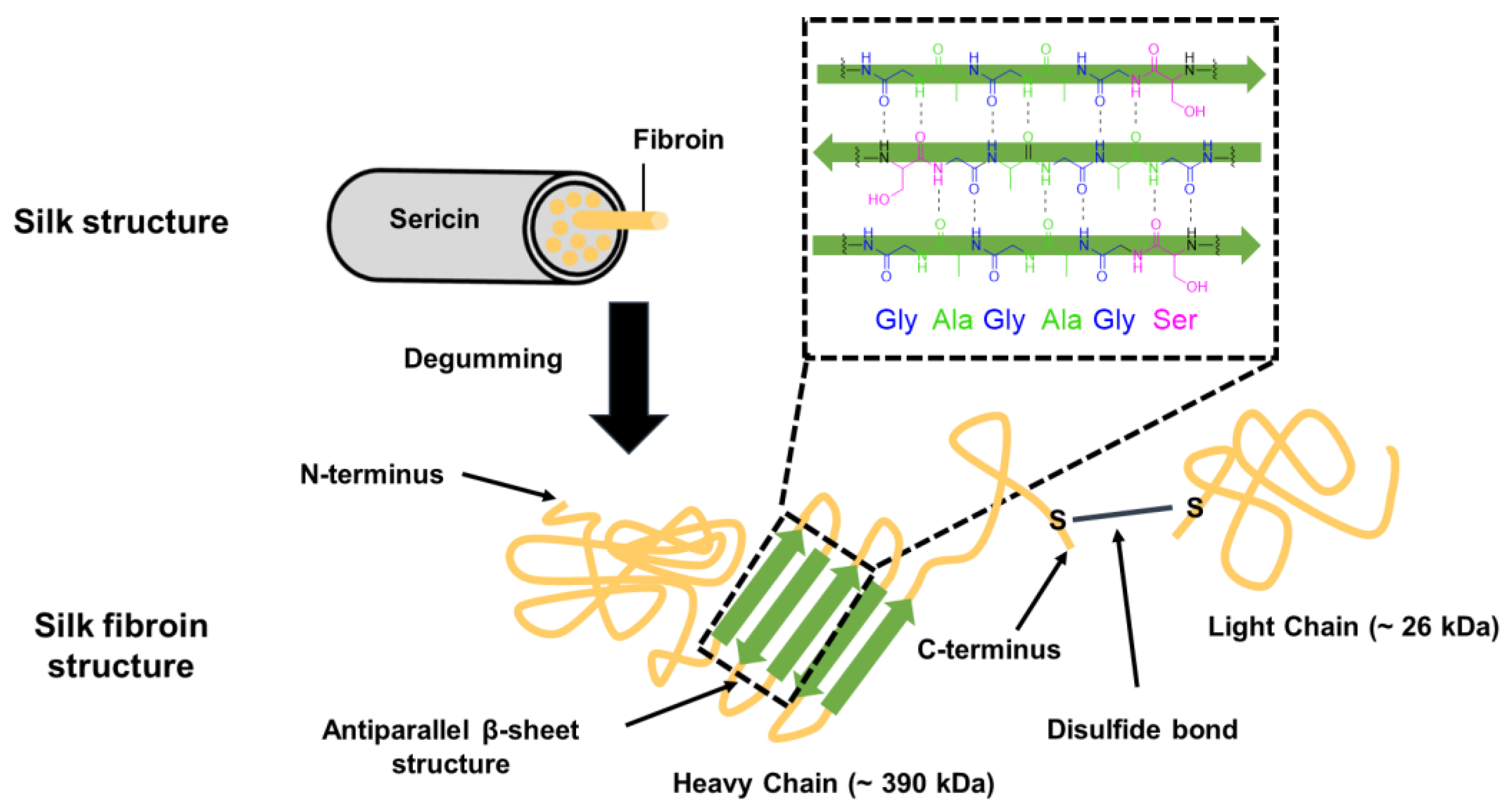
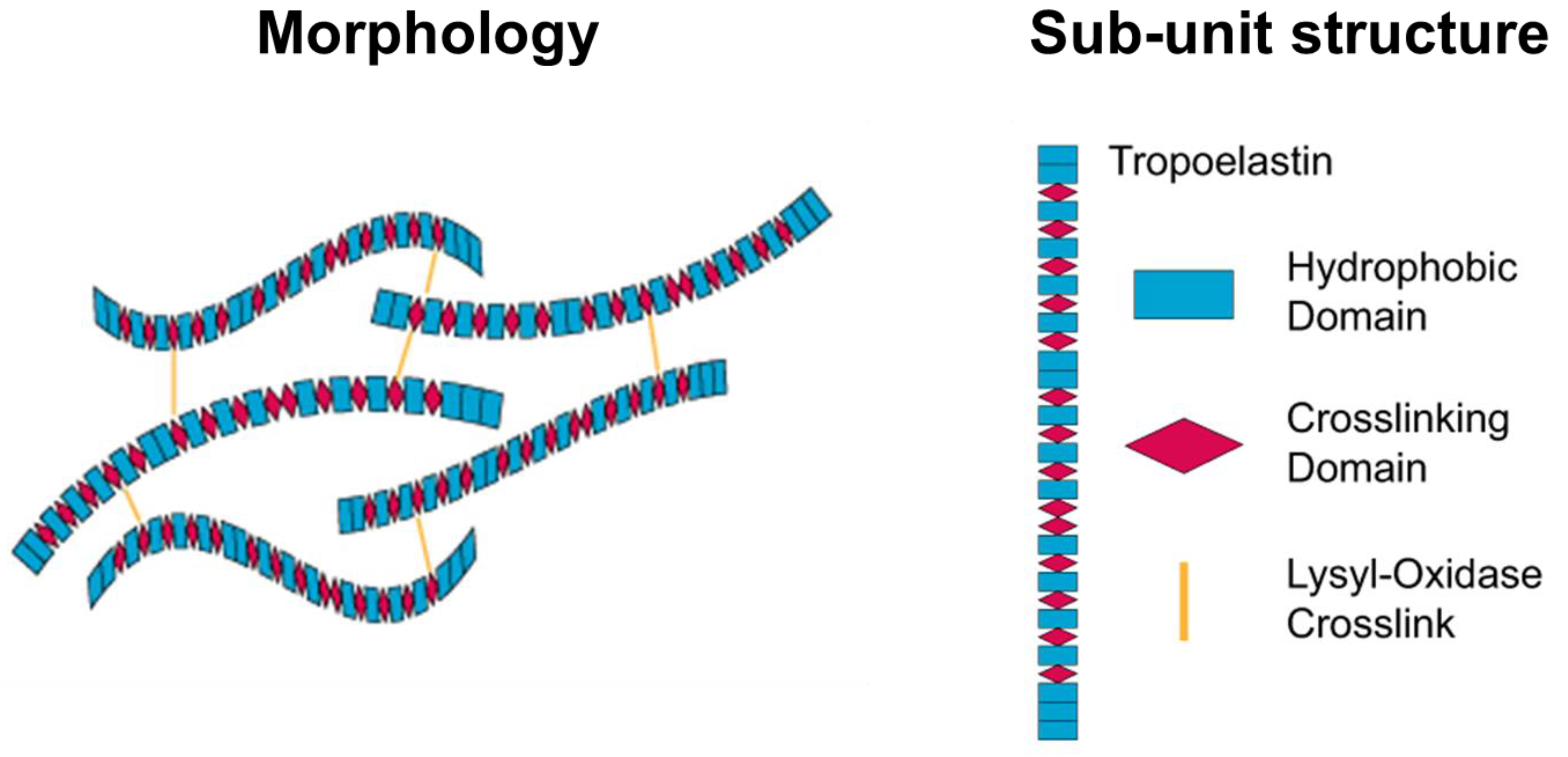
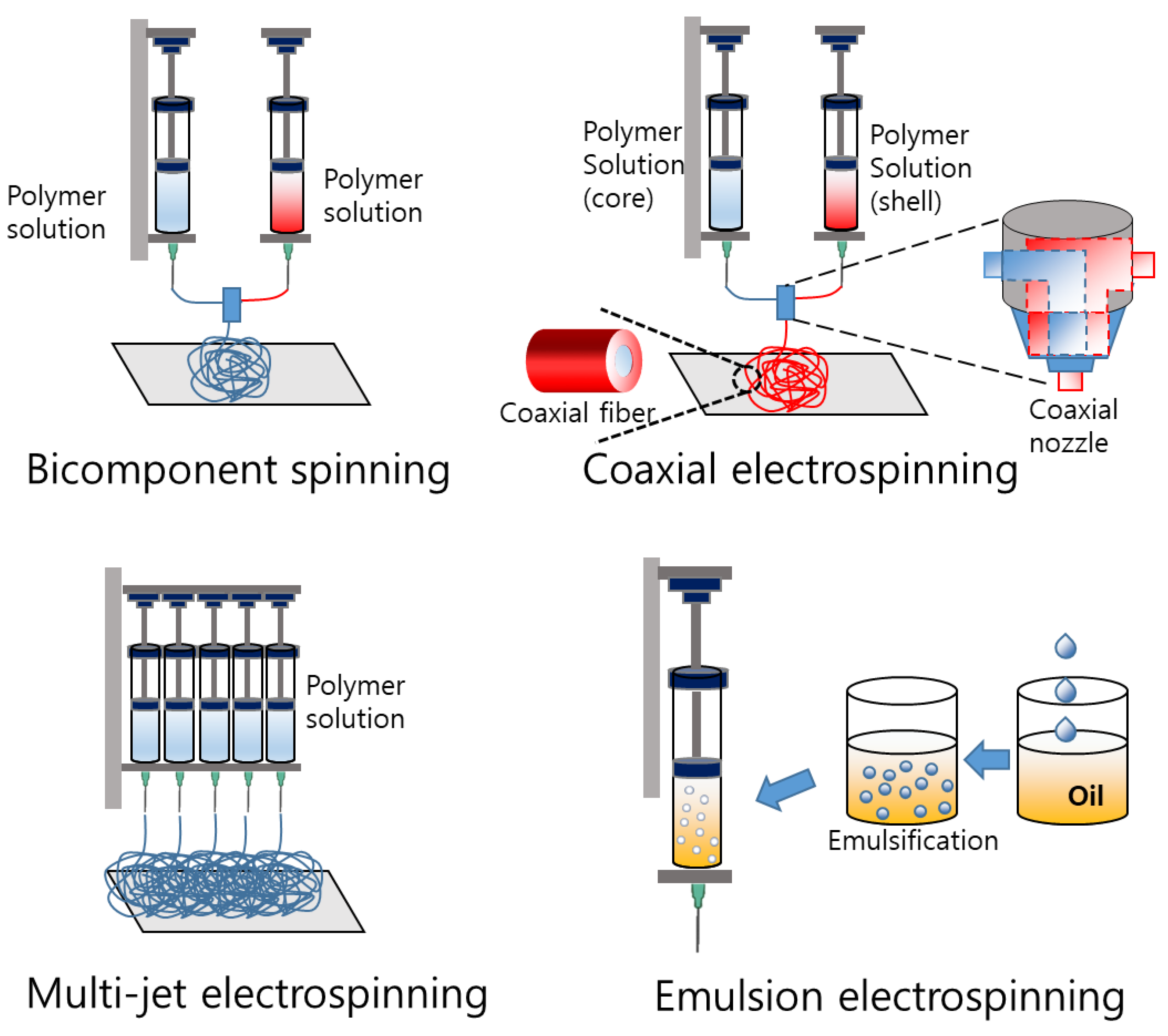
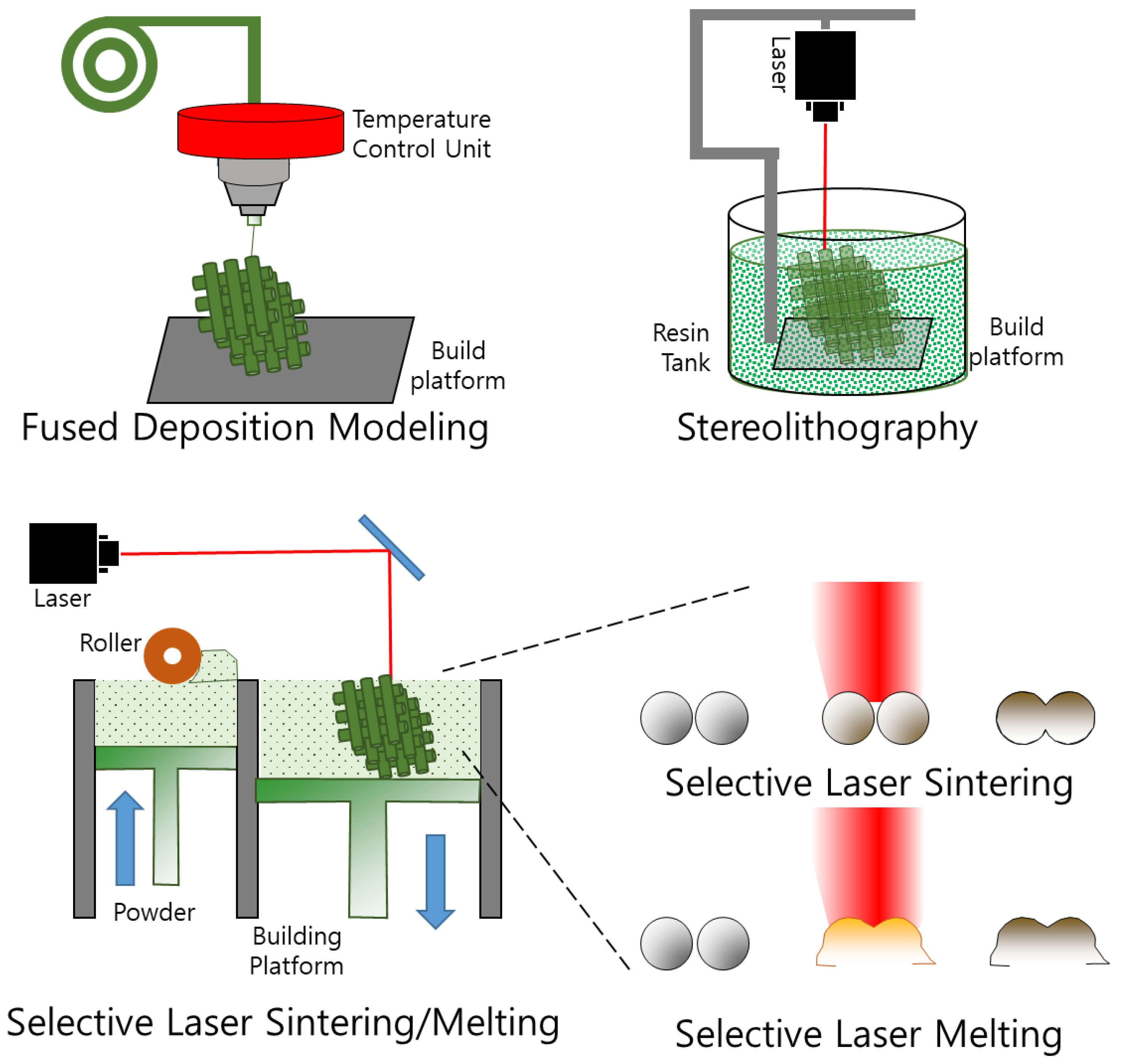
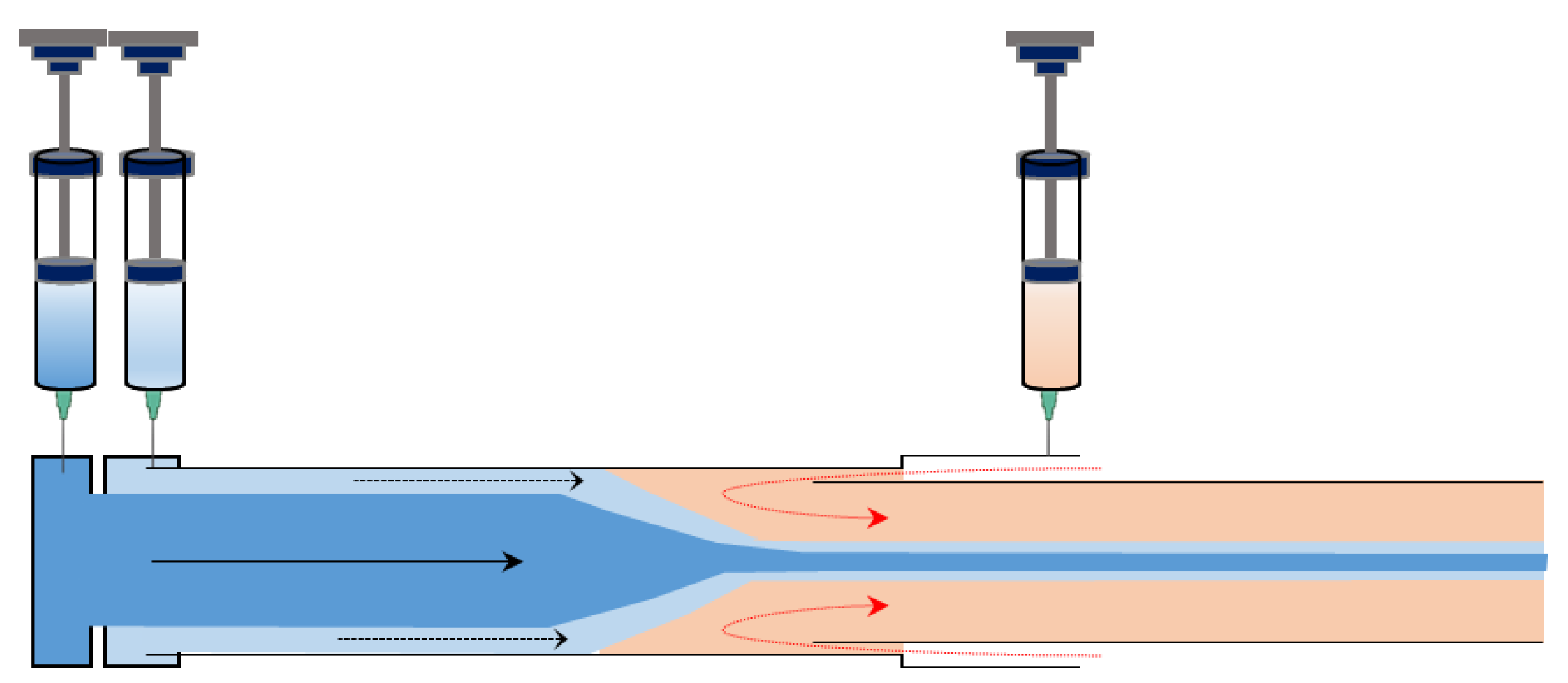
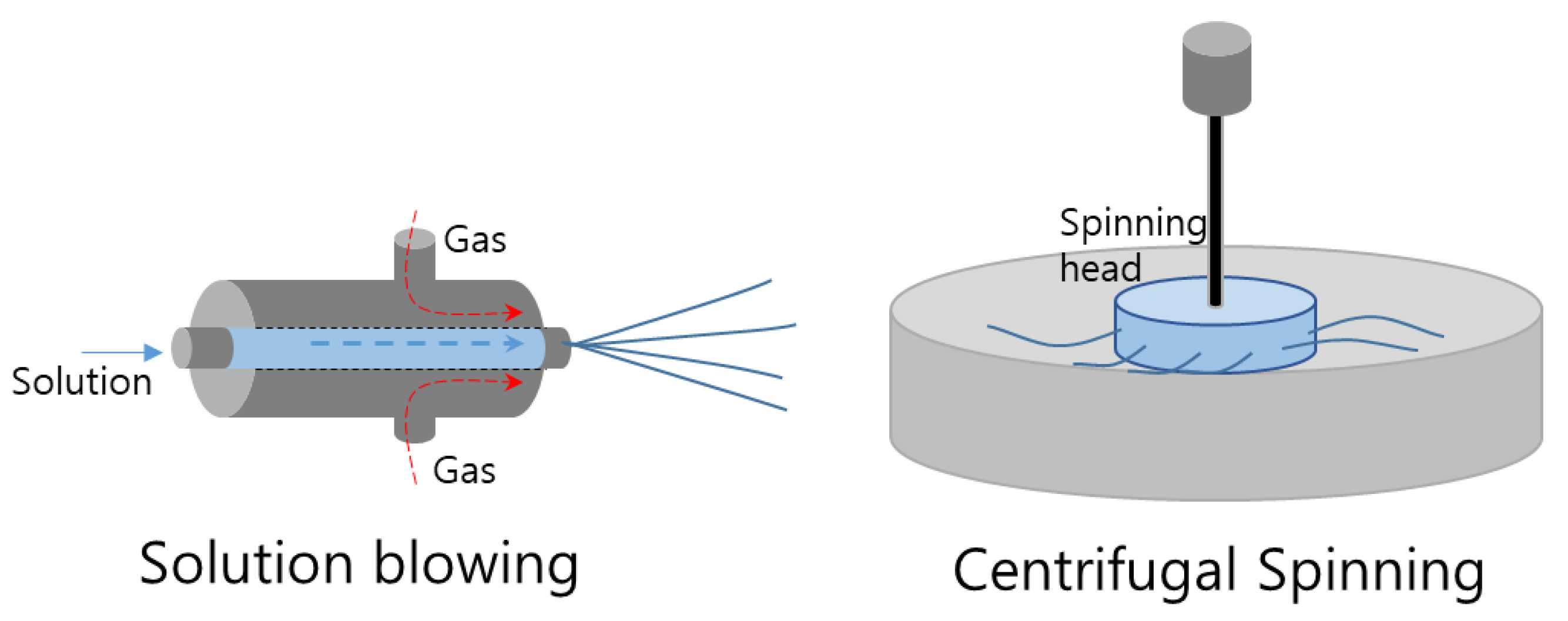
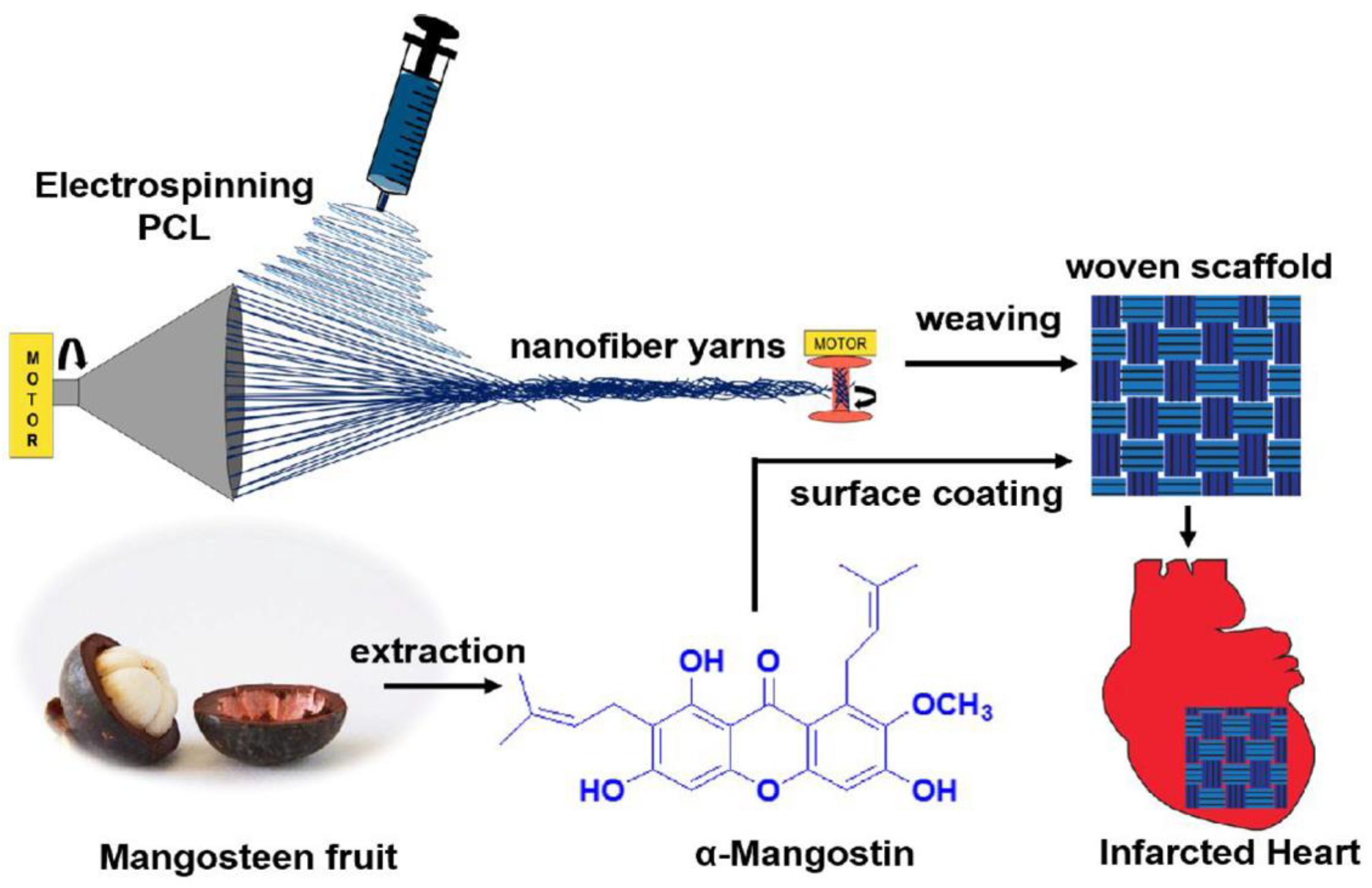
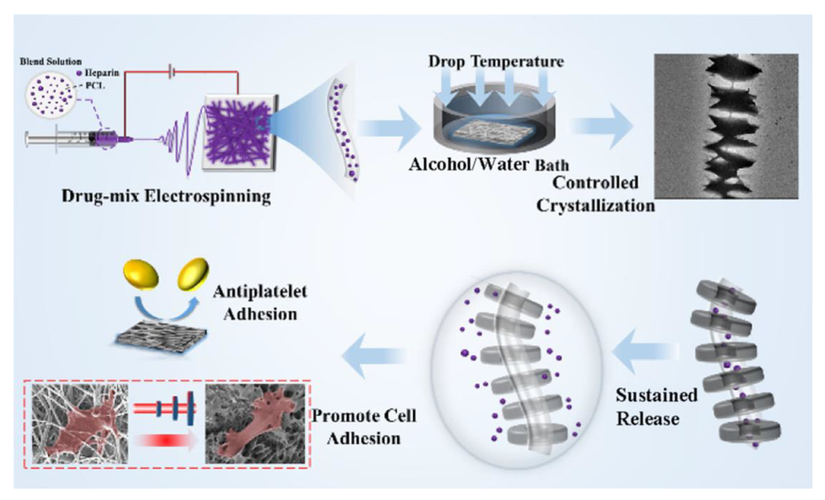
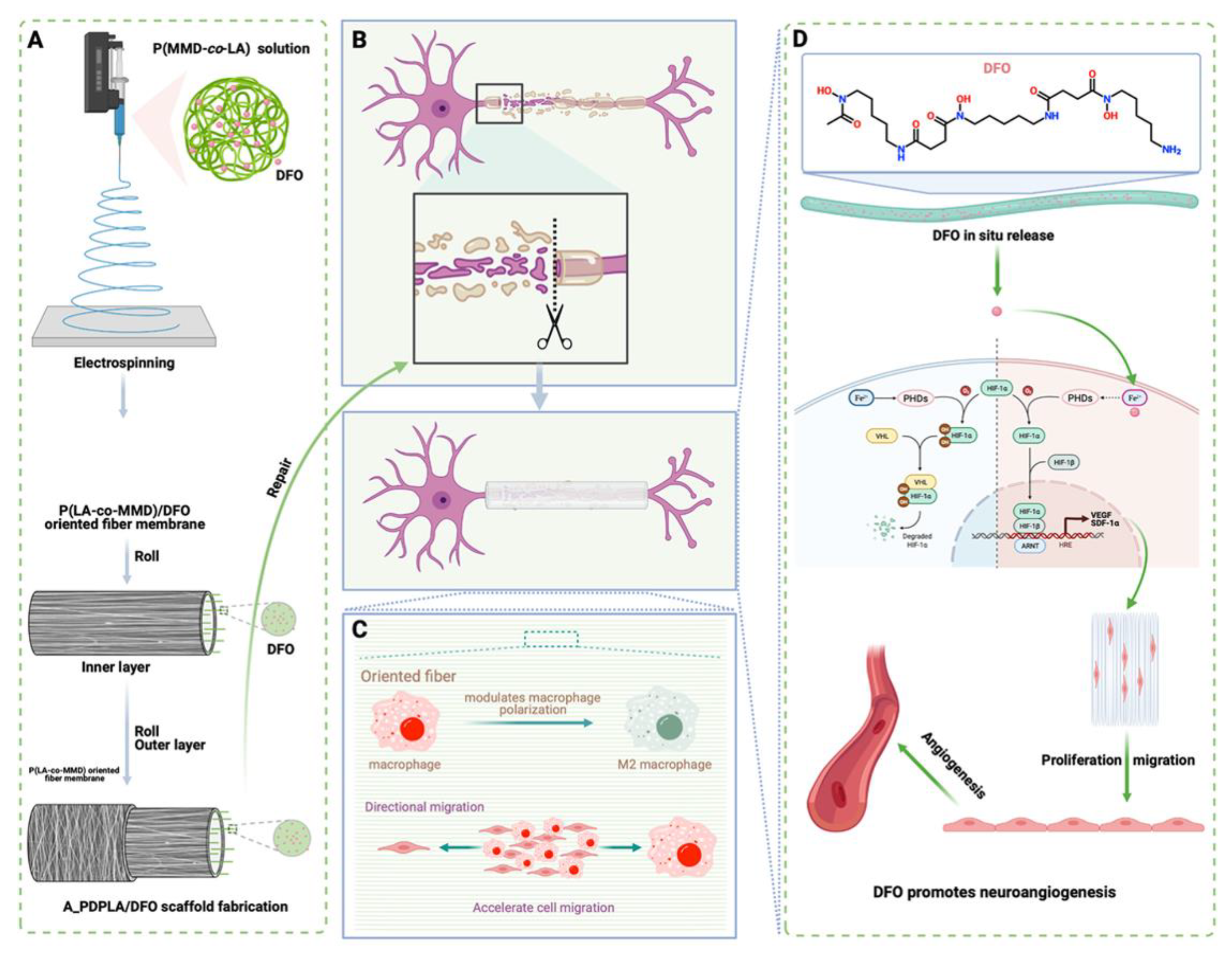
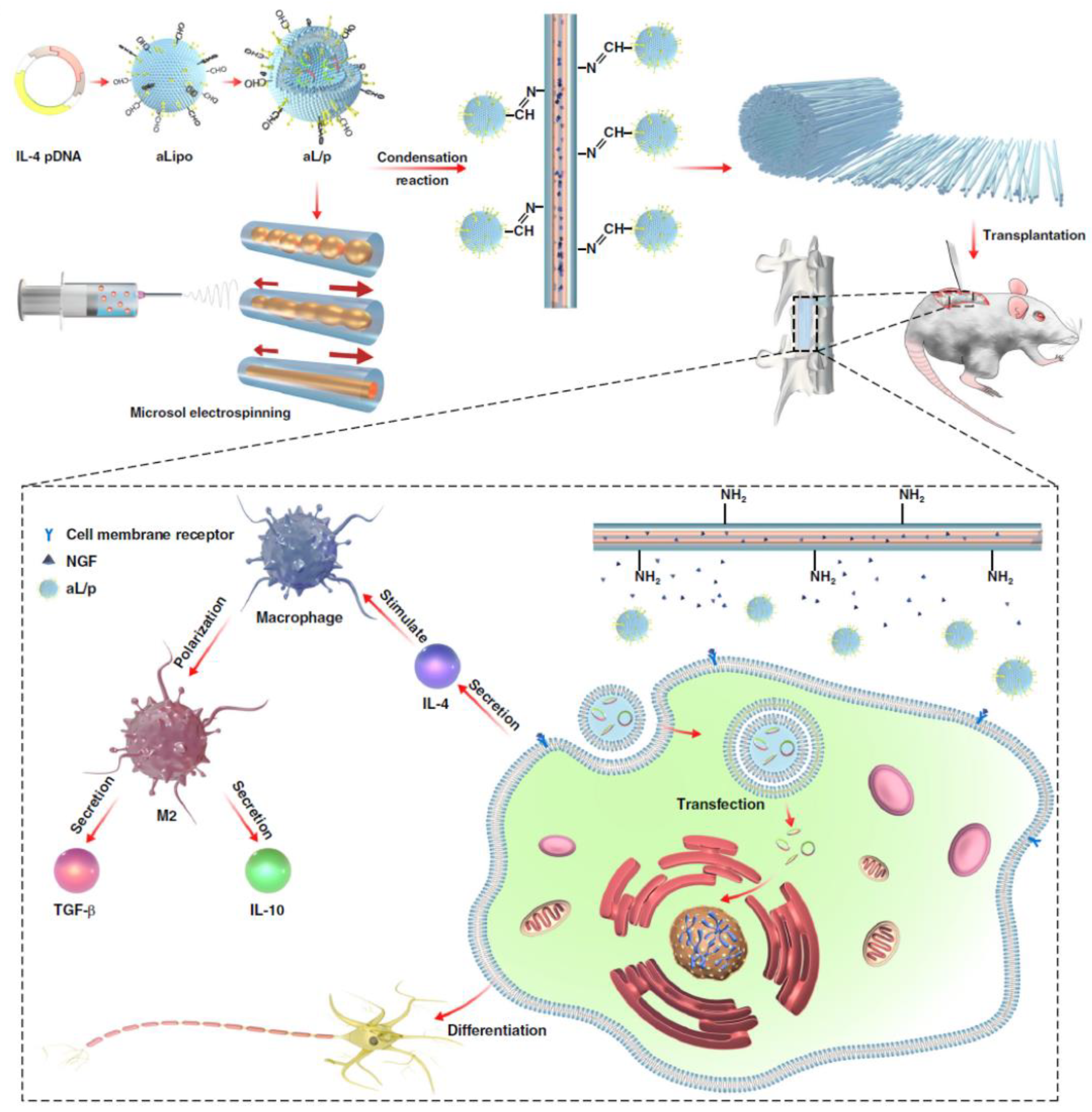
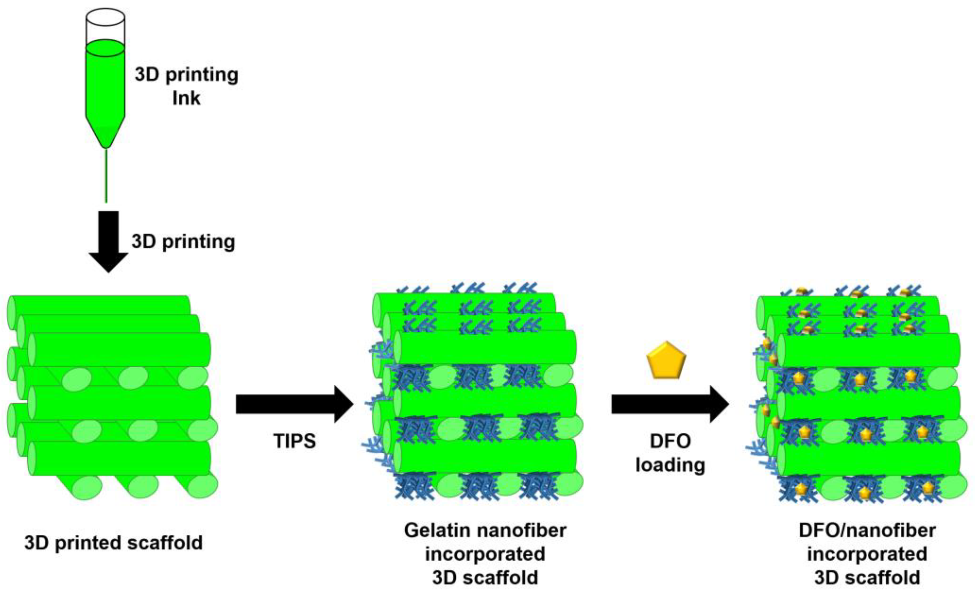
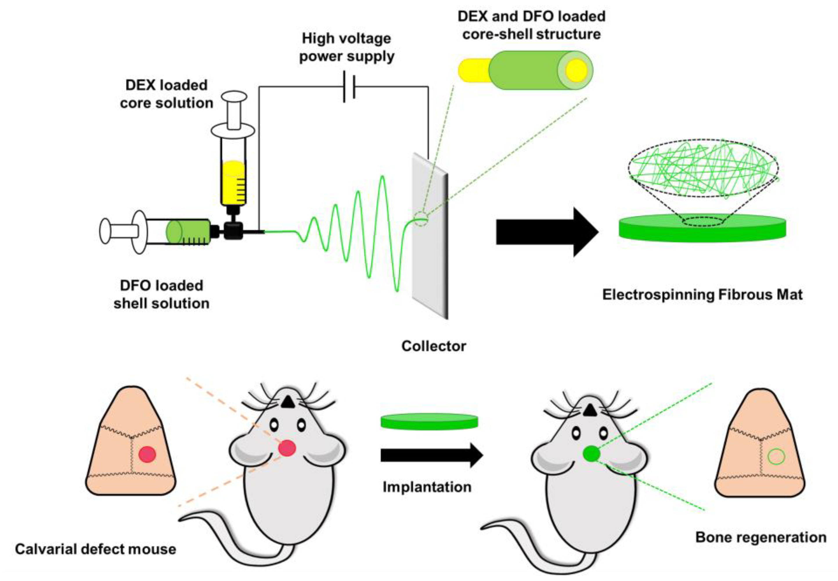
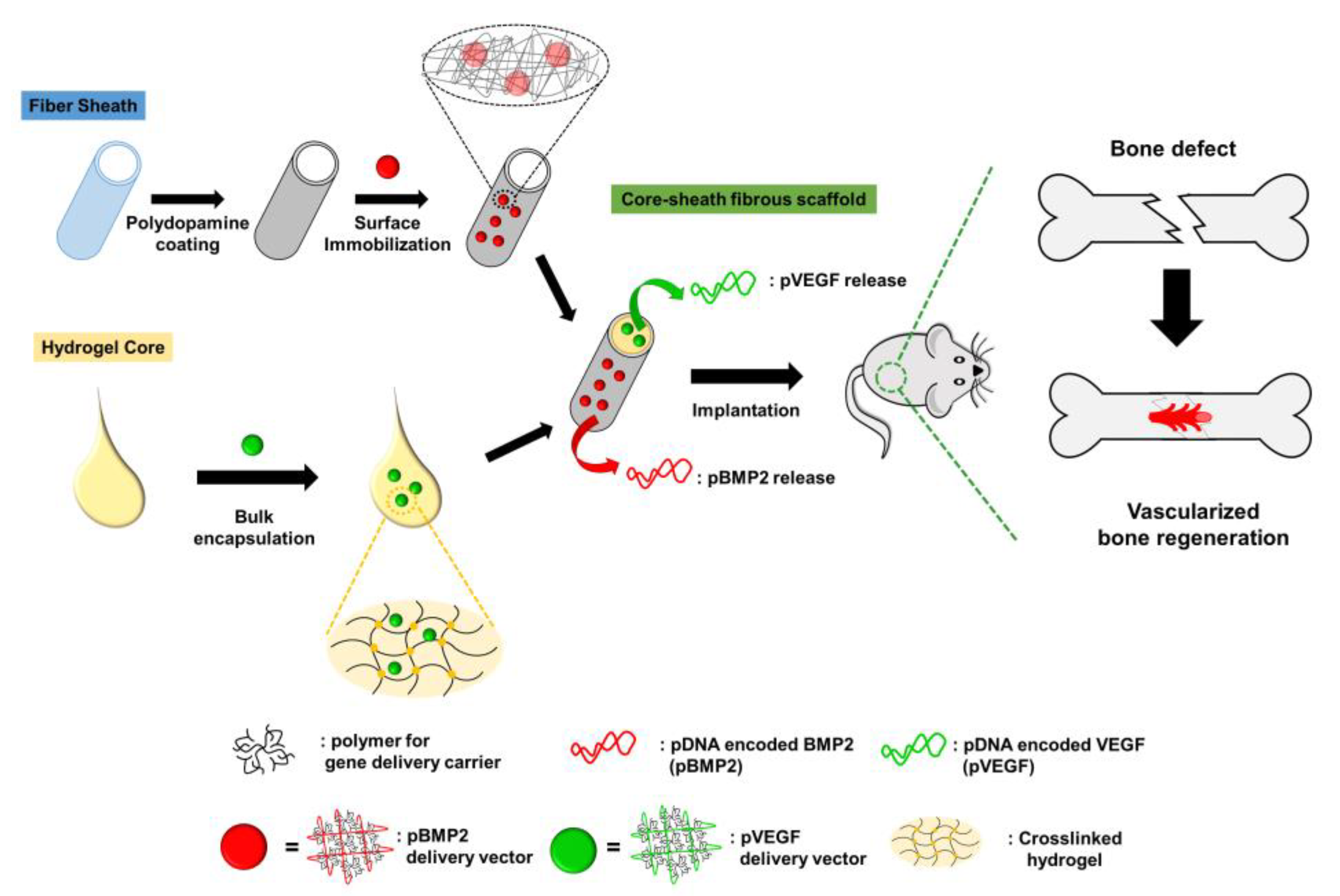
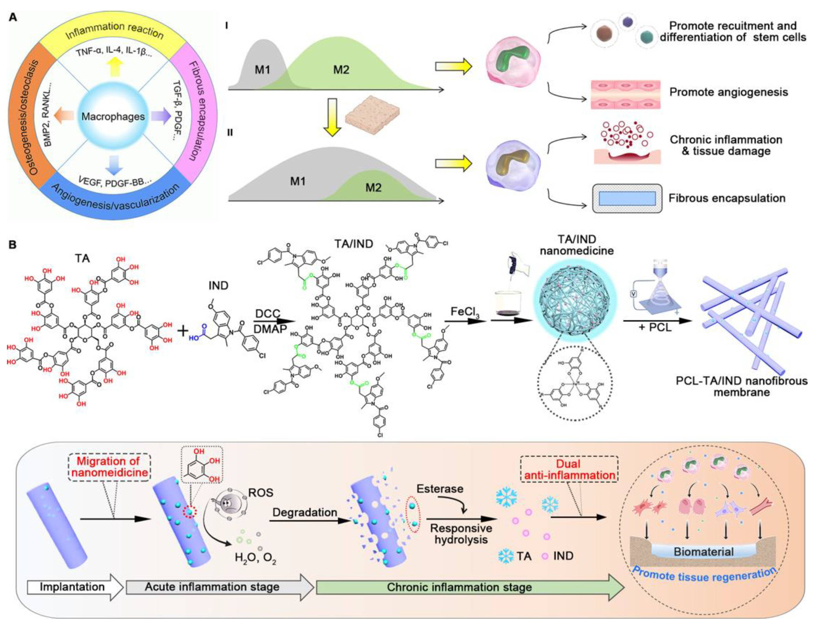
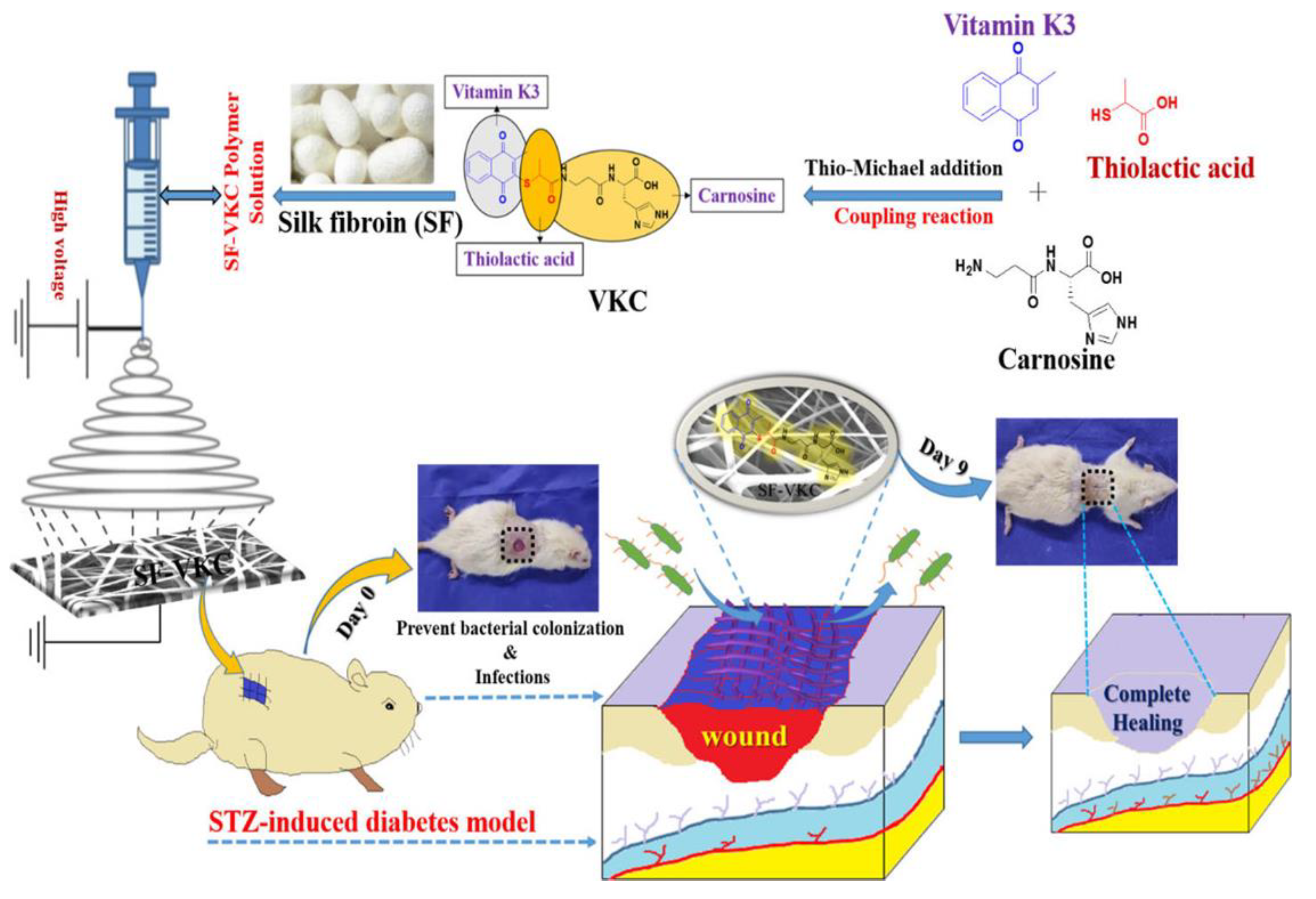
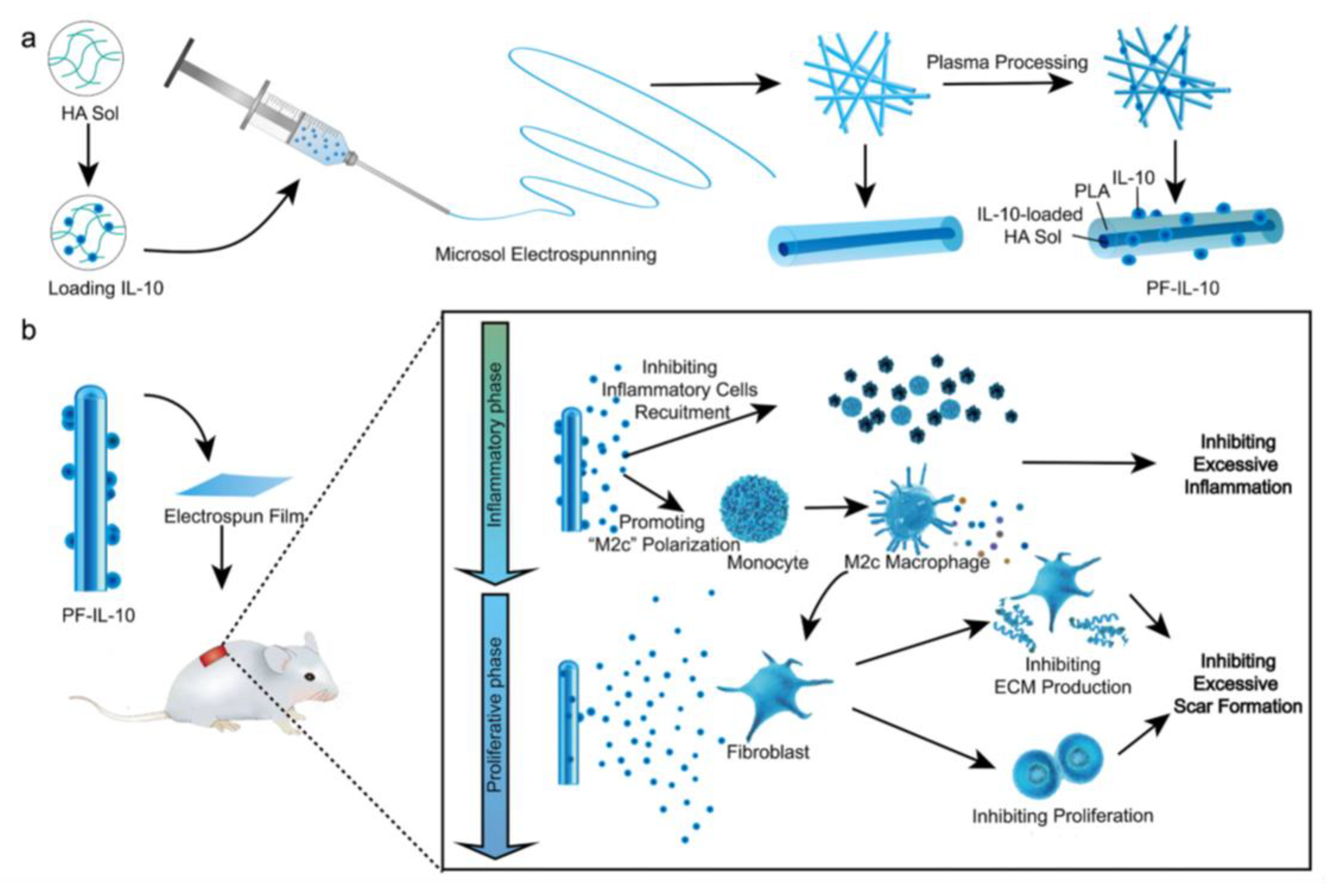
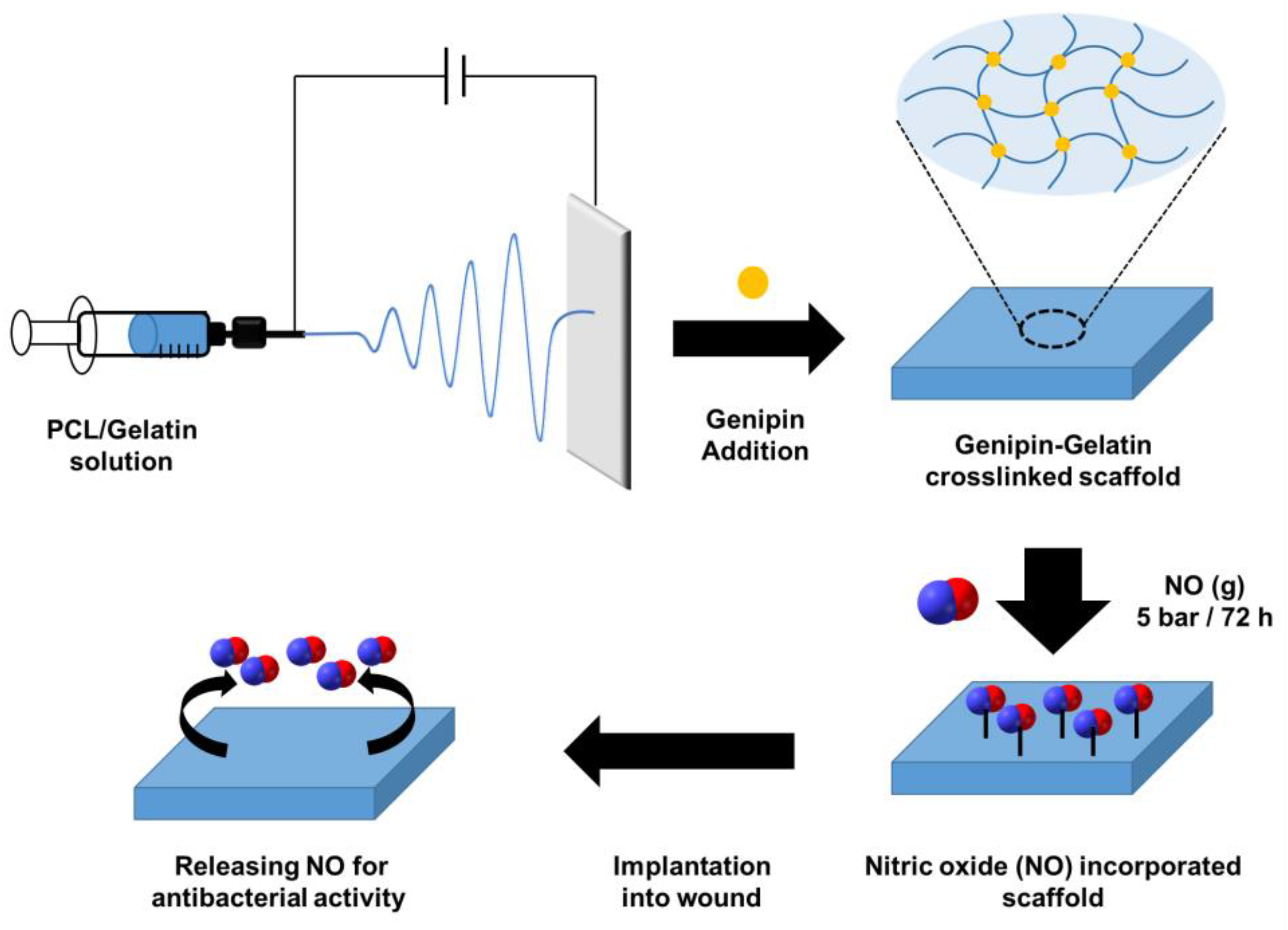
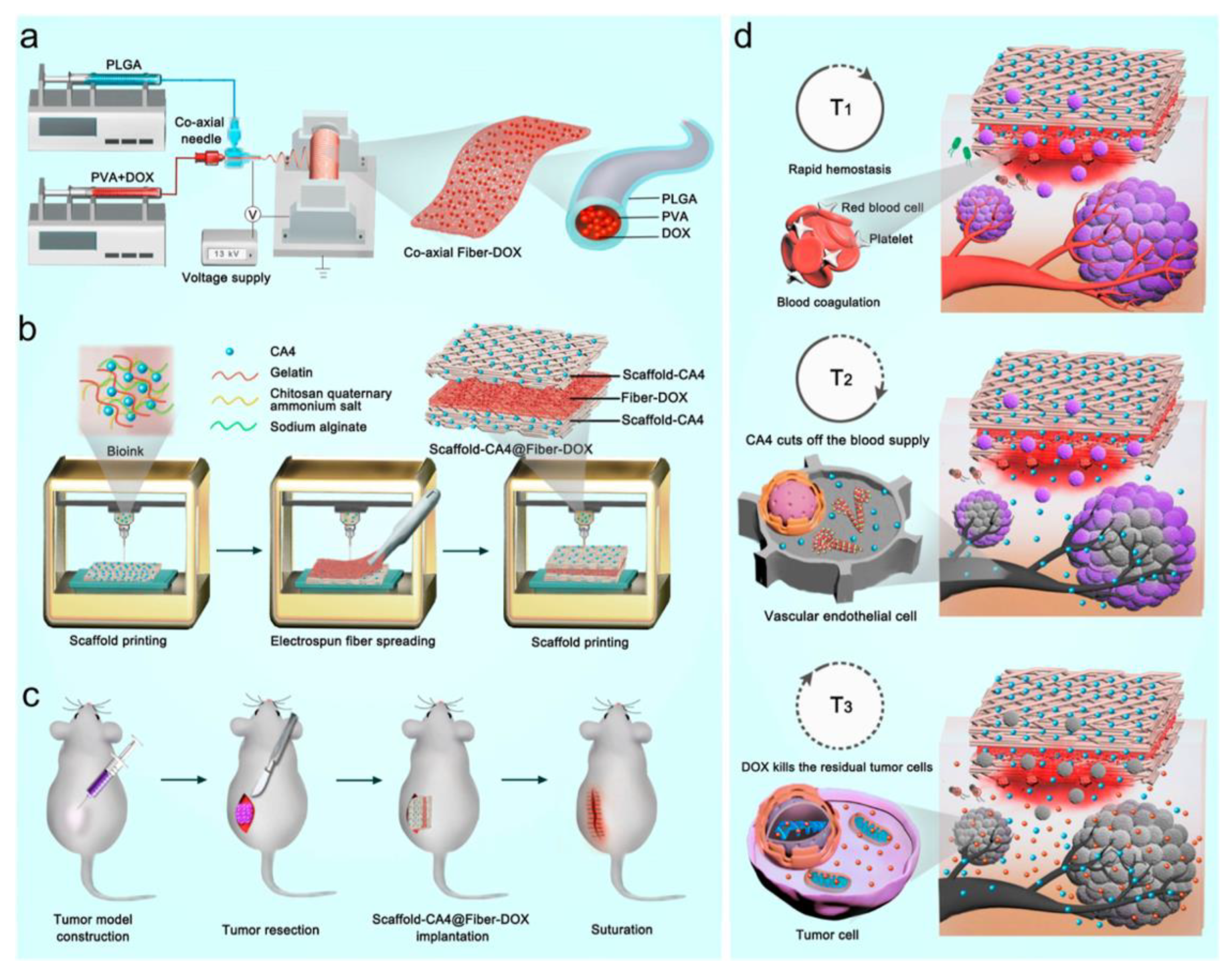
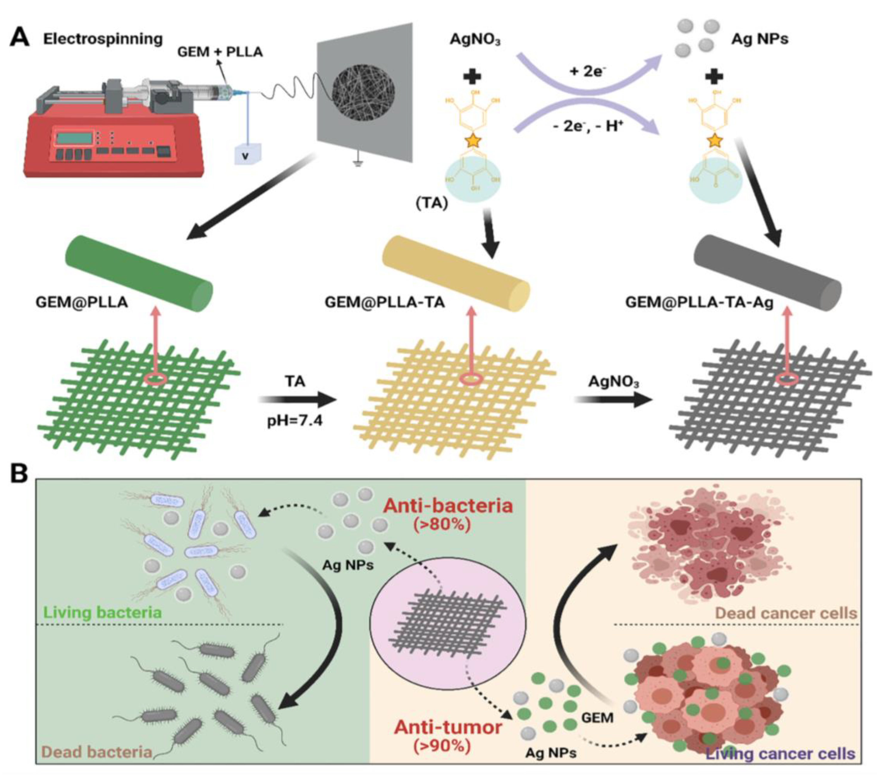
Table 1.
Comparison of electrospinning methods.
| Spinning Method | Key Features | Applications |
|---|---|---|
| Bicomponent spinning [79,80] | Two polymers are spun together to create a core–sheath or side-by-side structure | Medical textiles, filtration, and protective clothing |
| Coaxial electrospinning [81,82,83] | Two or more fluids are spun together in a coaxial arrangement, producing fibers with a core–shell structure | Drug delivery, tissue engineering, and energy storage |
| Multijet electrospinning [83,84,85] | Multiple spinning nozzles are used simultaneously to produce a large volume of fibers in a short amount of time | Tissue engineering, drug delivery, and energy storage |
| Emulsion electrospinning [86,87,88] | An emulsion of two immiscible liquids is spun to create fibers with a polymer and a liquid core | Drug delivery, tissue engineering, and food packaging |
Table 2.
Comparison of methods of 3D printing.
| Methods | Principles | Materials Used | Advantages | Disadvantages |
|---|---|---|---|---|
| Fused deposition modeling [89,90] | Thermoplastic filaments are melted and deposited layer by layer using a heated nozzle | ABS, PLA, Nylon, TPU, etc. | Low cost, user-friendly, wide material compatibility, good for prototyping | Lower resolution, visible layer lines, limited to certain geometries, weaker mechanical properties |
| Stereolithography (SLA) [91,92] | Uses a UV laser to selectively cure a liquid photopolymer resin layer by layer | Resin materials (acrylic, epoxy, polyurethane, etc.) | High resolution, good surface finish, suitable for small and intricate parts, good for prototyping and production | Limited material selection, expensive, post-processing required, not suitable for large parts |
| Selective laser sintering (SLS) [93,94,95] | Uses a laser to selectively fuse a powdered material (such as nylon) layer by layer | Nylon, TPU, polycarbonate, etc. | No support structures required, can produce complex geometries, good mechanical properties, good for low-volume production | Limited surface finish, expensive, post-processing required, not suitable for small parts |
| Selective laser melting [96] | Similar to SLS, but uses a laser to fully melt the powdered material instead of just fusing it, resulting in fully dense metal parts | Metals such as titanium, aluminum, steel, etc. | High strength, good for small and complex parts, wide range of materials available | Expensive, limited build size, slow printing speed, post-processing required |
Table 3.
Parameters and considerations for each fabrication method for fibrous scaffolds.
| Fabrication Methods | Parameters | Considerations |
|---|---|---|
| Electrospinning [119,120,121] | Solution viscosity | Higher viscosity results in larger polymer beads and thicker fibers |
| Solution conductivity | Higher conductivity improves the quality of fibers | |
| Applied voltage | Higher voltage results in thinner fibers | |
| Distance between the collector and the needle | Shorter distance results in denser fibers | |
| Flow rate of the solution | Higher flow rate leads to larger fiber diameter | |
| Needle diameter | Smaller diameter results in thinner fibers | |
| Solution concentration | Higher concentration results in thicker fibers | |
| Humidity and temperature of the environment | Affects fiber morphology and diameter | |
| 3D Printing [89,90,91,92,93,94,95,96] | Material properties | Affects printability and mechanical properties |
| Printing speed | Affects print quality and resolution | |
| Layer height | Affects resolution and mechanical properties | |
| Temperature of the printing environment | Affects material properties and print quality | |
| Extruder speed | Affects flow rate and print quality | |
| Nozzle diameter | Affects resolution and print speed | |
| Printing bed temperature | Affects adhesion and warping | |
| Printing orientation | Affects mechanical properties | |
| Microfluidic method [100,106,107,108,109,110,111] | Flow rate of the fluid | Affects fiber diameter and porosity |
| Viscosity of the fluid | Affects flow rate and fiber morphology | |
| Channel geometry | Affects fiber diameter and alignment | |
| Material properties | Affects fiber morphology and mechanical properties | |
| Surface properties | Affects fiber adhesion | |
| Temperature and pressure of the system | Affects fiber morphology and diameter | |
| Solution blowing [113,114,115] | Solution viscosity | Higher viscosity results in thicker fibers |
| Gas flow rate | Affects fiber diameter and morphology | |
| Distance between the nozzle and the collector | Affects fiber alignment and density | |
| Solution concentration | Higher concentration results in thicker fibers | |
| Temperature and humidity of the environment | Affects fiber morphology and diameter | |
| Centrifugal spinning [116,117,118] | Polymer concentration | Higher concentration results in thicker fibers |
| Centrifugal force | Affects fiber diameter and alignment | |
| Distance between the collector and the nozzle | Affects fiber alignment and density | |
| Solution viscosity | Higher viscosity results in thicker fibers | |
| Solution flow rate | Affects fiber diameter and morphology | |
| Polymer molecular weight | Affects mechanical properties | |
| Temperature and humidity of the environment | Affects fiber morphology and diameter |
Table 4.
Parameters that should be considered for each drug-loading method.
| Method | Parameters | Considerations |
|---|---|---|
| Blending [122,123,124,125,126,127] | Solubility of drug and polymer | Drug and polymer should have similar solubilities |
| Drug and polymer concentration | Higher concentrations lead to higher drug loading | |
| Mixing time and speed | Affects drug–polymer interaction and drug distribution | |
| Chemical immobilization [128,129] | Type of chemical reaction | Reaction should be specific to the drug and polymer |
| Concentration of reactants | Higher concentrations lead to higher drug loading | |
| Reaction time and temperature | Affects the efficiency of the reaction and drug loading | |
| Physical adsorption [130,131,132] | Surface area of the scaffold | Larger surface area leads to higher drug loading |
| Solution concentration and pH | Affects drug–polymer interaction and adsorption efficiency | |
| Adsorption time and temperature | Affects the efficiency of drug adsorption | |
| Emulsion electrospinning [133,134,135] | Polymer and drug solubility | Solubility should be matched for codissolving |
| Emulsifier concentration | Affects droplet size and stability | |
| Electrospinning parameters | Affects fiber diameter and drug distribution | |
| Emulsion stability | Emulsion should be stable for efficient drug loading | |
| Coaxial electrospinning [137,138,139] | Core and shell polymer solubility | Solubility should be matched for codissolving |
| Core and shell polymer concentration | Higher concentrations lead to higher drug loading | |
| Coaxial electrospinning parameters | Affects fiber diameter, shell thickness, and drug distribution | |
| Core–shell fiber stability | Core and shell should remain stable during processing | |
| Electrospray [140,141] | Solution concentration and pH | Affects drug–polymer interaction and encapsulation efficiency |
| Flow rate and voltage | Affects droplet size and drug distribution | |
| Solvent type | Affects the solubility of the drug and polymer | |
| Post-processing conditions | Can affect drug loading and release kinetics |
Table 5.
Applications of fibrous scaffolds.
| Biomedical Applications | Fabrication Methods of Scaffold | Therapeutic Agent (with its Function) | Drug-Loading Methods | References |
|---|---|---|---|---|
| Cardiovascular regeneration | Electrospinning method | α-Mangostin (antioxidant, cardioprotective activity) | Dip coating | [144] |
| Cardiovascular regeneration | Electrospinning method with crystallization | Heparin (antithrombosis activity) | Drug-mixing (blending) electrospinning method | [151] |
| Vascular regeneration | Coelectrospinning method | IL-4 (immune modulation for vascular stabilization) | Surface modification of the scaffold | [239] |
| Cardiac regeneration | Electrospinning method | Resveratrol (inducing cardioprotective effect) | Drug-mixing electrospinning method | [240] |
| Nerve regeneration | Electrospinning method | MicroRNAs (miRNAs, genetic modulation for nerve regeneration) | Passive loading after scaffold fabrication | [167] |
| Nerve regeneration | Electrospinning method with rolling | Deferoxamine (promoting angiogenesis with anti-inflammation) | Drug-mixing electrospinning method | [169] |
| Nerve regeneration | Microsol electrospinning | IL-4 encoding plasmid DNA (IL-4 generation for immune suppression) | Chemical modification of pDNA-loaded liposome | [176] |
| Nerve regeneration | Electrospinning method | Magnetic nanoparticles (MgO, inhibiting nerve cell apoptosis), purmorphamine and retinoic acid (Pur/RA, inducing neuronal differentiation) | Pur/RA-loaded MgO-mixing electrospinning method | [241] |
| Nerve regeneration | Electrospinning method | Acidic fibroblast growth factor (aFGF, signaling molecule for axon growth) | Coaxial electrospraying of aFGF-loaded nanoparticles | [242] |
| Nerve regeneration | Microsol electrospinning method | Brain-derived neurotrophic factor (BDNF, improving differentiation of bone marrow mesenchymal stem cells into neurons) | Drug-mixing electrospinning method | [243] |
| Bone regeneration | 3D printing with TIPS technique | Deferoxamine (DFO, inducing angiogenesis for vascularization) | Passive drug loading after scaffold fabrication | [182] |
| Bone regeneration | Coaxial electrospinning method | DFO (inducing vascularization), dexamethasone (DEX, modulating osteogenic differentiation) | Drug-mixing electrospinning method | [15] |
| Bone regeneration | Electrospinning method | Vascular endothelial growth factor (VEGF) encoding pDNA (inducing vascularization), bone morphogenetic protein 2 (BMP2, promoting growth of bone tissue) | Surface immobilization of drug-loaded vector and embedding drug containing hydrogel inside the scaffold | [186] |
| Bone regeneration | Electrospinning method | Tannic acid (TA) and indomethacin (IND) (agents for anti-inflammation) | TA/IND nanomedicine-mixing electrospinning method | [190] |
| Bone regeneration | Coaxial electrospinning method | Tauroursodeoxycholic acid (TUDCA, inducing vascularization), BMP2 (promoting growth of bone tissue) | Drug-mixing electrospinning method | [244] |
| Bone regeneration | Electrospinning method | Recombinant human vein endothelial growth factor (rhVEGF, inducing vascularization), recombinant human bone morphogenetic protein 2 (rhBMP2), and calcium phosphates (inducing bone tissue regeneration) | Drug-mixing electrospinning method | [245] |
| Bone regeneration | Electrospinning method with crosslinking | IL-4 (immunomodulation for vascular maturation and osteogenesis) | Drug-mixing electrospinning method | [246] |
| Wound healing | Electrospinning method | Vitamin K3 carnosine peptide (agent for antibacterial activity) | Drug-mixing electrospinning method | [198] |
| Wound healing | Microsol electrospinning method | IL-10 (agents for anti-inflammatory effect) | Drug-mixing electrospinning method | [210] |
| Wound healing | Electrospinning method | Nitric oxide (NO, agent for antibacterial activity) | Surface modification after scaffold fabrication | [214] |
| Wound healing | Electrospinning method | Cordia myxa (agent for antibacterial activity with antioxidant effect) | Immobilization after scaffold fabrication | [247] |
| Wound healing | Electrospinning method | Anemoside B4 (ANE, agent for anti-inflammation) | Incorporation of ANE after scaffold fabrication | [248] |
| Wound healing | 3D printing | Vascular endothelial growth factor (VEGF, inducing vascularization), gentamicin (antibiotic agent), silver nanoparticle (antibacterial agent) | Drug loading in 3D printing ink | [249] |
| Wound healing | Physical coassembling to construct fibrous hydrogel | Phycocyanin (anti-inflammatory activity), gallic acid (antibacterial activity and anti-inflammatory activity) | Mixing drugs for assembled fibrous scaffold fabrication | [250] |
| Wound healing | Electrospinning method | Manganese dioxide (MnO2, agent for assuaging oxidative stress), curcumin (agent for anti-inflammation) | Curcumin-loaded MnO2 nanoparticle-mixing electrospinning method | [251] |
| Wound healing | Electrospinning method | Pyrogallol and saikosaponin (reducing reactive oxygen species (ROS)) | Drug-mixing electrospinning method | [252] |
| Wound healing | Electrospinning method | Curcumin (agent for anti-inflammation), cerium nitrate (reducing ROS for antiscar wound healing) | Drug-mixing electrospinning method | [253] |
| Wound healing | Layer by layer electrospinning method | Silver (I) sulfadiazine (agent for antibacterial activity) | Drug-mixing electrospinning method | [254] |
| Tumor inhibition | 3D printing with electrospinning method for multiple-layered scaffold fabrication | Combretastin A4 (CA4, inhibiting tumor growth) and doxorubicin (DOX, inhibiting metastasis and recurrence of cancer) | DOX: drug-mixing electrospinning method, CA4: drug-mixing in bioink before 3D printing | [225] |
| Tumor inhibition with antibacterial activity | Electrospinning method | Gemicitabine (GEM, inhibits tumor growth), silver nanoparticle (antibacterial agent for enhancing tumor therapy) | GEM: drug mixing electrospinning method, silver nanoparticle: dipping process after scaffold fabrication with coating | [228] |
| Tumor inhibition | Sol–gel process with electrospinning method | Glucose oxidase (GOx, inducing starvation activity around tumor tissue), hyaluronidase (HAase, destroying tumor extracellular matrix (ECM) to penetrate nanomedicine), banoxantrone (AQ4N, antitumor agent) | Incorporation of HAase and GOx/AQ4N-loaded nanoparticles after scaffold fabrication | [255] |
| Tumor inhibition | Coaxial 3D printing | DOX (antitumor agent), polydopamine (photothermal agent for thermal-induced tumor therapy) | Mixing drugs in ink and fabricating scaffold | [256] |
| Tumor inhibition with wound healing | Coaxial electrospinning method | Fluorouracil (5-FU, agent for chemotherapy of melanoma) | 5-FU-loaded nanoparticle-mixing electrospinning method | [257] |
| Periodontal regeneration | Electrospinning method | Dimethyloxalylglycine (DMOG, inhibitor of prolyl hydroxylases to promote VEGF) | Drug-mixing electrospinning method | [231] |
| Tendon regeneration | Electrospinning method | Nitric oxide (NO, agent for vascularization) | Use of NO-loaded metal–organic framework-mixing electrospinning method | [237] |
Disclaimer/Publisher’s Note: The statements, opinions and data contained in all publications are solely those of the individual author(s) and contributor(s) and not of MDPI and/or the editor(s). MDPI and/or the editor(s) disclaim responsibility for any injury to people or property resulting from any ideas, methods, instructions or products referred to in the content. |
© 2023 by the authors. Licensee MDPI, Basel, Switzerland. This article is an open access article distributed under the terms and conditions of the Creative Commons Attribution (CC BY) license (https://creativecommons.org/licenses/by/4.0/).
Share and Cite
MDPI and ACS Style
Park, D.; Lee, S.J.; Choi, D.K.; Park, J.-W. Therapeutic Agent-Loaded Fibrous Scaffolds for Biomedical Applications. Pharmaceutics 2023, 15, 1522. https://doi.org/10.3390/pharmaceutics15051522
AMA Style
Park D, Lee SJ, Choi DK, Park J-W. Therapeutic Agent-Loaded Fibrous Scaffolds for Biomedical Applications. Pharmaceutics. 2023; 15(5):1522. https://doi.org/10.3390/pharmaceutics15051522
Chicago/Turabian StylePark, Dongsik, Su Jin Lee, Dong Kyu Choi, and Jee-Woong Park. 2023. "Therapeutic Agent-Loaded Fibrous Scaffolds for Biomedical Applications" Pharmaceutics 15, no. 5: 1522. https://doi.org/10.3390/pharmaceutics15051522
Note that from the first issue of 2016, this journal uses article numbers instead of page numbers. See further details here.