
Phytochemicals from Indian Ethnomedicines: Promising Prospects for the Management of Oxidative Stress and Cancer


 , , , and
Abstract
:1. Introduction
2. Relation between Oxidative Stress and Cancer
3. Indian Ethnomedicinal Plants as a Potential Source of Anticancer Phytochemicals

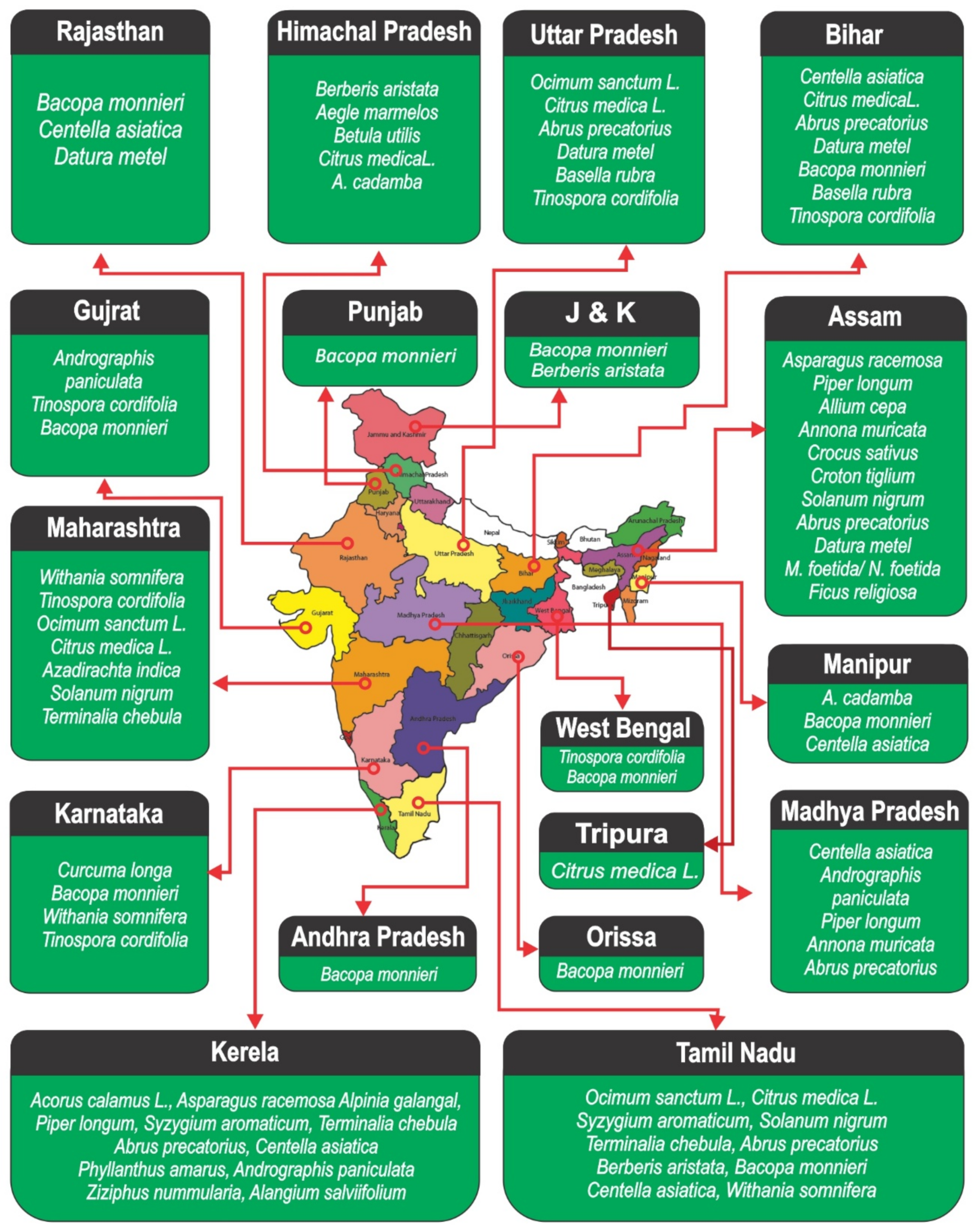
| Ayurvedic Name | Scientific Name | Family | Active/Antioxidant/Anticancer Phytochemicals | References |
|---|---|---|---|---|
| Amrita/Guduchi/Giloya | Tinospora cordifolia | Menispermaceae | Palmative, new clerodane furanoditerene glycoside, arabinogalactan, berberine, phenolic compounds, and epoxy cleodane diterpene | [19] |
| Amritavallari | Basella rubra | Basellaceae | p-Coumaric acid, caffeic acid, diosmetin | [20] |
| Amruta, Kalgur, or Narkya | Mappia foetida/Nothapodytes foetida | Icacinaceae | Camptothecin | [21] |
| Anlkol | Alangium salviifolium | Cornaceae | Deoxytubulosine, alangimarckine, dehydroprotoemetine, salviifosides A-C, kaempferol, salicin, and kaempferol 3-O-b-D-glucopyranoside | [22] |
| Ashwagandha | Withania somnifera | Solanaceae | Withanolides, sitoindosides | [23] |
| Ashwatha | Ficus religiosa | Moraceae | Quercetin, myricetin, kaempeferol, β-sitosteryl-D-glucoside | [24] |
| Bhukamtaka sukhsharanphala | Ziziphus nummularia | Rhamnaceae | Rutin, chlorogenic acid, quercetin, pyrogallol, mandelic acid, morin | [25] |
| Bhumyamalaki | Phyllanthus amarus | Phyllanthaceae | Nirtetralin (NIRT), niranthrin (NIRA), phyllanthin (PHYLLA), phyltetralin | [26] |
| Bhunimba and kalmegha | Andrographis paniculata | Acanthaceae | Andrographolide | [27] |
| Bhurjapatra | Betula utilis | Betulaceae | Betulinic acid | [28] |
| Bilva | Aegle marmelos | Rutaceae | Skimmianine, marmesin, imperatorin, cuminaldehyde, xanthotoxol | [29] |
| Brahmamanduki | Centella asiatica | Acanthaceae | Asiaticoside | [30] |
| Brahmi | Bacopa monnieri | Scrophulariaceae | Brahmine, herpestine, nicotinine, bacosides A and B, betulinic acid, wogonin, and oroxindin | [31,32] |
| Daruharidra | Berberis aristata | Berberidaceae | Berberine, berbamine, oxyberberine, aromoline, a protoberberine alkaloid karachine, palmatine, taxilamine, and oxycanthine | [33] |
| Dhatura | Datura metel | Solanaceae | Pterodontriol B, scopolamine, adenosine, disciferitriol, thymidine, dioscoroside D, and ilekudinoside C | [34] |
| Gunja | Abrus precatorius | Leguminosae | Abrin, Cycloartenol, Luteolin, Isoorientin, Trigonelline | [35] |
| Haridra | Curcuma longa | Zingiberaceae | Curcumin, Curcuminoid, Desmethoxycurcumin, Bisdemethoxycurcumin, Curdione, Bisacurone | [36] |
| Haritaki | Terminalia chebula | Combretaceae | Arjunglucoside I, chebulosides I and II, arjungenin, chebulin, 2,4-chebulyl-ß-D-glucopyranose, chebulic acid, chebulinic acid, terchebin | [37] |
| Kadamba, Vrattapuspa, Sisupala | Anthocephalus cadamba | Rubiaceae | Cadamine, isocadambine, isocadambine | [38] |
| Kakamachi | Solanum nigrum | Solanaceae | Solamargine, Solasonine, Solasodine, Solanidine | [39] |
| Kumbhinī | Croton tiglium | Euphorbiaceae | Corydine and salutaridine | [40] |
| Kumkuma/Ghusrun/Agneeshekhar | Crocus sativus | Iridaceae | Crocin, crocetin, picrocrocin, and safranal | [41] |
| Lakshamanap hala | Annona muricata | Annonaceae | Annonacin, Isoquinoline, Anonaine, Bullatacin, Annonamine, Gentisic acid | [42] |
| Lavanga | Syzygium aromaticum | Myrtaceae | Eugenol, Bicornin, Caryophyllene, Tellimagrandin II | [43] |
| Neem | Azadirachta indica | Meliaceae | Azadirachtin, Nimbin, Gedunin, epoxyazadiradione, Oleic acid | [44] |
| Nimbuka | Citrus medica | Rutaceae | Citronellal, Undecanal, Bisabolene | [45] |
| Palandu | Allium cepa | Liliaceae | Quercetin, Diallyl disulfide, Allicin | [46] |
| Pippali | Piper longum | Piperaceae | Piperine, Chavicine, Piperlongumine, Piperlonguminine | [47] |
| Rasna | Alpinia galangal | Zingiberaceae | Galangin, Kaempferide, Cadinene, fenchyl acetate | [48] |
| Rasona | Allium sativum | Amaryllidaceae | Alliin, allicin alliin, alliinase | [49] |
| Revandachini | Rheum emodi | Polygonaceae | Emodin, aloe-emodin, chrysophanol, physcion, rhein, emodin glycoside, and chrysophanol glycoside | [50] |
| Shatavari | Asparagus racemosa | Liliaceae | Shatavaroside A and B, shatavarins, filiasparoside C, immunoside, and schidigerasaponin | [51] |
| Sunthi | Zingiber officinalis | Zingiberaceae | Gingerol, Zingiberene, Shogaol, Paradol, Zingerone | [52] |
| Tulsi | Ocimum sanctum | Labiatae | Eugenol, Caryophyllene, Linalool, Methyl eugenol, Estragole, Carvacrol, Cadinene, Farnesene | [53] |
| Vacha, Ugargandha, Chhadgrantha | Acorus calamus | Acoraceae | Asarone, Cadinene, Methyl isoeugenol | [54] |
| Ethnomedicinal/Ayurvedic Remedy | Plant Species | Description | Benefits and Medicinal Uses | References |
|---|---|---|---|---|
| Jatamansi | Nardostachys jatamansi DC | Ayurvedic Powder | Use as brain tonic that help to improve memory and brain functions by preventing cell damage due to its antioxidant property | [55] |
| Bhootkeshi | Selinum vaginatum (Edgew) Cl | Ayurvedic Powder | Sleep and mental disorders, high blood pressure | [56] |
| Haridra/Daruharidra | Curcuma longa | Ayurvedic Powder | Chronic anterior uveitis, rheumatoid arthritis, conjunctivitis, small pox, skin cancer, chicken pox, urinary tract infections, wound healing, and liver ailments | [57] |
| Drakshasava | Vitis Vinifera, Woodfordia Fruticosa, Piper Cubeba, Cinnamomum Tamala, Mesua Ferrea, Syzygium Aromaticum Myristica Fragrans, Piper Nigrum, Plumbago Zeylanica, Piper Retrofractum Piper Longum, Vitex Negundo | Ayurvedic rejuvenators | Weakness, lethargy and heat exhaustion, curing haemorrhoids and cardiac disorders | [58] |
| Kanchanar Guggulu | Bauhinia variegata L. (BV), Zingiber officinale, Piper nigrum, Piper longum, Terminalia chebula, Terminalia bellerica, Embelica officinalis, Crataeva nurvala, Cinnamomum tamala, Elletaria cardemomum, Cinnamomum zeylanicum, Commiphora muku | Ayurvedic Formulation | Hormonal imbalance, PCOS, hypothyroidism, and joint pains. weight loss, lipoma, tumor, cysts, cancer, goiter, fistula, boils and skin related diseases | [59] |
| Hingvastaka | Zingiber officinale, Piper nigrum, Piper longum, Trachyspermum ammi, Carum carvi, Cuminum Cyminum, Ferula asafoetida | Ayurvedic churna | Indigestion, Anorexia and all Vata Disorders, relieve flatulence | [60] |
| Triphala | Phyllanthus emblica,Terminalia bellirica, Terminalia chebula | Polyherbal Ayurvedic medicine | Indigestion, weight loss,Reducing inflammation, regulate blood sugar levels, Lower cholesterol, reduce tumors, prevents cancer, inhibit HIV | [61] |
| Aswagandharishtam | Withania somnifera, Rubia cordifolia, Chlorophytum tuberosum, Terminalia chebula Curcuma longa, Berberis aristata, Glycyrrhiza glabra, Pluchea lanceolata, Pueraria tuberose Terminalia arjuna, Cyperus rotundus, Ipomoea turpethum, Hemidesmus indicus, Santalum album Acorus calamus, Plumbago indica | Liquid Ayurveda medicine | Anti-anxiety, anti-stress, antidepressant | [62] |
| Kasisadi Tailam | Leucaena Leucocephala, Ficus Religiosa, Dry Ginger, and Plumbago Zeylanica | Polyherbal Ayurvedic oil | External application on corns, piles and warts | [63] |
| Pusyanuga curna | Cissampelaos pareira, Syzygium cumini, Mangifera indica, Bergenia lingulata, Berberis aristata, Ambastha (Patha)- Cissampelos pareira, Mochrasa- Salmalia malabarica, Mimosa pudica, Holarrhena antidysentrica, Crocus sativus, Aconitum heterophylum, Aegle marmelos, Cyperus rotundus, Symplocos racemosa, Ochre or Haematite, Ailanthus excels, Piper nigrum, Zingiber officinale, Vitis vinifera, Pterocarpus santalinus, Myrica esculenta, Holarrhena antidysentrica, Hemidesmus indicus, Woodfordia fruticosa, Glycyrrhiza glabra, Terminalia arjuna | Herbo-mineral formulation | Conditions involving menstrual irregularities such as menorrhagia,metrorrhagia, dysmenorrhoea and endometriosis, piles, diarrhoea, bloody stools and different types of discharges from vaginal tract | [64] |
| Lashuna taila | Allium Sativum linn, Sesamum indicum linn | Ayurvedic oil | Fungal infections, warts, and corns, hair loss and thrush | [65] |
4. Antioxidant Phytochemicals in the Management of Cancer
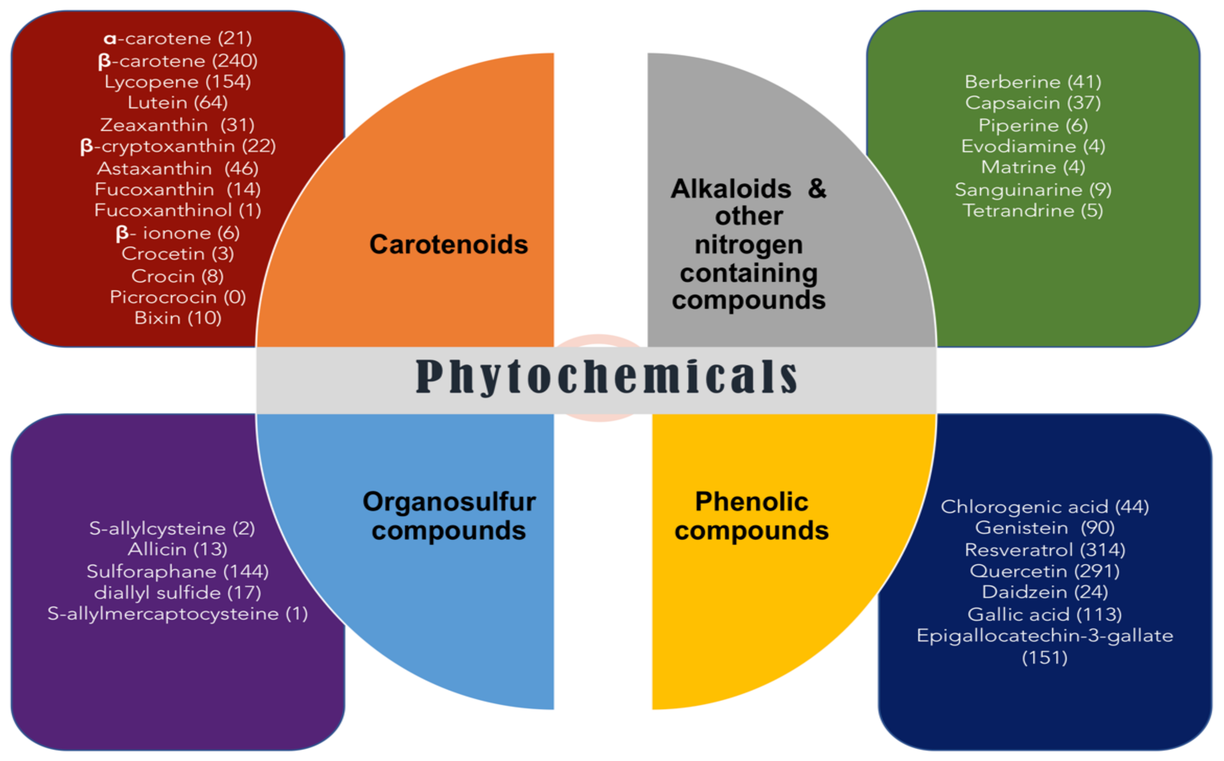
5. Classification of Dietary Antioxidant Phytochemicals in the Management of Oxidative Stress and Cancer
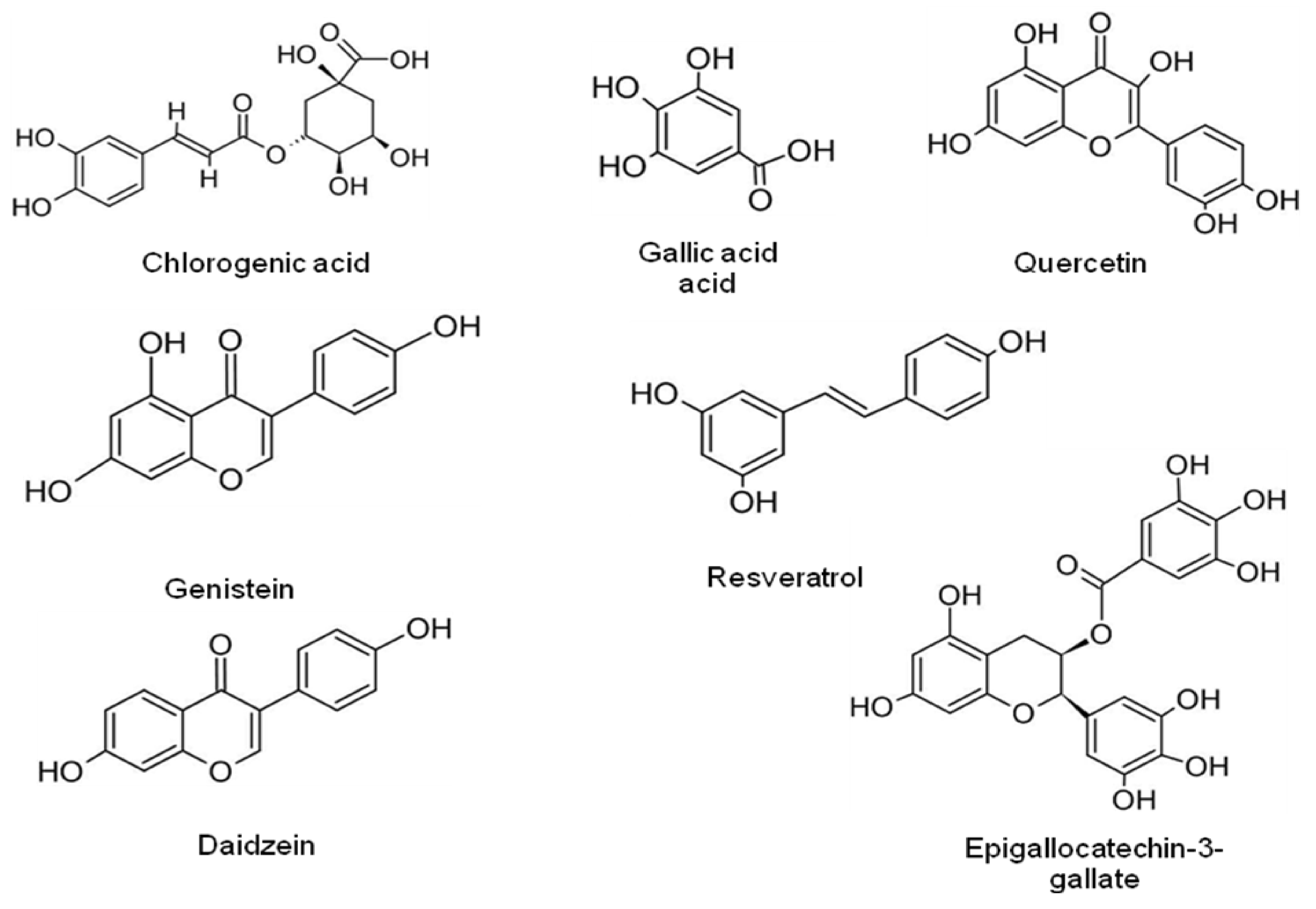
5.1. Phenolic Compounds
| Bioactive Molecules | Indian Ethnomedicinal Plant Source | Chemical Classification | Associated Condition | Cell Line Tested | Biological Approach (In Vitro/In Vivo) | Mechanism of Action | References |
|---|---|---|---|---|---|---|---|
| Chlorogenic acid | Jjatamansi (Nardostachys jatamansi) | Ester of caffeic acid and (−)-quinic acid | Osteosarcoma | U2OS, Saos-2, and MG-63 OS | In vitro | Activates extracellular-signal-regulated kinase1/2 (ERK1/2) | [97] |
| Epigallocatechin-3-gallate | Syamaparni (Camellia sinensis) | Ester of epigallocatechin and gallic acid | Oral squamous cell carcinoma | KBV200 | In vivo | Inhibition of angiogenesis via VEGF down-regulation | [98] |
| Genistein | Rajapatha (Stephania glabra) | Isoflavones | Kidney cancer | In vitro | Induces apoptosis and inhibit the proliferation via regulating CDKN2a methylation | [99] | |
| Resveratrol | Drakshasava (Vitis vinifera L.) | Stilbenoid | Renal cell carcinoma | In vitro/in vivo | Depressing activity of NLRP3, and NLRP3 | [100] | |
| Quercetin | Tulsi (Ocimum sanctum) | Flavonol | Malignant melanoma | B16 | In vitro | Reduced the proportion of cells in the S and G2/M stages of the cell cycle | [101] |
| Daidzein | Vidari (Pueraria tuberosa) | Isoflavones | Colorectal cancer | In vivo | Lessened the protein expression of p-ERK/ERK and p-AKT/AKT | [102] | |
| Gallic acid | Amla (Emblica officinalis ) | Phenolic acid | Prostate cancer | PC-3 | In vitro/in vivo | Inhibits HDAC1 and 2 expression | [103] |
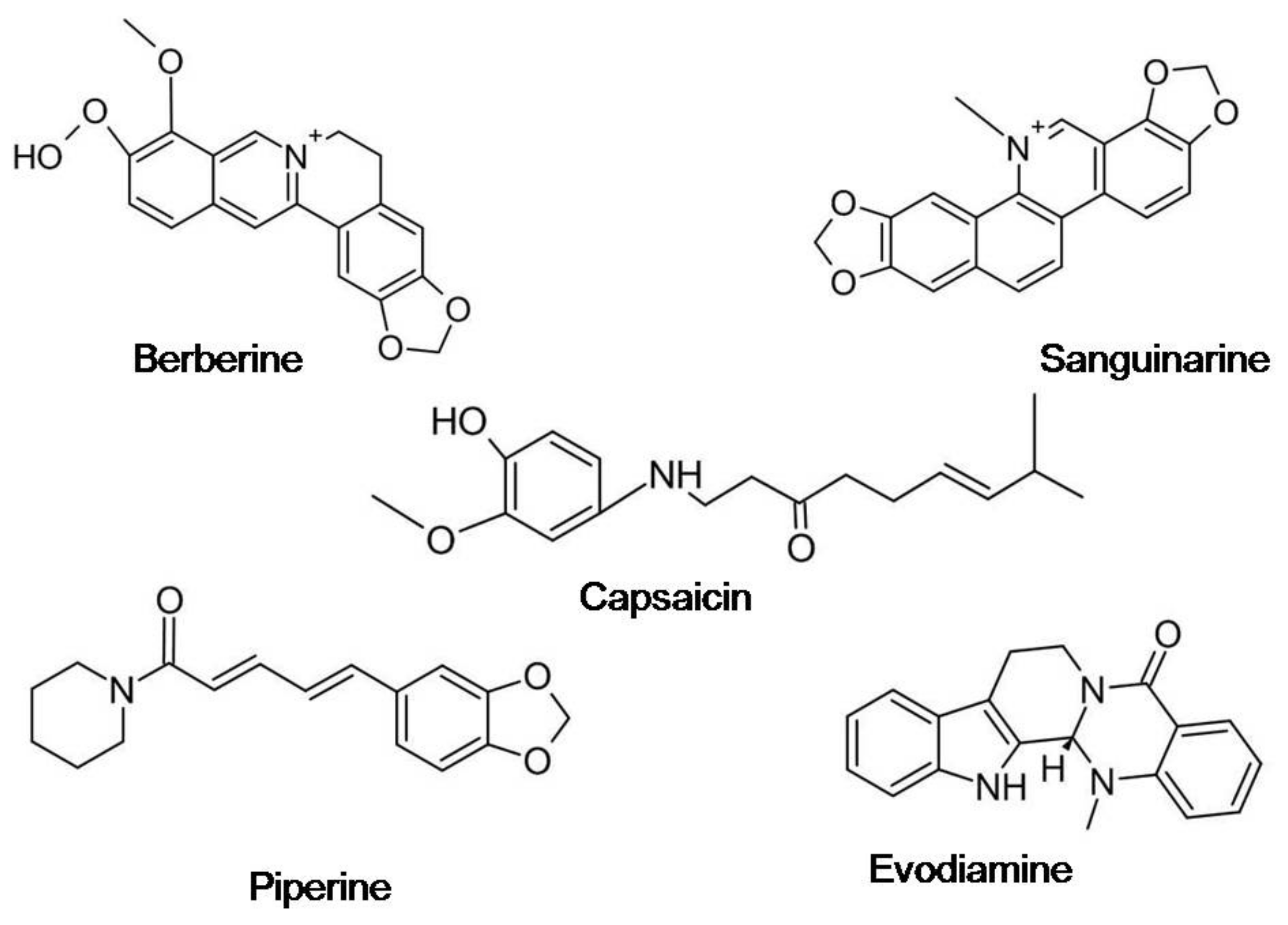
5.2. Alkaloids
| Bioactive Molecules | Indian Ethnomedicinal Plant Source | Chemical Classification | Associated Condition | Cell Line Tested | Biological Approach (In Vitro/In Vivo) | Mechanism of Action | References |
|---|---|---|---|---|---|---|---|
| Berberine | Daruharidra (Beberis aristata) | Isoquinoline alkaloid | Colon cancer | In vitro | Targets SCAP/SREBP-1 pathway driving lipogenesis | [114] | |
| Chronic lymphocytic leukemia | Clinical trial/human patient | Induces apoptosis by decreasing ROR1, Bcl-2, and mir-21 | [115] | ||||
| Capsaicin | Twak (Cinnamomum verum) | Capsaicinoids | Prostate cancer | PC-3, DU145 | In vitro | Suppressionof prostate cancer stem cells via inhibition of Wnt/β-catenin pathway | [116] |
| Piperine | Pipali (Piper nigrum) | N-acylpiperidine | Human melanoma cells | A375P, A375SM | In vitro and in vivo | Increased the expression of BCL2-associated X, apoptosis regulator (BAX), cleaved poly (ADP-ribose) polymerase, cleaved caspase-9, phospho-c-Jun N-terminal kinase and phospho-p38, and reduced that of B-cell lymphoma 2 (BCL2) | [117] |
| Sanguinarine | Svarnakshiri (Argemone mexicana) | Benzophenanthridine alkaloid | Multiple myeloma | U266, IM9, MM1S, and RPMI-8226 | In vitro | Induces mitochondrial/caspase-dependent apoptosis, produces oxidative stress, and suppresses proliferation cancer cell lines | [118] |
| Tetrandrine | Patha (Cissampelos pareira) | Bis-benzylisoquinoline alkaloid | Breast cancer | MDA-MB-231 | In vivo | TET-upregulated Caspase-3, Bid, Bax, and, downregulated by Bcl-2, Survivin, and PARP | [119,120] |
5.3. Organosulfur Compounds
| Bioactive Molecules | Indian Ethnomedicinal Plant Source | Chemical Classification | Associated Condition | Cell Line Tested | Biological Approach (In Vitro/In Vivo) | Mechanism of Action | References |
|---|---|---|---|---|---|---|---|
| S-allylcysteine | Lasuna (Fresh garlic) | Derivative of the amino acid cysteine | Lung cancer | A549 | In vitro/in silico | Reduced expression of PD-L1 and HIF-1α, showed efficient docking with PD-L1 immune checkpoint target | [135] |
| Allicin | Lasuna (Crushed or chopped garlic) | Sulfoxide | Skin cancer/melanoma | A375 | In vitro | Reduction of MMP-9 mRNA expression | [136] |
| Cholangiocarcinoma | MCF-7, HCC-70 | In vitro/in vivo | Overpowers cell proliferation and invasion through STAT3 signaling | [137] | |||
| S-allylmercaptocysteine | Lasuna (Aged garlic) | Thioallyl compound | Lung cancer | A549 | In vitro | Suppression of cell proliferation, regulation of cell cycle, attenuation of ROS formation, inhibition of DNA damage, increase of SOD activity and inhibition of nuclear factor-kappa B (NF-κB) activity | [138] |
| Sulforaphane | Shatavari (Asparagus racemosus) | Isothiocyanate | Small-cell lung cancer | SCLC cell lines NCI-H69, NCI-H69AR and NCI-H82 | In vitro | Induced cell death was mediated via ferroptosis and Inhibition of the mRNA and protein expression levels | [139] |
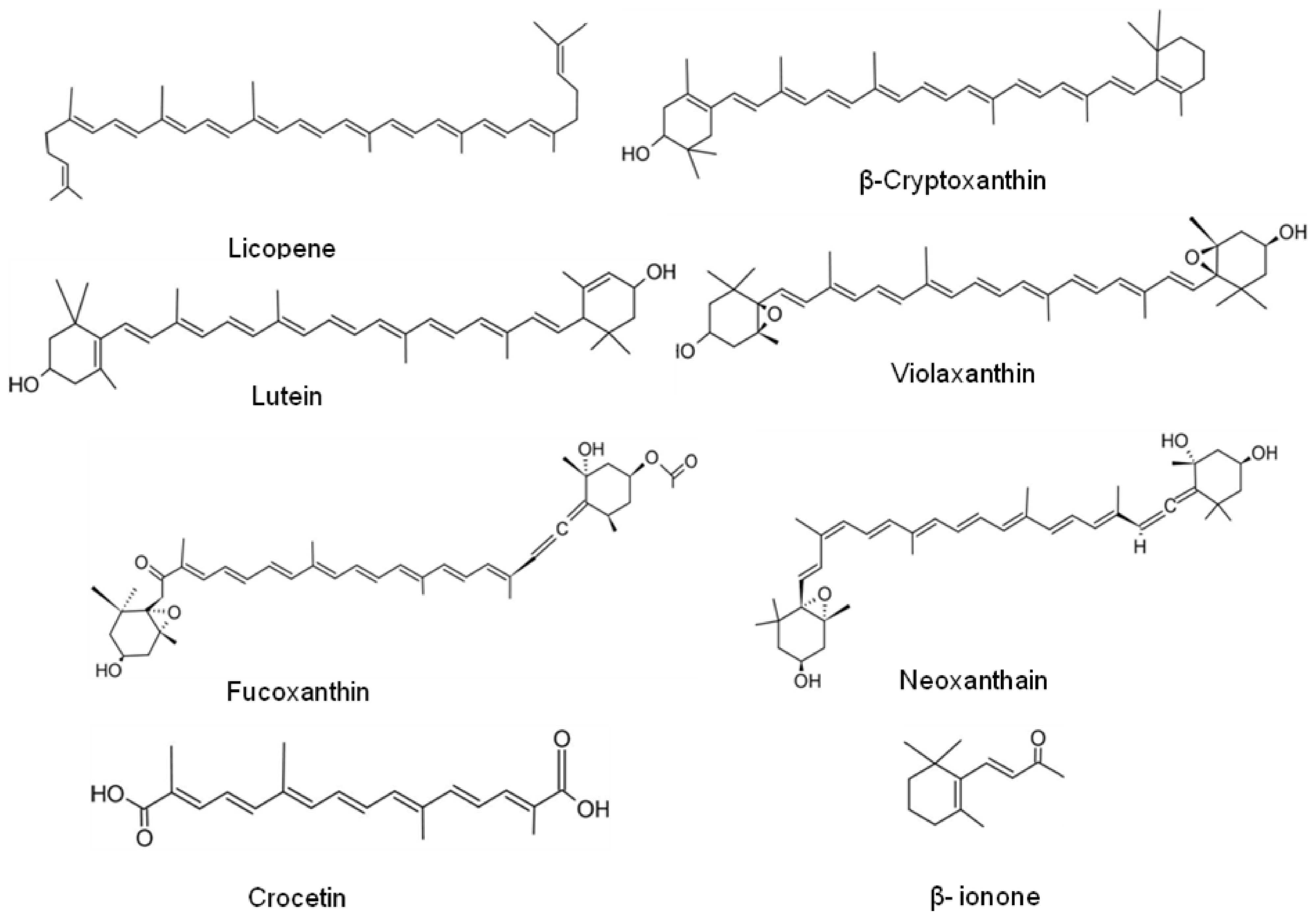
5.4. Carotenoids
6. Conclusions and Future Prospects
Author Contributions
Funding
Acknowledgments
Conflicts of Interest
References
- Croce, C.M. Oncogenes and cancer. N. Engl. J. Med. 2008, 358, 502–511. [Google Scholar] [CrossRef] [Green Version]
- Wang, L.H.; Wu, C.F.; Rajasekaran, N.; Shin, Y.K. Loss of Tumor Suppressor Gene Function in Human Cancer: An Overview. Cell Physiol. Biochem. 2018, 51, 2647–2693. [Google Scholar] [CrossRef]
- Bhattacharya, S.; Ghosh, M.K. Cell death and deubiquitinases: Perspectives in cancer. BioMed Res. Int. 2014, 2014, 435197. [Google Scholar] [CrossRef] [PubMed] [Green Version]
- Redza-Dutordoir, M.; Averill-Bates, D.A. Activation of apoptosis signalling pathways by reactive oxygen species. Biochim. Biophys. Acta 2016, 1863, 2977–2992. [Google Scholar] [CrossRef]
- Goodman, M. Managing the side effects of chemotherapy. Semin. Oncol. Nurs. 1989, 5 (Suppl. 2), 29–52. [Google Scholar] [CrossRef]
- Di Gioia, F.; Tzortzakis, N.; Rouphael, Y.; Kyriacou, M.C.; Sampaio, S.L.; CFR Ferreira, I.; Petropoulos, S.A. Grown to Be Blue—Antioxidant Properties and Health Effects of Colored Vegetables. Part II: Leafy, Fruit, and Other Vegetables. Antioxidants 2020, 9, 97. [Google Scholar] [CrossRef] [PubMed] [Green Version]
- Shin, S.A.; Moon, S.Y.; Kim, W.Y.; Paek, S.M.; Park, H.H.; Lee, C.S. Structure-Based Classification and Anti-Cancer Effects of Plant Metabolites. Int. J. Mol. Sci. 2018, 19, 2651. [Google Scholar] [CrossRef] [Green Version]
- Reuter, S.; Gupta, S.C.; Chaturvedi, M.M.; Aggarwal, B.B. Oxidative stress, inflammation, and cancer: How are they linked? Free Radic. Biol. Med. 2010, 49, 1603–1616. [Google Scholar] [CrossRef] [Green Version]
- Visconti, R.; Grieco, D. New insights on oxidative stress in cancer. Curr. Opin. Drug Discov. Dev. 2009, 12, 240–245. [Google Scholar]
- Ďuračková, Z. Some current insights into oxidative stress. Physiol. Res. 2010, 59, 459–469. [Google Scholar] [CrossRef]
- Kurutas, E.B. The importance of antioxidants which play the role in cellular response against oxidative/nitrosative stress: Current state. Nutr. J. 2016, 15, 71. [Google Scholar] [CrossRef] [PubMed] [Green Version]
- Noda, N.; Wakasugi, H. Cancer and oxidative stress. Jpn. Med. Assoc. J. 2001, 44, 535–539. [Google Scholar]
- Sen, S.; Chakraborty, R. Revival, modernization and integration of Indian traditional herbal medicine in clinical practice: Importance, challenges and future. J. Tradit. Complement. Med. 2017, 7, 234–244. [Google Scholar] [CrossRef]
- Vaidya, A.D.; Devasagayam, T.P. Current status of herbal drugs in India: An overview. J. Clin. Biochem. Nutr. 2007, 41, 1–11. [Google Scholar] [CrossRef] [PubMed] [Green Version]
- Samal, J. Medicinal plants and related developments in India: A peep into 5-year plans of India. Indian J. Health Sci. Biomed. Res. (KLEU) 2016, 9, 14–19. [Google Scholar] [CrossRef]
- Balachandran, P.; Govindarajan, R. Cancer—An ayurvedic perspective. Pharmacol. Res. 2005, 51, 19–30. [Google Scholar] [CrossRef]
- Hussein, R.; El-Anssary, A. Plants Secondary Metabolites: The Key Drivers of the Pharmacological Actions of Medicinal Plants. Herb. Med. 2019, 1, 13. [Google Scholar]
- Pan, L.; Chai, H.; Kinghorn, A.D. The continuing search for antitumor agents from higher plants. Phytochem. Lett. 2010, 3, 1–8. [Google Scholar] [CrossRef] [Green Version]
- Ansari, J.A.; Rastogi, N.; Ahmad, M.K.; Mahdi, A.A.; Khan, A.R.; Thakur, R.; Srivastava, V.K.; Mishra, D.P.; Fatima, N.; Khan, H.J.; et al. ROS mediated pro-apoptotic effects of Tinospora cordifolia on breast cancer cells. Front. Biosci. (Elite Ed.) 2017, 9, 89–100. [Google Scholar]
- Kumar, B.R.; Anupam, A.; Manchikanti, P.; Rameshbabu, A.P.; Dasgupta, S.; Dhara, S. Identification and characterization of bioactive phenolic constituents, anti-proliferative, and anti-angiogenic activity of stem extracts of Basella alba and rubra. J. Food Sci. Technol. 2018, 55, 1675–1684. [Google Scholar] [CrossRef]
- Khan, N.; Tamboli, E.; Sharma, V.K.; Kumar, S. Phytochemical and pharmacological aspects of Nothapodytes nimmoniana. An overview. Herba Pol. 2013, 59, 1. [Google Scholar] [CrossRef] [Green Version]
- Tran, M.H.; Nguyen, H.D.; Kim, J.C.; Choi, J.S.; Lee, H.K.; Min, B.S. Phenolic glycosides from Alangium salviifolium leaves with inhibitory activity on LPS-induced NO, PGE(2), and TNF-alpha production. Bioorg. Med. Chem. Lett. 2009, 19, 4389–4393. [Google Scholar] [PubMed]
- Gavande, K.; Jain, K.; Jain, B.; Mehta, R. Comprehensive Report on Phytochemistry and Pharmacological Prominence of Withania somnifera. UK J. Pharm. Biosci. 2015, 3, 15. [Google Scholar] [CrossRef]
- Devanesan, E.B.; Anand, A.V.; Kumar, P.S.; Vinayagamoorthy, P.; Basavaraju, P. Phytochemistry and Pharmacology of Ficus religiosa. Syst. Rev. Pharm. 2018, 9, 45–48. [Google Scholar] [CrossRef]
- Uddin, N.; Ali, N.; Uddin, Z.; Nazir, N.; Zahoor, M.; Rashid, U.; Ullah, R.; Alqahtani, A.S.; Alqahtani, A.M.; Nasr, F.A.; et al. Evaluation of Cholinesterase Inhibitory Potential of Different Genotypes of Ziziphus nummularia, Their HPLC-UV, and Molecular Docking Analysis. Molecules 2020, 25, 5011. [Google Scholar] [CrossRef]
- Devi, S.; Rashid, R.; Kumar, M. Phytochemistry and pharmacological properties of Phyllanthus amarus Schum: A review. Pharma Innov. J. 2017, 6, 169–172. [Google Scholar]
- Jayakumar, T.; Hsieh, C.Y.; Lee, J.J.; Sheu, J.R. Experimental and Clinical Pharmacology of Andrographis paniculata and Its Major Bioactive Phytoconstituent Andrographolide. Evid.-Based Complement. Altern. Med. eCAM 2013, 2013, 846740. [Google Scholar] [CrossRef] [PubMed] [Green Version]
- Pandey, S.; Phulara, S.C.; Mishra, S.K.; Bajpai, R.; Kumar, A.; Niranjan, A.; Lehri, A.; Upreti, D.K.; Chauhan, P.S. Betula utilis extract prolongs life expectancy, protects against amyloid-β toxicity and reduces Alpha Synuclien in Caenorhabditis elegans via DAF-16 and SKN-1. Comp. Biochem. Physiol. Toxicol. Pharmacol. CBP 2020, 228, 108647. [Google Scholar] [CrossRef]
- Seemaisamy, R.; Faruck, L.H.; Gattu, S.; Neelamegam, R.; Bakshi, H.A.; Rashan, L.; Al-Buloshi, M.; Hasson, S.S.A.A.; Nagarajan, K. Anti-Microbial and Anti-Cancer Activity of Aegle Marmelos and Gas Chromatography Coupled Spectrometry Analysis of Their Chemical Constituents. Int. J. Pharm. Sci. Res. 2019, 10, 373–380. [Google Scholar]
- Prakash, V.; Jaiswal, N.; Srivastava, M. A Review on Medicinal Properties of Centella Asiatica. Asian J. Pharm. Clin. Res. 2017, 10, 69–74. [Google Scholar] [CrossRef] [Green Version]
- Jeyasri, R.; Muthuramalingam, P.; Suba, V.; Ramesh, M.; Chen, J.T. Bacopa monnieri and Their Bioactive Compounds Inferred Multi-Target Treatment Strategy for Neurological Diseases: A Cheminformatics and System Pharmacology Approach. Biomolecules 2020, 10, 536. [Google Scholar] [CrossRef] [Green Version]
- Koczurkiewicz, P.; Łojewski, M.; Piska, K.; Michalik, M.; Wójcik-Pszczoła, K.; Szewczyk, A.; Hałaszuk, P.; Pękala, E.; Muszyńska, B. Chemopreventive and Anticancer Activities of Bacopa monnieri Extracted from Artificial Digestive Juices. Nat. Prod. Commun. 2017, 12, 337–342. [Google Scholar] [CrossRef] [Green Version]
- Aswal, J.; Dobhal, R.; Uniyal, D.P.; Nautiyal, V. A review on Pharmacological potential of Berberine; an active component of Himalayan Berberis aristata. J. Phytopharmacol. 2017, 6, 53–58. [Google Scholar]
- Al-Snafi, A. Medical importance of Datura fastuosa (syn: Datura metel) and Datura stramonium—A review. IOSR J. Pharm. 2017, 7, 43–58. [Google Scholar] [CrossRef]
- Okoro, E.E.; Osoniyi, O.R.; Jabeen, A.; Shams, S.; Choudhary, M.I.; Onajobi, F.D. Anti-proliferative and immunomodulatory activities of fractions from methanol root extract of Abrus precatorius L. Clin. Phytoscience 2019, 5, 45. [Google Scholar] [CrossRef]
- Tomeh, M.A.; Hadianamrei, R.; Zhao, X. A Review of Curcumin and Its Derivatives as Anticancer Agents. Int. J. Mol. Sci. 2019, 20, 1033. [Google Scholar] [CrossRef] [PubMed] [Green Version]
- Rathinamoorthy, R.; Thilagavathi, G. Terminalia chebula-review on pharmacological and biochemical studies. Int. J. Pharm.Tech. Res. 2014, 6, 97–116. [Google Scholar]
- Fatima, N.; Ahmad, M.; Ansari, J.; Khan, H.; Rastogi, N.; Srivastava, S.; Ahmad, S.; Ali, Z. Antiproliferative and Antioxidant Studies of Anthocephalus cadamba Rox. Miq. Bark. Indian J. Pharm. Sci. 2016, 78, 525–531. [Google Scholar] [CrossRef] [Green Version]
- Khan, H.J.; Ahmad, M.K.; Khan, A.R.; Rastogi, N.; Mahdi, A.A.; Ansari, J.A.; Fatima, N.; Satyanarayan, G.N.V. Identification of Anticancer and Antioxidant phytoconstituents from chloroform fraction of Solanum nigrum L. berries using GC-MS/MS analysis. Indian J. Exp. Biol. 2016, 54, 774–782. [Google Scholar] [PubMed]
- Pal, P.K.; Nandi, M.K.; Singh, N.K. Detoxification of Croton tiglium L. seeds by Ayurvedic process of Śodhana. Anc. Sci. Life 2014, 33, 157. [Google Scholar] [PubMed]
- Bukhari, S.I.; Manzoor, M.; Dhar, M.K. A comprehensive review of the pharmacological potential of Crocus sativus and its bioactive apocarotenoids. Biomed. Pharmacother. 2018, 98, 733–745. [Google Scholar] [CrossRef]
- Coria-Téllez, A.V.; Montalvo-Gónzalez, E.; Yahia, E.M.; Obledo-Vázquez, E.N. Annona muricata: A comprehensive review on its traditional medicinal uses, phytochemicals, pharmacological activities, mechanisms of action and toxicity. Arab. J. Chem. 2018, 11, 662–691. [Google Scholar] [CrossRef] [Green Version]
- El-Saber Batiha, G.; Alkazmi, L.M.; Wasef, L.G.; Beshbishy, A.M.; Nadwa, E.H.; Rashwan, E.K. Syzygium aromaticum L. (Myrtaceae): Traditional Uses, Bioactive Chemical Constituents, Pharmacological and Toxicological Activities. Biomolecules 2020, 10, 202. [Google Scholar] [CrossRef] [PubMed] [Green Version]
- Braga, T.M.; Rocha, L.; Chung, T.Y.; Oliveira, R.F.; Pinho, C.; Oliveira, A.I.; Morgado, J.; Cruz, A. Azadirachta indica A. Juss In Vivo Toxicity—An Updated Review. Molecules 2021, 26, 252. [Google Scholar] [CrossRef]
- Chhikara, N.; Kour, R.; Jaglan, S.; Gupta, P.; Gat, Y.; Panghal, A. Citrus medica: Nutritional, phytochemical composition and health benefits—A review. Food Funct. 2018, 9, 1978–1992. [Google Scholar] [CrossRef]
- Marrelli, M.; Amodeo, V.; Statti, G.; Conforti, F. Biological Properties and Bioactive Components of Allium cepa L.: Focus on Potential Benefits in the Treatment of Obesity and Related Comorbidities. Molecules 2019, 24, 119. [Google Scholar] [CrossRef] [Green Version]
- Yadav, V.; Krishnan, A.; Vohora, D. A systematic review on Piper longum L.: Bridging traditional knowledge and pharmacological evidence for future translational research. J. Ethnopharmacol. 2020, 247, 112255. [Google Scholar] [CrossRef]
- Anirban, C.; Santanu, P. A Review on Phytochemical and Pharmacological Potential of Alpinia galanga. Pharmacogn. J. 2018, 10, 9–15. [Google Scholar]
- Alam, K.; Hoq, O.; Uddin, S. Medicinal plant Allium sativum: A Review. J. Med. Plants Stud. 2016, 4, 72–79. [Google Scholar]
- Zargar, B.A.; Masoodi, M.H.; Ahmed, B.; Ganie, S.A. Phytoconstituents and therapeutic uses of Rheum emodi wall. ex Meissn. Food Chem. 2011, 128, 585–589. [Google Scholar] [CrossRef]
- Alok, S.; Jain, S.K.; Verma, A.; Kumar, M.; Mahor, A.; Sabharwal, M. Plant profile, phytochemistry and pharmacology of Asparagus racemosus (Shatavari): A review. Asian Pac. J. Trop. Dis. 2013, 3, 242–251. [Google Scholar] [CrossRef]
- Ansari, J.A.; Ahmad, M.K.; Khan, A.R.; Fatima, N.; Khan, H.J.; Rastogi, N.; Mishra, D.P.; Mahdi, A.A. Anticancer and Antioxidant activity of Zingiber officinale Roscoe rhizome. Indian J. Exp. Biol. 2016, 54, 767–773. [Google Scholar] [PubMed]
- Joshi, R.K.; Setzer, W.N.; Da Silva, J.K. Phytoconstituents, traditional medicinal uses and bioactivities of Tulsi (Ocimum sanctum Linn.): A review. Am. J. Essent. Oils Nat. Prod. 2017, 5, 18–21. [Google Scholar]
- Kami, S.S.; Hameed, I.H.; Hamza, L.F. Acorus calamus: Parts used, insecticidal, anti-fungal, antitumour and anti-inflammatory activity: A review. Int. J. Pharm. Clin. Res. 2017, 8, 153–157. [Google Scholar]
- Razack, S.; Kumar, K.H.; Nallamuthu, I.; Naika, M.; Khanum, F. Antioxidant, Biomolecule Oxidation Protective Activities of Nardostachys jatamansi DC and Its Phytochemical Analysis by RP-HPLC and GC-MS. Antioxidants 2015, 4, 185–203. [Google Scholar] [CrossRef] [Green Version]
- Pandey, M.M.; Katara, A.; Pandey, G.; Rastogi, S.; Rawat, A.K.S. An Important Indian Traditional Drug of Ayurveda Jatamansi and Its Substitute Bhootkeshi: Chemical Profiling and Antioxidant Activity. Evid.-Based Complement. Altern. Med. 2013, 2013, 142517. [Google Scholar] [CrossRef] [Green Version]
- Dixit, V.; Jain, P.; Joshi, S. Hypolipidaemic effects of Curcuma longa Linn., and Nardostachys jatamansi DC, in triton-induced hyperlipidaemic rats. Indian J. Physiol. Pharmacol. 1988, 32, 299–304. [Google Scholar]
- Pillai, D.; Pandita, N. Determination of Quality Standards for Draksharishta, a Polyherbal Ayurvedic Formulation. Indian J. Pharm. Sci. 2016, 78, 129–135. [Google Scholar]
- Sathiya, M. Scientific Evaluation of Antioxidant and Anti Cancer Activity of Kanchanara Guggulu Vati by Invitro Methods. Ph.D. Thesis, Madras Medical College, Chennai, India, 2017. [Google Scholar]
- Saraswathy, A.; Sundaresan, R.; Joy, S.; Gopal, R.H. Effect of Container on Ayurvedic Drugs—A Select Study. Anc. Sci. Life 2004, 24, 11. [Google Scholar]
- Peterson, C.T.; Denniston, K.; Chopra, D. Therapeutic uses of triphala in ayurvedic medicine. J. Altern. Complement. Med. 2017, 23, 607–614. [Google Scholar] [CrossRef] [Green Version]
- Kotteswari, M.; Rao, M.; Kumar, S.; Prabhu, K.; Sundaram, R.L.; Dinakar, S. GC MS Analysis of One Ayurvedic Preparation ‘Aswagandharishtam’. Biomed. Pharmacol. J. 2018, 11, 1061–1072. [Google Scholar] [CrossRef]
- Patel, V.; Parmar, N. A Comparative Clinical Study of Kasisadi Taila and Jatyadi Taila in the Management of Arsha. J. Ayurveda Holist. Med. 2017, 5, 16–24. [Google Scholar]
- Jadhav, A.N.; Bhutani, K. Ayurveda and gynecological disorders. J. Ethnopharmacol. 2005, 97, 151–159. [Google Scholar] [CrossRef]
- Govardhan, B.; Manjunatha, A.; Kumar, S.S. Standardization of Lashuna taila: An ayurvedic oil based medicine. J Pharmacogn. Phytochem. 2018, 7, 28338. [Google Scholar]
- Ferguson, L.R.; Chen, H.; Collins, A.R.; Connell, M.; Damia, G.; Dasgupta, S.; Malhotra, M.; Meeker, A.K.; Amedei, A.; Amin, A.; et al. Genomic instability in human cancer: Molecular insights and opportunities for therapeutic attack and prevention through diet and nutrition. Semin. Cancer Biol. 2015, 35, S5–S24. [Google Scholar] [CrossRef] [PubMed]
- Forni, C.; Facchiano, F.; Bartoli, M.; Pieretti, S.; Facchiano, A.; D’Arcangelo, D.; Norelli, S.; Valle, G.; Nisini, R.; Beninati, S.; et al. Beneficial Role of Phytochemicals on Oxidative Stress and Age-Related Diseases. BioMed Res. Int. 2019, 2019, 8748253. [Google Scholar] [CrossRef] [PubMed] [Green Version]
- Zhang, Y.-J.; Gan, R.-Y.; Li, S.; Zhou, Y.; Li, A.-N.; Xu, D.-P.; Li, H.-B. Antioxidant Phytochemicals for the Prevention and Treatment of Chronic Diseases. Molecules 2015, 20, 21138–21156. [Google Scholar] [CrossRef] [PubMed]
- Mileo, A.M.; Miccadei, S. Polyphenols as Modulator of Oxidative Stress in Cancer Disease: New Therapeutic Strategies. Oxidative Med. Cell. Longev. 2016, 2016, 6475624. [Google Scholar] [CrossRef] [Green Version]
- Choi, D.G.; Venkatesan, J.; Shim, M.S. Selective Anticancer Therapy Using Pro-Oxidant Drug-Loaded Chitosan–Fucoidan Nanoparticles. Int. J. Mol. Sci. 2019, 20, 3220. [Google Scholar] [CrossRef] [Green Version]
- Kocyigit, A.; Guler, E.M.; Dikilitas, M. Role of antioxidant phytochemicals in prevention, formation and treatment of cancer. In Reactive Oxygen Species (ROS) in Living Cells; InterchOpen: London, UK, 2018; pp. 21–45. [Google Scholar]
- Swallah, M.S.; Sun, H.; Affoh, R.; Fu, H.; Yu, H. Antioxidant potential overviews of secondary metabolites (polyphenols) in fruits. Int. J. Food Sci. 2020, 2020, 9081686. [Google Scholar] [CrossRef]
- Giordano, A.; Tommonaro, G. Curcumin and Cancer. Nutrients 2019, 11, 2376. [Google Scholar] [CrossRef] [Green Version]
- Shahidi, F.; Ambigaipalan, P. Phenolics and polyphenolics in foods, beverages and spices: Antioxidant activity and health effects—A review. J. Funct. Foods 2015, 18, 820–897. [Google Scholar] [CrossRef]
- Vafadar, A.; Shabaninejad, Z.; Movahedpour, A.; Fallahi, F.; Taghavipour, M.; Ghasemi, Y.; Akbari, M.; Shafiee, A.; Hajighadimi, S.; Moradizarmehri, S.; et al. Quercetin and cancer: New insights into its therapeutic effects on ovarian cancer cells. Cell Biosci. 2020, 10, 32. [Google Scholar] [CrossRef] [Green Version]
- Batiha, G.E.-S.; Beshbishy, A.M.; Ikram, M.; Mulla, Z.S.; El-Hack, M.E.A.; Taha, A.E.; Algammal, A.M.; Elewa, Y.H.A. The Pharmacological Activity, Biochemical Properties, and Pharmacokinetics of the Major Natural Polyphenolic Flavonoid: Quercetin. Foods 2020, 9, 374. [Google Scholar] [CrossRef] [Green Version]
- Sharmila, G.; Athirai, T.; Kiruthiga, B.; Senthilkumar, K.; Elumalai, P.; Arunkumar, R.; Arunakaran, J. Chemopreventive effect of quercetin in MNU and testosterone induced prostate cancer of Sprague-Dawley rats. Nutr. Cancer 2014, 66, 38–46. [Google Scholar] [CrossRef]
- Nair, M.P.; Mahajan, S.; Reynolds, J.L.; Aalinkeel, R.; Nair, H.; Schwartz, S.A.; Kandaswami, C. The Flavonoid Quercetin Inhibits Proinflammatory Cytokine (Tumor Necrosis Factor Alpha) Gene Expression in Normal Peripheral Blood Mononuclear Cells via Modulation of the NF-κβ System. Clin. Vaccine Immunol. 2006, 13, 319–328. [Google Scholar] [CrossRef] [Green Version]
- Jing, Z.; Wang, Z.; Li, X.; Li, X.; Cao, T.; Bi, Y.; Zhou, J.; Chen, X.; Yu, D.; Zhu, L.; et al. Protective Effect of Quercetin on Posttraumatic Cardiac Injury. Sci. Rep. 2016, 6, 30812. [Google Scholar] [CrossRef] [Green Version]
- Vaid, M.; Katiyar, S.K. Molecular mechanisms of inhibition of photocarcinogenesis by silymarin, a phytochemical from milk thistle (Silybum marianum L. Gaertn.). Int. J. Oncol. 2010, 36, 1053–1060. [Google Scholar] [PubMed] [Green Version]
- Udenigwe, C.C.; Ramprasath, V.R.; Aluko, R.E.; Jones, P.J. Potential of resveratrol in anticancer and anti-inflammatory therapy. Nutr. Rev. 2008, 66, 445–454. [Google Scholar] [CrossRef] [PubMed]
- Storniolo, C.E.; Moreno, J.J. Resveratrol Analogs with Antioxidant Activity Inhibit Intestinal Epithelial Cancer Caco-2 Cell Growth by Modulating Arachidonic Acid Cascade. J. Agric. Food Chem. 2019, 67, 819–828. [Google Scholar] [CrossRef] [PubMed]
- Gupta, M.; Panizai, M.; Tareen, M.F.; Ortega-Martinez, S.; Doreulee, N. An Overview on Novel Antioxidant and Anti-cancer Properties of Lycopene: A Comprehensive Review. GMJ Med. 2018, 2, 45–50. [Google Scholar] [CrossRef] [Green Version]
- Palozza, P.; Serini, S.; Di Nicuolo, F.; Calviello, G. Modulation of apoptotic signalling by carotenoids in cancer cells. Arch. Biochem. Biophys. 2004, 430, 104–109. [Google Scholar] [CrossRef]
- Mordi, R.C.; Ademosun, O.T.; Ajanaku, C.O.; Olanrewaju, I.O.; Walton, J.C. Free Radical Mediated Oxidative Degradation of Carotenes and Xanthophylls. Molecules 2020, 25, 1038. [Google Scholar] [CrossRef] [Green Version]
- Lamoral-Theys, D.; Pottier, L.; Dufrasne, F.; Nève, J.; Dubois, J.; Kornienko, A.; Kiss, R.; Ingrassia, L. Natural polyphenols that display anticancer properties through inhibition of kinase activity. Curr. Med. Chem. 2010, 17, 812–825. [Google Scholar] [CrossRef] [Green Version]
- Anantharaju, P.G.; Gowda, P.C.; Vimalambike, M.G.; Madhunapantula, S.V. An overview on the role of dietary phenolics for the treatment of cancers. Nutr. J. 2016, 15, 99. [Google Scholar] [CrossRef] [PubMed] [Green Version]
- Shaito, A.; Posadino, A.M.; Younes, N.; Hasan, H.; Halabi, S.; Alhababi, D.; Al-Mohannadi, A.; Abdel-Rahman, W.M.; Eid, A.H.; Nasrallah, G.K.; et al. Potential Adverse Effects of Resveratrol: A Literature Review. Int. J. Mol. Sci. 2020, 21, 2084. [Google Scholar] [CrossRef] [PubMed] [Green Version]
- Tuorkey, M.J. Curcumin a potent cancer preventive agent: Mechanisms of cancer cell killing. Interv. Med. Appl. Sci. 2014, 6, 139–146. [Google Scholar] [CrossRef] [PubMed] [Green Version]
- Balasubramanian, S.; Eckert, R.L. Curcumin suppresses AP1 transcription factor-dependent differentiation and activates apoptosis in human epidermal keratinocytes. J. Biol. Chem. 2007, 282, 6707–6715. [Google Scholar] [CrossRef] [PubMed] [Green Version]
- Ali, B.H.; Blunden, G.; Tanira, M.O.; Nemmar, A. Some phytochemical, pharmacological and toxicological properties of ginger (Zingiber officinale Roscoe): A review of recent research. Food Chem. Toxicol. Int. J. Publ. Br. Ind. Biol. Res. Assoc. 2008, 46, 409–420. [Google Scholar] [CrossRef]
- Mao, Q.-Q.; Xu, X.-Y.; Cao, S.-Y.; Gan, R.-Y.; Corke, H.; Beta, T.; Li, H.-B. Bioactive Compounds and Bioactivities of Ginger (Zingiber officinale Roscoe). Foods 2019, 8, 185. [Google Scholar] [CrossRef] [Green Version]
- De Lima, R.M.T.; Dos Reis, A.C.; de Menezes, A.P.M.; Santos, J.V.O.; Filho, J.; Ferreira, J.R.O.; de Alencar, M.; da Mata, A.; Khan, I.N.; Islam, A.; et al. Protective and therapeutic potential of ginger (Zingiber officinale) extract and [6]-gingerol in cancer: A comprehensive review. Phytother. Res. PTR 2018, 32, 1885–1907. [Google Scholar] [CrossRef] [PubMed]
- Ju, J.; Picinich, S.C.; Yang, Z.; Zhao, Y.; Suh, N.; Kong, A.N.; Yang, C.S. Cancer-preventive activities of tocopherols and tocotrienols. Carcinogenesis 2010, 31, 533–542. [Google Scholar] [CrossRef] [Green Version]
- Jiang, Q. Natural forms of vitamin E and metabolites-regulation of cancer cell death and underlying mechanisms. IUBMB Life 2019, 71, 495–506. [Google Scholar] [CrossRef]
- Ashoori, M.; Saedisomeolia, A. Riboflavin (vitamin B2) and oxidative stress: A review. Br. J. Nutr. 2014, 111, 1985–1991. [Google Scholar] [CrossRef] [PubMed] [Green Version]
- Sapio, L.; Salzillo, A.; Illiano, M.; Ragone, A.; Spina, A.; Chiosi, E.; Pacifico, S.; Catauro, M.; Naviglio, S. Chlorogenic acid activates ERK1/2 and inhibits proliferation of osteosarcoma cells. J. Cell. Physiol. 2020, 235, 3741–3752. [Google Scholar] [CrossRef] [PubMed]
- Chen, L.; Guo, X.; Hu, Y.; Li, L.; Liang, G.; Zhang, G. Epigallocatechin-3-gallate sensitises multidrug-resistant oral carcinoma xenografts to vincristine sulfate. FEBS Open Bio 2020, 10, 1403–1413. [Google Scholar] [CrossRef] [PubMed]
- Ji, Z.; Huo, C.; Yang, P. Genistein inhibited the proliferation of kidney cancer cells via CDKN2a hypomethylation: Role of abnormal apoptosis. Int. Urol. Nephrol. 2020, 52, 1049–1055. [Google Scholar] [CrossRef] [PubMed]
- Tian, X.; Zhang, S.; Zhang, Q.; Kang, L.; Ma, C.; Feng, L.; Li, S.; Li, J.; Yang, L.; Liu, J.; et al. Resveratrol inhibits tumor progression by down-regulation of NLRP3 in renal cell carcinoma. J. Nutr. Biochem. 2020, 85, 108489. [Google Scholar] [CrossRef]
- Soll, F.; Ternent, C.; Berry, I.M.; Kumari, D.; Moore, T.C. Quercetin Inhibits Proliferation and Induces Apoptosis of B16 Melanoma Cells In Vitro. Assay Drug Dev. Technol. 2020, 18, 261–268. [Google Scholar] [CrossRef]
- Salama, A.A.A.; Allam, R.M. Promising targets of chrysin and daidzein in colorectal cancer: Amphiregulin, CXCL1, and MMP-9. Eur. J. Pharmacol. 2021, 892, 173763. [Google Scholar] [CrossRef]
- Jang, Y.G.; Ko, E.B.; Choi, K.C. Gallic acid, a phenolic acid, hinders the progression of prostate cancer by inhibition of histone deacetylase 1 and 2 expression. J. Nutr. Biochem. 2020, 84, 108444. [Google Scholar] [CrossRef]
- Habli, Z.; Toumieh, G.; Fatfat, M.; Rahal, O.N.; Gali-Muhtasib, H. Emerging Cytotoxic Alkaloids in the Battle against Cancer: Overview of Molecular Mechanisms. Molecules 2017, 22, 250. [Google Scholar] [CrossRef] [PubMed]
- Srivastava, S.; Rawat, A.K. Quality evaluation of ayurvedic crude drug daruharidra, its allied species, and commercial samples from herbal drug markets of India. Evid.-Based Complement. Altern. Med. eCAM 2013, 2013, 472973. [Google Scholar] [CrossRef] [PubMed] [Green Version]
- Liu, B.; Li, W.; Chang, Y.; Dong, W.; Ni, L. Extraction of berberine from rhizome of Coptis chinensis Franch using supercritical fluid extraction. J. Pharm. Biomed. Anal. 2006, 41, 1056–1060. [Google Scholar] [CrossRef] [PubMed]
- Singh, R.K.; Ranjan, A.; Srivastava, A.K.; Singh, M.; Shukla, A.K.; Atri, N.; Mishra, A.; Singh, A.K.; Singh, S.K. Cytotoxic and apoptotic inducing activity of Amoora rohituka leaf extracts in human breast cancer cells. J. Ayurveda Integr. Med. 2020, 11, 383–390. [Google Scholar] [CrossRef] [PubMed]
- Harmon, A.D.; Weiss, U.; Silverton, J. The structure of rohitukine, the main alkaloid of Amoora rohituka (syn. Aphanamixis polystachya)(Meliaceae). Tetrahedron Lett. 1979, 20, 721–724. [Google Scholar] [CrossRef]
- Isah, T. Anticancer Alkaloids from Trees: Development into Drugs. Pharmacogn. Rev. 2016, 10, 90–99. [Google Scholar] [CrossRef] [PubMed] [Green Version]
- Mahajan, V.; Sharma, N.; Kumar, S.; Bhardwaj, V.; Ali, A.; Khajuria, R.K.; Bedi, Y.S.; Vishwakarma, R.A.; Gandhi, S.G. Production of rohitukine in leaves and seeds of Dysoxylum binectariferum: An alternate renewable resource. Pharm. Biol. 2015, 53, 446–450. [Google Scholar] [CrossRef] [Green Version]
- Saisomboon, S.; Kariya, R.; Vaeteewoottacharn, K.; Wongkham, S.; Sawanyawisuth, K.; Okada, S. Antitumor effects of flavopiridol, a cyclin-dependent kinase inhibitor, on human cholangiocarcinoma in vitro and in an in vivo xenograft model. Heliyon 2019, 5, e01675. [Google Scholar] [CrossRef] [Green Version]
- Wiernik, P.H. Alvocidib (flavopiridol) for the treatment of chronic lymphocytic leukemia. Expert Opin. Investig. Drugs 2016, 25, 729–734. [Google Scholar] [CrossRef]
- Safia; Kamil, M.; Jadiya, P.; Sheikh, S.; Haque, E.; Nazir, A.; Lakshmi, V.; Mir, S.S. The Chromone Alkaloid, Rohitukine, Affords Anti-Cancer Activity via Modulating Apoptosis Pathways in A549 Cell Line and Yeast Mitogen Activated Protein Kinase (MAPK) Pathway. PLoS ONE 2015, 10, e0137991. [Google Scholar] [CrossRef]
- Liu, Y.; Hua, W.; Li, Y.; Xian, X.; Zhao, Z.; Liu, C.; Zou, J.; Li, J.; Fang, X.; Zhu, Y. Berberine suppresses colon cancer cell proliferation by inhibiting the SCAP/SREBP-1 signaling pathway-mediated lipogenesis. Biochem. Pharmacol. 2020, 174, 113776. [Google Scholar] [CrossRef]
- Mohammadlou, M.; Abdollahi, M.; Hemati, M.; Baharlou, R.; Doulabi, E.M.; Pashaei, M.; Ghahremanfard, F.; Faranoush, M.; Kokhaei, P. Apoptotic effect of berberine via Bcl-2, ROR1, and mir-21 in patients with B-chronic lymphocytic leukemia. Phytother. Res. PTR 2021, 35, 2025–2033. [Google Scholar] [CrossRef]
- Zhu, M.; Yu, X.; Zheng, Z.; Huang, J.; Yang, X.; Shi, H. Capsaicin suppressed activity of prostate cancer stem cells by inhibition of Wnt/β-catenin pathway. Phytother. Res. PTR 2020, 34, 817–824. [Google Scholar] [CrossRef]
- Yoo, E.S.; Choo, G.S.; Kim, S.H.; Woo, J.S.; Kim, H.J.; Park, Y.S.; Kim, B.S.; Kim, S.K.; Park, B.K.; Cho, S.D.; et al. Antitumor and Apoptosis-inducing Effects of Piperine on Human Melanoma Cells. Anticancer. Res. 2019, 39, 1883–1892. [Google Scholar] [CrossRef]
- Akhtar, S.; Achkar, I.W.; Siveen, K.S.; Kuttikrishnan, S.; Prabhu, K.S.; Khan, A.Q.; Ahmed, E.I.; Sahir, F.; Jerobin, J.; Raza, A.; et al. Sanguinarine Induces Apoptosis Pathway in Multiple Myeloma Cell Lines via Inhibition of the JaK2/STAT3 Signaling. Front. Oncol. 2019, 9, 285. [Google Scholar] [CrossRef] [PubMed] [Green Version]
- Wang, C.H.; Yang, J.M.; Guo, Y.B.; Shen, J.; Pei, X.H. Anticancer Activity of Tetrandrine by Inducing Apoptosis in Human Breast Cancer Cell Line MDA-MB-231 In Vivo. Evid.-Based Complementary Altern. Med. eCAM 2020, 2020, 6823520. [Google Scholar]
- Bhagya, N.; Chandrashekar, K.R.; Prabhu, A.; Rekha, P.D. Tetrandrine isolated from Cyclea peltata induces cytotoxicity and apoptosis through ROS and caspase pathways in breast and pancreatic cancer cells. In Vitro Cell. Dev. Biol. Anim. 2019, 55, 331–340. [Google Scholar]
- Portenoy, R.K. Opioid therapy for chronic nonmalignant pain: A review of the critical issues. J. Pain Symptom Manag. 1996, 11, 203–217. [Google Scholar] [CrossRef]
- Practice guidelines for chronic pain management. A report by the American Society of Anesthesiologists Task Force on Pain Management, Chronic Pain Section. Anesthesiology 1997, 86, 995–1004.
- Gutstein, H.; Akil, H. Opioid Analgesics in the Pharmacological Basis of Therapeutics; McGraw-Hill: New York, NY, USA, 2001. [Google Scholar]
- Boeing, H.; Bechthold, A.; Bub, A.; Ellinger, S.; Haller, D.; Kroke, A.; Leschik-Bonnet, E.; Müller, M.J.; Oberritter, H.; Schulze, M.; et al. Critical review: Vegetables and fruit in the prevention of chronic diseases. Eur. J. Nutr. 2012, 51, 637–663. [Google Scholar] [CrossRef] [Green Version]
- Corzo-Martínez, M.; Corzo, N.; Villamiel, M. Biological properties of onions and garlic. Trends Food Sci. Technol. 2007, 18, 609–625. [Google Scholar] [CrossRef]
- Goncharov, N.; Belinskaia, D.; Ukolov, A.; Jenkins, R.; Avdonin, P. Organosulfur Compounds as Nutraceuticals; Academic Press: New York, NY, USA, 2021; pp. 911–924. [Google Scholar]
- Farhat, Z.; Hershberger, P.A.; Freudenheim, J.L.; Mammen, M.J.; Hageman Blair, R.; Aga, D.S.; Mu, L. Types of garlic and their anticancer and antioxidant activity: A review of the epidemiologic and experimental evidence. Eur. J. Nutr. 2021, 60, 3585–3609. [Google Scholar] [CrossRef]
- Joshi, V.; Joshi, A. Garlic in Traditional Indian Medicine (Ayurveda) for Health and Healing; IntechOpen: London, UK, 2021. [Google Scholar] [CrossRef]
- Rosas-González, V.C.; Téllez-Bañuelos, M.C.; Hernández-Flores, G.; Bravo-Cuellar, A.; Aguilar-Lemarroy, A.; Jave-Suárez, L.F.; Haramati, J.; Solorzano-Ibarra, F.; Ortiz-Lazareno, P.C. Differential effects of alliin and allicin on apoptosis and senescence in luminal A and triple-negative breast cancer: Caspase, ΔΨm, and pro-apoptotic gene involvement. Fundam. Clin. Pharmacol. 2020, 34, 671–686. [Google Scholar] [CrossRef]
- Li, C.; Jing, H.; Ma, G.; Liang, P. Allicin induces apoptosis through activation of both intrinsic and extrinsic pathways in glioma cells. Mol. Med. Rep. 2018, 17, 5976–5981. [Google Scholar] [CrossRef] [Green Version]
- Miękus, N.; Marszałek, K.; Podlacha, M.; Iqbal, A.; Puchalski, C.; Świergiel, A.H. Health Benefits of Plant-Derived Sulfur Compounds, Glucosinolates, and Organosulfur Compounds. Molecules 2020, 25, 3804. [Google Scholar] [CrossRef]
- Das, A.; Banik, N.L.; Ray, S.K. Garlic compounds generate reactive oxygen species leading to activation of stress kinases and cysteine proteases for apoptosis in human glioblastoma T98G and U87MG cells. Cancer 2007, 110, 1083–1095. [Google Scholar] [CrossRef]
- Wang, H.C.; Yang, J.H.; Hsieh, S.C.; Sheen, L.Y. Allyl sulfides inhibit cell growth of skin cancer cells through induction of DNA damage mediated G2/M arrest and apoptosis. J. Agric. Food Chem. 2010, 58, 7096–7103. [Google Scholar] [CrossRef]
- Kanamori, Y.; Via, L.D.; Macone, A.; Canettieri, G.; Greco, A.; Toninello, A.; Agostinelli, E. Aged garlic extract and its constituent, S-allyl-L-cysteine, induce the apoptosis of neuroblastoma cancer cells due to mitochondrial membrane depolarization. Exp. Ther. Med. 2020, 19, 1511–1521. [Google Scholar] [CrossRef] [Green Version]
- Khan, F.; Pandey, P.; Mishra, R.; Arif, M.; Kumar, A.; Jafri, A.; Mazumder, R. Elucidation of S-Allylcysteine Role in Inducing Apoptosis by Inhibiting PD-L1 Expression in Human Lung Cancer Cells. Anti-Cancer Agents Med. Chem. 2021, 21, 532–541. [Google Scholar] [CrossRef]
- Jobani, B.M.; Najafzadeh, N.; Mazani, M.; Arzanlou, M.; Vardin, M.M. Molecular mechanism and cytotoxicity of allicin and all-trans retinoic acid against CD44(+) versus CD117(+) melanoma cells. Phytomed. Int. J. Phytother. Phytopharm. 2018, 48, 161–169. [Google Scholar] [CrossRef] [PubMed]
- Chen, H.; Zhu, B.; Zhao, L.; Liu, Y.; Zhao, F.; Feng, J.; Jin, Y.; Sun, J.; Geng, R.; Wei, Y. Allicin Inhibits Proliferation and Invasion in Vitro and in Vivo via SHP-1-Mediated STAT3 Signaling in Cholangiocarcinoma. Cell. Physiol. Biochem. 2018, 47, 641–653. [Google Scholar] [CrossRef] [PubMed]
- Wang, K.; Wang, Y.; Qi, Q.; Zhang, F.; Zhang, Y.; Zhu, X.; Liu, G.; Luan, Y.; Zhao, Z.; Cai, J.; et al. Inhibitory effects of S-allylmercaptocysteine against benzo(a)pyrene-induced precancerous carcinogenesis in human lung cells. Int. Immunopharmacol. 2016, 34, 37–43. [Google Scholar] [CrossRef] [PubMed]
- Iida, Y.; Okamoto-Katsuyama, M.; Maruoka, S.; Mizumura, K.; Shimizu, T.; Shikano, S.; Hikichi, M.; Takahashi, M.; Tsuya, K.; Okamoto, S.; et al. Effective ferroptotic small-cell lung cancer cell death from SLC7A11 inhibition by sulforaphane. Oncol. Lett. 2021, 21, 71. [Google Scholar] [CrossRef] [PubMed]
- Fernandes, A.S.; do Nascimento, T.C.; Jacob-Lopes, E.; De Rosso, V.V.; Zepka, L.Q. Carotenoids: A brief overview on its structure, biosynthesis, synthesis, and applications. In Progress in Carotenoid Research; Zepka, L., Jacob-Lopes, E., De Rosso, V.V., Eds.; IntechOpen: London, UK, 2018; pp. 1–15. [Google Scholar] [CrossRef] [Green Version]
- Giuliano, G.; Al-Babili, S.; von Lintig, J. Carotenoid oxygenases: Cleave it or leave it. Trends Plant Sci. 2003, 8, 145–149. [Google Scholar] [CrossRef]
- Young, A.J.; Lowe, G.L. Carotenoids—Antioxidant properties. Antioxidants 2018, 7, 28. [Google Scholar] [CrossRef] [Green Version]
- Ramel, F.; Birtic, S.; Cuiné, S.; Triantaphylidès, C.; Ravanat, J.-L.; Havaux, M. Chemical Quenching of Singlet Oxygen by Carotenoids in Plants. Plant Physiol. 2012, 158, 1267–1278. [Google Scholar] [CrossRef] [Green Version]
- Black, H.S.; Boehm, F.; Edge, R.; Truscott, T.G. The Benefits and Risks of Certain Dietary Carotenoids that Exhibit both Anti- and Pro-Oxidative Mechanisms—A Comprehensive Review. Antioxidants 2020, 9, 264. [Google Scholar] [CrossRef] [Green Version]
- Niranjana, R.; Gayathri, R.; Nimish Mol, S.; Sugawara, T.; Hirata, T.; Miyashita, K.; Ganesan, P. Carotenoids modulate the hallmarks of cancer cells. J. Funct. Foods 2015, 18, 968–985. [Google Scholar] [CrossRef]
- Toti, E.; Chen, C.O.; Palmery, M.; Villaño Valencia, D.; Peluso, I. Non-Provitamin A and Provitamin A Carotenoids as Immunomodulators: Recommended Dietary Allowance, Therapeutic Index, or Personalized Nutrition? Oxidative Med. Cell. Longev. 2018, 2018, 4637861. [Google Scholar] [CrossRef]
- IARC Working Group on the Evaluation of Cancer-Preventive Agents. IARC Handbooks of Cancer Prevention: Carotenoids; IARC: Lyon, France, 1998. [Google Scholar]
- Omenn, G.S.; Goodman, G.E.; Thornquist, M.D.; Balmes, J.; Cullen, M.R.; Glass, A.; Keogh, J.P.; Meyskens, F.L.; Valanis, B.; Williams, J.H.; et al. Effects of a combination of beta carotene and vitamin A on lung cancer and cardiovascular disease. N. Engl. J. Med. 1996, 334, 1150–1155. [Google Scholar] [CrossRef] [Green Version]
- Sathasivam, R.; Ki, J.-S. A Review of the Biological Activities of Microalgal Carotenoids and Their Potential Use in Healthcare and Cosmetic Industries. Marine Drugs 2018, 16, 26. [Google Scholar] [CrossRef] [Green Version]
- Takata, Y.; Xiang, Y.-B.; Yang, G.; Li, H.; Gao, J.; Cai, H.; Gao, Y.-T.; Zheng, W.; Shu, X.-O. Intakes of fruits, vegetables, and related vitamins and lung cancer risk: Results from the Shanghai Men’s Health Study (2002–2009). Nutr. Cancer 2013, 65, 51–61. [Google Scholar] [CrossRef] [PubMed]
- Tanaka, T.; Shnimizu, M.; Moriwaki, H. Cancer chemoprevention by carotenoids. Molecules 2012, 17, 3202–3242. [Google Scholar] [CrossRef] [PubMed]
- Liu, Y.Z.; Yang, C.M.; Chen, J.Y.; Liao, J.W.; Hu, M.L. Alpha-carotene inhibits metastasis in Lewis lung carcinoma in vitro, and suppresses lung metastasis and tumor growth in combination with taxol in tumor xenografted C57BL/6 mice. J. Nutr. Biochem. 2015, 26, 607–615. [Google Scholar] [CrossRef] [PubMed]
- Kim, D.; Lim, J.W.; Kim, H. β-carotene Inhibits Expression of c-Myc and Cyclin E in Helicobacter pylori-infected Gastric Epithelial Cells. J. Cancer Prev. 2019, 24, 192–196. [Google Scholar] [CrossRef] [PubMed] [Green Version]
- Cui, L.; Xu, F.; Wu, K.; Li, L.; Qiao, T.; Li, Z.; Chen, T.; Sun, C. Anticancer effects and possible mechanisms of lycopene intervention on N-methylbenzylnitrosamine induced esophageal cancer in F344 rats based on PPARγ1. Eur. J. Pharmacol. 2020, 881, 173230. [Google Scholar] [CrossRef] [PubMed]
- Aktepe, O.H.; Şahin, T.K.; Güner, G.; Arik, Z.; Yalçin, Ş. Lycopene sensitizes the cervical cancer cells to cisplatin via targeting nuclear factor- kappa B (NF-κB) pathway. Turk. J. Med. Sci. 2021, 51, 368–374. [Google Scholar]
- Gansukh, E.; Mya, K.K.; Jung, M.; Keum, Y.-S.; Kim, D.H.; Saini, R.K. Lutein derived from marigold (Tagetes erecta) petals triggers ROS generation and activates Bax and caspase-3 mediated apoptosis of human cervical carcinoma (HeLa) cells. Food Chem. Toxicol. 2019, 127, 11–18. [Google Scholar] [CrossRef] [PubMed]
- Sheng, Y.-N.; Luo, Y.-H.; Liu, S.-B.; Xu, W.-T.; Zhang, Y.; Zhang, T.; Xue, H.; Zuo, W.-B.; Li, Y.-N.; Wang, C.-Y.; et al. Zeaxanthin Induces Apoptosis via ROS-Regulated MAPK and AKT Signaling Pathway in Human Gastric Cancer Cells. Onco Targets Ther. 2020, 13, 10995–11006. [Google Scholar] [CrossRef]
- Gansukh, E.; Nile, A.; Sivanesan, I.; Rengasamy, K.R.R.; Kim, D.-H.; Keum, Y.-S.; Saini, R.K. Chemopreventive Effect of β-Cryptoxanthin on Human Cervical Carcinoma (HeLa) Cells Is Modulated through Oxidative Stress-Induced Apoptosis. Antioxidants 2020, 9, 28. [Google Scholar] [CrossRef] [Green Version]
- Wu, H.L.; Fu, X.Y.; Cao, W.Q.; Xiang, W.Z.; Hou, Y.J.; Ma, J.K.; Wang, Y.; Fan, C.D. Induction of Apoptosis in Human Glioma Cells by Fucoxanthin via Triggering of ROS-Mediated Oxidative Damage and Regulation of MAPKs and PI3K-AKT Pathways. J. Agric. Food Chem. 2019, 67, 2212–2219. [Google Scholar] [CrossRef] [PubMed]
- Shukla, M.; Varalakshmi, K.N. Apoptosis induction in cancer cell lines by the carotenoid Fucoxanthinol from Pseudomonas stutzeri JGI 52. Indian J. Pharmacol. 2018, 50, 116. [Google Scholar] [PubMed]
- Abd-Elbaset, M.; Mansour, A.M.; Ahmed, O.M.; Abo-Youssef, A.M. The potential chemotherapeutic effect of β-ionone and/or sorafenib against hepatocellular carcinoma via its antioxidant effect, PPAR-γ, FOXO-1, Ki-67, Bax, and Bcl-2 signaling pathways. Naunyn-Schmiedeberg’s Arch. Pharmacol. 2020, 393, 1611–1624. [Google Scholar] [CrossRef] [PubMed]
- Li, S.; Qu, Y.; Shen, X.Y.; Ouyang, T.; Fu, W.B.; Luo, T.; Wang, H.Q. Multiple Signal Pathways Involved in Crocetin-Induced Apoptosis in KYSE-150 Cells. Pharmacology 2019, 103, 263–272. [Google Scholar] [CrossRef]
- Hire, R.R.; Srivastava, S.; Davis, M.B.; Kumar Konreddy, A.; Panda, D. Antiproliferative Activity of Crocin Involves Targeting of Microtubules in Breast Cancer Cells. Sci. Rep. 2017, 7, 44984. [Google Scholar] [CrossRef] [PubMed]
- Yu, L.; Li, J.; Xiao, M. Picrocrocin exhibits growth inhibitory effects against SKMEL- 2 human malignant melanoma cells by targeting JAK/ STAT5 signaling pathway, cell cycle arrest and mitochondrial mediated apoptosis. J. B.U.ON. Off. J. Balk. Union Oncol. 2018, 23, 1163–1168. [Google Scholar]
- De Oliveira Júnior, R.G.; Bonnet, A.; Braconnier, E.; Groult, H.; Prunier, G.; Beaugeard, L.; Grougnet, R.; da Silva Almeida, J.R.G.; Ferraz, C.A.A.; Picot, L. Bixin, an apocarotenoid isolated from Bixa orellana L., sensitizes human melanoma cells to dacarbazine-induced apoptosis through ROS-mediated cytotoxicity. Food Chem. Toxicol. 2019, 125, 549–561. [Google Scholar] [CrossRef]
- Posadzki, P.; Watson, L.K.; Ernst, E. Adverse effects of herbal medicines: An overview of systematic reviews. Clin. Med. 2013, 13, 7–12. [Google Scholar] [CrossRef]


| Bioactive Molecules | Indian Ethnomedicinal Plant Source | Chemical Classification | Associated Condition | Cell Line Tested | Biological Approach (In Vitro/ In Vivo) | Mode of Action | References |
|---|---|---|---|---|---|---|---|
| α-carotene | Tulsi (Ocimum tenuiflorum) | Carotenes | Lung carcinoma | LLC | In vitro, In vivo | Inhibits Lewis lung carcinoma metastasis and suppresses lung metastasis | [152] |
| β-carotene | Gajara (Daucus carota subsp. sativus) | Carotenes | Gastric cancer | gastric epithelial cells | In vitro | β-catenin signaling and oncogene expression. | [153] |
| Lycopene | Devataruni (Rose hips-Rosa canina L.) | Carotenes | Esophageal cancer | In vivo | Pro-apoptosis | [154] | |
| Human cervical cancer | HeLa cells | In vitro | Inhibition of cell viability, upregulation of Bax expression, and downregulation of Bcl-2 expression | [155] | |||
| Lutein | Sthulapushpa- Marigold (Tagetes erecta) petals | Xanthophylls | Human cervical cancer | HeLa cells | In vitro | Induce apoptosis by increasing ROS production, interaction with mitochondrial factors, and upregulation of caspase-3-mediated pathway resulting in fragmentation of nuclei DNA | [156] |
| Zeaxanthin | Kumkuma (Crocus sativus) | Xanthophylls | Gastric cancer | AGS, KATO-3, MKN-45, MKN-28, NCI-N87, YCC-1, YCC-6, SUN-5, SUN-216, YCC-16, SUN-668, SUN-484 | In vitro | Upregulating intracellular ROS levels, and regulating AKT, MAPK, NF-KB, and STAT3 signaling pathways | [157] |
| β-cryptoxanthin | Tulsi (Ocimum tenuiflorum) | Xanthophylls | Human cervical cancer | HeLa | In vitro | Activated nuclear condensation and disruption of the integrity of the mitochondrial membrane, upregulation of caspase-3, -7, and -9 mRNA, and increased activation of caspase-3 proteins leading to apoptosis and nuclei DNA damage | [158] |
| Fucoxanthin | Kelp (seaweed) | Xanthophylls | Brain and spinal cord cancer | U251-human-glioma-cell | In vitro | Apoptosis by triggering ROS-mediated oxidative damage and dysfunction of MAPKs and PI3K-AKT pathways | [159] |
| Fucoxanthinol | Kelp (seaweed) | Xanthophylls | Human cervical cancer | HeLa, HepG2, and Jurkat cells | In vitro | Induces apoptosis by cleavage of DNA, LDH release, activation of caspase-3, and decrease in cell count | [160] |
| β-ionone | Shatapattri (Rosa Centifolia) | Apocarotenoids/cyclic isoprenoid | Hepatocellular carcinoma | In vivo | Apoptogenic signal induction mediated by downregulation of Bcl-2 and upregulation of Bax, PPAR-γ, and FOXO-1 expressions | [161] | |
| Crocetin | Kumkuma (Crocus sativus-flower) and Gandhraj (Gardenia jasminoides-fruits) | Apocarotenoids | Esophageal squamous cell carcinoma | KYSE-150 | In vitro | Activated mitochondrial-mediated apoptosis pathway with an eventual disruption of MMP, increased levels of Bax and cleaved caspase-3, and decreased levels of Bcl-2 | [162] |
| Crocin | Kumkuma (Crocus sativus-flower) | Apocarotenoids | Breast cancer | HCC70, HCC1806, HeLa and CCD1059sk | In vitro | Inhibited cell proliferation mainly by disrupting the microtubule network | [163] |
| Picrocrocin | Kumkuma (Crocus sativus-flower) | Apocarotenoids | Skin cancer | SK-MEL-2 | In vitro | Targeting signaling pathway of JAK/STAT5, cell cycle arrest and mitochondrial assisted apoptosis | [164] |
| Bixin | Sinduri (Bixa orellana L.) | Apocarotenoids | Cutaneous melanoma | A2058 | In vitro | ROS-mediated cytotoxicity | [165] |
Publisher’s Note: MDPI stays neutral with regard to jurisdictional claims in published maps and institutional affiliations. |
© 2021 by the authors. Licensee MDPI, Basel, Switzerland. This article is an open access article distributed under the terms and conditions of the Creative Commons Attribution (CC BY) license (https://creativecommons.org/licenses/by/4.0/).
Share and Cite
Fatima, N.; Baqri, S.S.R.; Alsulimani, A.; Fagoonee, S.; Slama, P.; Kesari, K.K.; Roychoudhury, S.; Haque, S. Phytochemicals from Indian Ethnomedicines: Promising Prospects for the Management of Oxidative Stress and Cancer. Antioxidants 2021, 10, 1606. https://doi.org/10.3390/antiox10101606
Fatima N, Baqri SSR, Alsulimani A, Fagoonee S, Slama P, Kesari KK, Roychoudhury S, Haque S. Phytochemicals from Indian Ethnomedicines: Promising Prospects for the Management of Oxidative Stress and Cancer. Antioxidants. 2021; 10(10):1606. https://doi.org/10.3390/antiox10101606
Chicago/Turabian StyleFatima, Nishat, Syed Shabihe Raza Baqri, Ahmad Alsulimani, Sharmila Fagoonee, Petr Slama, Kavindra Kumar Kesari, Shubhadeep Roychoudhury, and Shafiul Haque. 2021. "Phytochemicals from Indian Ethnomedicines: Promising Prospects for the Management of Oxidative Stress and Cancer" Antioxidants 10, no. 10: 1606. https://doi.org/10.3390/antiox10101606