Article

Chemical Constituents and Antimicrobial Activity of a Ganoderma lucidum (Curtis.) P. Karst. Aqueous Ammonia Extract

1
Department of Agricultural and Forestry Engineering, ETSIIAA, University of Valladolid, Avenida de Madrid 44, 34004 Palencia, Spain
2
Department of Biology, School of Sciences, University of Minho, Campus de Gualtar, 4710-057 Braga, Portugal
3
Centre of Molecular and Environmental Biology (CBMA), University of Minho, Campus de Gualtar, 4710-057 Braga, Portugal
*
Author to whom correspondence should be addressed.
Plants 2023, 12(12), 2271; https://doi.org/10.3390/plants12122271
Submission received: 28 April 2023 / Revised: 5 June 2023 / Accepted: 9 June 2023 / Published: 11 June 2023
(This article belongs to the Special Issue Novel Biocontrol Tools and Resources for Plant Protection)

Abstract

:
Mushroom extracts have shown potential as a source of new antimicrobial agents. This study investigates the chemical profile of an aqueous ammonia extract obtained from the carpophores of Ganoderma lucidum, which grows on Quercus ilex trees, and explores its valorization as a biorational. The major chemical constituents of the extract, identified through gas chromatography–mass spectrometry, include acetamide, oleic acid, 1,2,3,4-butanetetrol, monomethyl azelate, undecane, and palmitic acid. The anti-oomycete and antifungal activity of G. lucidum extract was evaluated against Phytophthora cinnamomi, the primary threat to Quercus spp. in the dehesa biome, as well as three Botryosphaeriaceae fungi. In vitro tests revealed minimum inhibitory concentration (MIC) values of 187.5 μg·mL−1 against P. cinnamomi and 187.5–1000 μg·mL−1 against the fungi. Furthermore, conjugation of the G. lucidum extract with chitosan oligomers (COS) synergistically enhanced its antimicrobial activity, resulting in MIC values of 78.12 and 375–500 μg·mL−1 against P. cinnamomi and the fungi, respectively. These MIC values are among the highest reported to date for natural products against these phytopathogens. Subsequent ex situ testing of the COS-G. lucidum conjugate complex on artificially inoculated Q. ilex excised stems resulted in high protection against P. cinnamomi at a dose of 782 µg·mL−1. These findings support the potential utilization of this resource from the dehesa ecosystem to protect the holm oak, aligning with sustainable and circular economy approaches.

1. Introduction

Medicinal mushrooms’ fruiting bodies, mycelium, and spores are valuable sources of bioactive products [1]. Ganoderma lucidum (Curtis.) P. Karst. is a dark, large fungus with a glossy exterior and a woody texture. It has been used for promoting health and longevity in Japan and China, where it is known as ‘reishi’ or ‘mannentake’, and ‘lingzhi’, respectively. The G. lucidum fruiting body has a tawny-to-russet-colored stipe (Figure 1). The context tissue, cinnamon-buff to pink-buff in color, shows concentric growth zones.
Several researchers have carried out the extraction of metabolites from G. lucidum using various solvents, namely, methanol, chloroform, acetone, or water [2,3]. Ganoderma lucidum extracts contain secondary metabolites such as phenols, steroids, terpenoids, nucleotides, glycoproteins, and polysaccharides [4]. Polysaccharides (ganoderans) and triterpenes (ganoderic acids, ganodermanondiol, ganodermanontriol, ganolucidic acid B, and lucidumol B) are the major bioactive chemical constituents [5,6].
The biological activity of G. lucidum has been investigated by Mizuno et al. [7] and Liu et al. [8]. Its polysaccharide composition significantly contributes to G. lucidum’s immunomodulatory, antioxidant, antitumor, and antibacterial properties [5,9]. On the other hand, its triterpene content is responsible for its antitumor, anti-inflammatory, antioxidant, anti-hepatitis, antimalarial, hypoglycemic, antimicrobial, and anti-inflammatory activity [10,11]. Furthermore, its polyphenol content plays a role in its antioxidant, antimicrobial, and anti-inflammatory properties, as well as its anti-tyrosinase activity [12,13].
The antimicrobial activity of G. lucidum extracts has been evaluated against bacteria such as Bacillus subtilis, Staphylococcus aureus, Klebsiella aerogenes, Corynebacterium diphtheriae, Escherichia coli, Salmonella spp., and Pseudomonas aeruginosa [2], as well as against fungi such as Aspergillus niger, Aspergillus fumigatus, Aspergillus flavus, Mucor indicus, Curvularia lunata, Fusarium oxysporum, Alternaria alternata, Drashelaria spp., and Penicillium spp. [3]. Yang et al. [14] demonstrated that G. lucidum polysaccharides combined with small amounts of chemical fungicides were successful in controlling plant diseases such as wheat brood, root rot, and corn stalk rot.
Concerning phytopathogens, Phytophthora spp. are a threat to global food security and the health, function, and biodiversity of native ecosystems [15]. The dehesa (semi-natural open woodlands) is a characteristic ecosystem of the Iberian Peninsula that is affected by one of these pathogens. The loss of trees due to the disease caused by the oomycete Phytophthora cinnamomi is one of the most significant problems that this biome faces, which is exacerbated by climate change [16]. Phytophthora cinnamomi is a globally distributed pathogen that can infect thousands of species and is considered to be the main biotic driver of Quercus spp. woodlands’ decline in Spain [17]. It is also one of the most threatening invasive pathogens in the world [18]. In addition to P. cinnamomi, the trees in the dehesa are also threatened by ascomycete fungi of the genus Botryosphaeria, including Botryosphaeria dothidea, Diplodia corticola, and Dothiorella iberica. These fungi cause cankers and dieback of twigs and have been associated with the decay of holm oaks and cork oaks, although B. dothidea has also been found in other species of the genus Quercus such as Quercus robur L. and Quercus rubra Michx. L. [19].
Taking into consideration that the use of fungicides is discouraged under the current new European Union forest strategy for 2030 (Sustainable Forest Management in Europe, 2022/2016(INI)) and that Article 14 of Directive 2009/128/EC promotes the use of formulations based on natural ingredients as new protection techniques, the study presented herein aims to study the chemical constituents present in G. lucidum aqueous ammonia extract by gas chromatography–mass spectrometry (GC–MS) and to explore opportunities for the valorization of this extract for the control of aforementioned phytopathogens. This second goal was addressed by first studying the in vitro antifungal and anti-oomycete activity of the aqueous ammonia extract, alone and upon conjugation with chitosan oligomers (COS), and by subsequent ex situ testing of the most effective treatment on Quercus ilex L. excised stems to confirm its anti-oomycete activity against P. cinnamomi.

2. Results

2.1. Infrared Vibrational Characterization

The primary absorption bands in the infrared spectra of G. lucidum carpophores powder are summarized in Table 1, alongside those of the commercial G. lucidum powder of Chinese origin. The identified functional groups are consistent with the presence of the chemical constituents identified in the aqueous ammonia extract by GC−MS (such as polyphenols, esters of organic acids, and alkaloids), together with non-extracted constituents, such as glucans (a characteristic β-glucan band appears at 1036 cm−1 and another band which represents (1→4) linked glucans is located at 1153 cm−1).

2.2. Extract Phytoconstituents Elucidation by GC−MS

The main components of the G. lucidum carpophore aqueous ammonia extract (Figure S1 and Figure 2, and Table 2) were: acetamide or ethanamide (28.3%); 9-octadecenoic acid and its methyl ester (8%); l-threitol (or 1,2,3,4-butanetetrol) (4.8%); nonanedioic acid, monomethyl ester (4.8%); undecane (4.5%); n-hexadecanoic acid (palmitic acid) and its methyl ester (4.6%); glycerin (3.9%); 2,6-dimethoxy-phenol (2.5%); 5-hydroxy-2(1H)-pyridinone (2.5%); mequinol or 4-hydroxyanisole (2.2%); N-methoxy-2-carbamino aziridine (2.2%); dodecanoic acid and its methyl ester (2.2%); 3-(acetyloxy)-N,N-dimethyl-2-propenethioamide (2.1%); and N,N-dimethylaceto acetamide (1.7%).

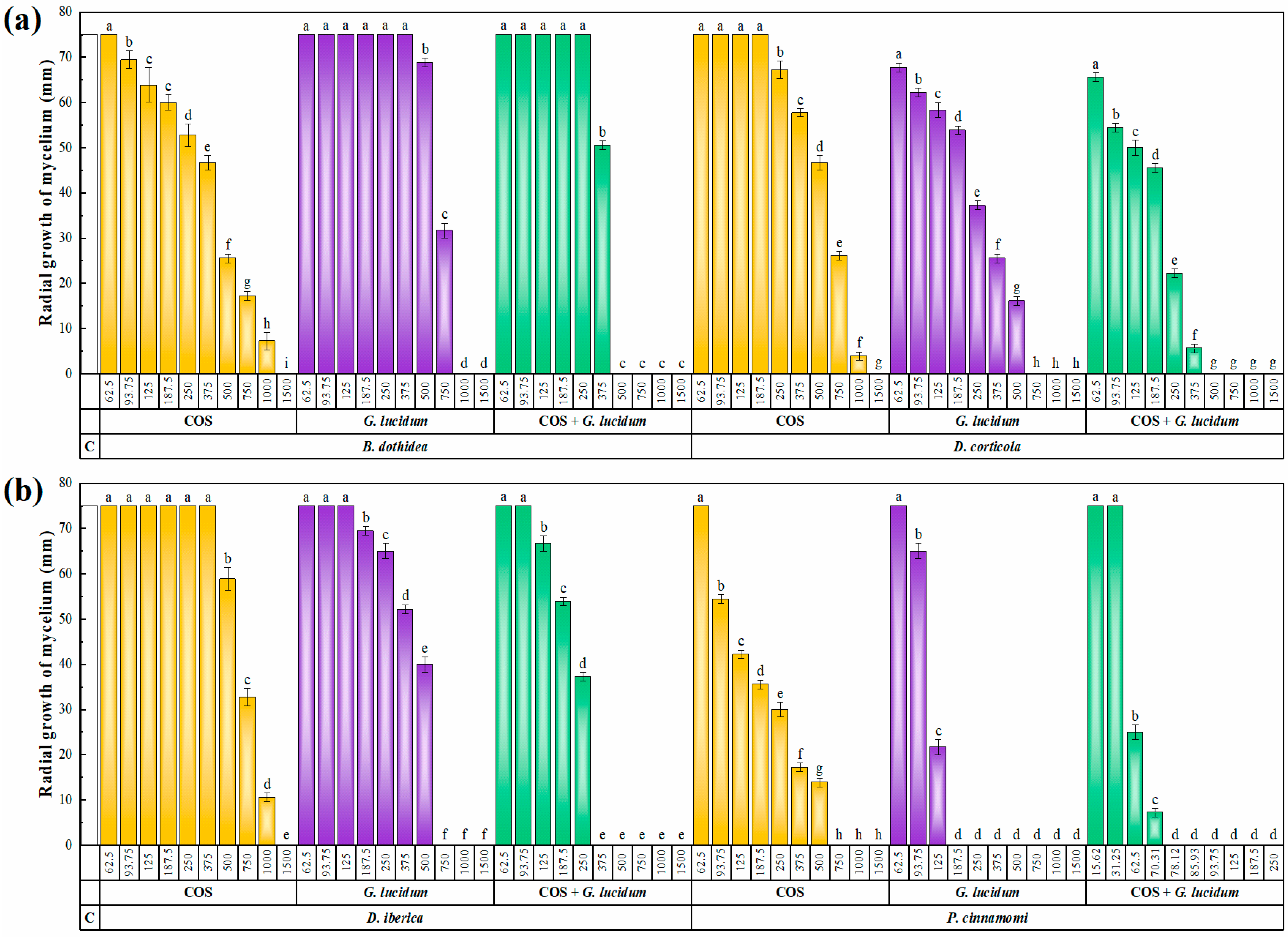
2.3. Antifungal and Anti-Oomycete Activity

The results of the antifungal/anti-oomycete susceptibility test are presented in Figure 3. An increase in concentration led to a decrease in the radial growth of the mycelium for all three tested products (COS, G. lucidum carpophore aqueous ammonia extract, and their conjugate complex), resulting in statistically significant differences. The aqueous ammonia extract of G. lucidum carpophores exhibited higher antifungal/anti-oomycete activity than COS, with minimum inhibitory concentrations (MICs) ranging from 187.5 to 1000 μg·mL−1 and from 750 to 1500 μg·mL−1, respectively. Phytophthora cinnamomi was the most sensitive phytopathogen in both cases, with MIC values of 187.5 and 750 μg·mL−1 for G. lucidum extract and COS, respectively. The formation of conjugate complexes improved the activity, with the COS–G. lucidum conjugate producing complete inhibition of Botryosphaeriaceae family pathogens at concentrations in the range of 375 to 500 μg·mL−1, while the inhibition value was as low as 78.12 μg·mL−1 for P. cinnamomi. The 50 and 90% effective concentrations (EC50 and EC90, respectively), presented in Table 3, allow for a clearer observation of this enhancement of the antifungal/anti-oomycete activity, which was quantified according to Wadley’s method. The synergy factor values were in the range of 1.98–3.63. As these values were higher than 1, a synergistic behavior can be inferred in all cases.
For the purpose of comparison, Fosetyl-Al, a conventional synthetic fungicide widely employed against Phytophthora spp. and fungi associated with grapevine trunk diseases (GTDs), was utilized as a positive control. As indicated in Table 4, when administered at the recommended dose of 2000 μg·mL−1 (equivalent to 2.5 g·L−1 for Fosbel®, fosetyl-Al 80%), complete inhibition of the four phytopathogens was observed. However, when applied at one-tenth of the recommended dose, a moderate inhibition was observed against B. dothidea and D. corticola, while a weak inhibition was observed in the case of P. cinnamomi, and no inhibition was detected against D. iberica.

2.4. Protection of Excised Stems against P. cinnamomi

The COS−G. lucidum conjugate complex was the most active product in the in vitro tests and was subsequently tested as a protective treatment against P. cinnamomi on holm-oak-excised stems. Three different concentrations were used, corresponding to the MIC, MIC × 5, and MIC × 10 (i.e., 78, 391, and 782 µg·mL−1, respectively). Results are presented in Figure S2, and a comparison of canker lengths is shown in Table 5. No protective effect was observed at the lowest dose tested (i.e., at the MIC value obtained in the in vitro tests), with canker lengths similar to those of the positive control (non-treated stems infected with the oomycete). At a dose equal to five times the MIC, significantly lower canker lengths were observed. However, it was necessary to increase the concentration up to 10 times the MIC to achieve effective protection, with no significant differences compared to the negative control. Nevertheless, at this dose, small cankers were still visible in four of the excised stems (out of fifteen replicates), indicating that higher doses may be required in field conditions.

3. Discussion

3.1. On the Chemical Profile

Among the list of compounds presented in Table 2, acetamide or ethanamide has been previously identified in red beetroots (Beta vulgaris var. rubra) and Clerodendrum infortunatum L. leaves [20]. It has also been found in extracts from Larrea divaricata Cav., Picea pungens Engelm., and Sequoiadendron giganteum (Lindl.) Buchholz. The presence of acetamide in the extract may be attributed to the partial hydrolysis of N,N-dimethylacetoacetamide, which was also identified in the extract. Alternatively, it could originate from N-(3-methylbutyl)acetamide or N(2-phenylethyl)acetamide, which are common components of fresh wild mushrooms [21]. However, it is worth noting that the presence of acetamide in the extract may be an artifact resulting from the extraction procedure, as it can also be formed through the decomposition of ammonium acetate. Ammonium acetate is generated by neutralizing excess ammonia in the extract with acetic acid. It is important to mention that acetamide-containing compounds are widely used as herbicides in agriculture [22], and several acetamide derivatives have been reported to act as antimicrobial agents [23].
Oleic acid, or 9-octadecenoic acid, has been identified in damask rose oil [24], Chenopodium album L. root methanolic extract [25], Allium sativum Regel L. [26], Sesuvium portulacastrum L. [27], Armeria maritima (Mill.) Willd. [28], Taxus baccata L. [29], and in small amounts in pomegranates, peas, cabbages [30], Foeniculum vulgare Mill. [31], and Landolphia owariensis Beauv. [32]. Its antifungal activity has been demonstrated against soil pathogens affecting the family Cucurbitaceae, namely, Fusarium equiseti, Fusarium oxysporum f. sp. niveum, Neocosmospora falciformis, Neocosmospora keratoplastica, Macrophomina phaseolina, and Sclerotinia sclerotiorum [28], corroborating the activity previously reported by Walters et al. [33] against Crinipellis perniciosa, a pathogen of the genera Theobroma and Herrania, responsible for witches’ broom, as well as against the oomycete Pythium ultimum, which affects flower bulbs, summer flowers, and perennials.
L-threitol, also known as 1,2,3,4-butanetetrol, is a non-cariogenic component found in Shiitake mushrooms [34] and is also the primary component of Thaumatococcus daniellii (Benn.) Benth. ex B.D.Jacks. leaves [35]. At present, there is no available information on the antimicrobial, antibacterial, or antifungal activity of this compound.
Nonanedioic acid (or 8-carbomethoxyoctanoic acid) monomethyl ester, also known as monomethyl azelate, is a dicarboxylic acid naturally produced by Malassezia furfur (C.P. Robin) Baill. and is also present in whole-grain cereals, rye, and barley. It is known to be effective in treating acne and various cutaneous disorders [36].
Undecane was previously identified as a major constituent of the extract of the stem bark of Symplocos crataegoides Buch.-Ham. ex D. Don (7.5%) [37], Opuntia ficus indica (L.) Mill (20%) [38], Seseli pallasii Besser (13.3%), T. baccata (12.2%) [29], and in smaller percentages in the essential oils of Hypericum hirsutum L. [39] and Lantana camara L. [40]. There is no clear information available on the mechanism of action of undecane as an antimicrobial agent.
n-Hexadecanoic acid (palmitic acid) and its methyl ester were identified in several plants such as Equisetum arvense L. (18.3%) [41], A. maritima (18%) [28], Limonium binervosum (G.E.Sm.) C.E. Salmon (15%) [42], Hibiscus syriacus L. (9.6%) [43], and Tamarix gallica L. (3.7%) [44]. Palmitic acid has been found to have nematicide and pesticide properties [45]. Moreover, it has demonstrated antifungal activity against various fungi, including Alternaria solani, F. oxysporum, Colletotrichum lagenaria, A. niger, Aspergillus terreus, Aspergillus nidulans, N. falciformis, N. keratoplastica, M. phaseolina, and S. sclerotiorum [28,46,47].
2,6-Dimethoxyphenol (syringol) has been identified in various extracts, including Macrotermes gilvus fungus combs (6.5%) [48], Uncaria tomentosa (Willd. ex Schult.) DC. [49], and T. gallica [44]. The antimicrobial effects of syringol isolated from Camelia japonica wood vinegar have been demonstrated against Globisporangium splendens, Ralstonia solanacearum, F. oxysporum, and Phytophthora capsici [50].
5-hydroxy-2(1H)-pyridinone is analogous to 6-hydroxy-2(1H)-pyridinone, the primary natural compound found in the wild berry Rubus fraxinifolius Poir. [51]. Although no information is currently available on the antimicrobial activity of 5-hydroxy-2(1H)-pyridinone, the 2(1H)-pyridone ring system is abundantly found in a wide variety of naturally occurring alkaloids and novel synthetic biologically active molecules. Heterocycles containing a 2(1H)-pyridone framework constitute a highly studied class of compounds due to their diverse biological activities, including anti-HIV, antibacterial, antifungal, and free radical scavengers [52].

3.2. On the Antimicrobial Activity Comparison of G. lucidum Extracts

The antibacterial and antifungal activity results reported for G. lucidum aqueous ammonia extract in this study are consistent with the previously reported antimicrobial activity of G. lucidum extracts in other solvents (Table S1) [3,12,53,54,55,56,57,58,59]. However, previous reports have primarily focused on human pathogens, with limited data on phytopathogens, thus making a direct comparison among extraction media unfeasible.

3.3. Comparison of Efficacy vs. Other Natural Compounds

The use of different isolates with distinct susceptibility profiles generally makes it difficult to accurately compare the activity of G. lucidum aqueous ammonia extract with that of other plant extracts reported in the literature (see Table 6). Nevertheless, it can be observed that G. lucidum-based treatments exhibit some of the highest activities against the four phytopathogens. Regarding B. dothidea, the efficacy of the pure extract is comparable to that of a compound herbal extract compound consisting of seven Chinese medicinal plants [60]. Meanwhile, the activity of the conjugate complex is intermediate between those of COS-U. dioica and COS-E. arvense conjugates [41], tested against the same isolate. Concerning D. corticola, the extract displays the highest activity. As for D. iberica, the data are only available for COS-U. dioica and COS-E. arvense conjugates [41] (tested against the same isolate), which exhibited lower activity, with MIC values at least twice that of the COS-G. lucidum conjugate complex. In terms of the activity against P. cinnamomi (MIC = 187.5 for G. lucidum extract), it is only lower than those reported for an aqueous ammonia extract of holm oak bark (MIC = 78.12 µg·mL−1) [61] and O. ficus-indica aqueous extract (EC90 = 121.7 µg·mL−1) [62], and comparable to those of Flourensia cernua DC. extract (EC90 = 193.4 µg·mL−1) [62] and Thymus vulgaris L. essential oil (MIC = 200 µg·mL−1) [63].

3.4. Comparison of Efficacy vs. Fosetyl-Al

Upon comparing the values of mycelial growth inhibition for Fosetyl-Al (as shown in Table 4) with the effective concentrations reported for G. lucidum extract and its conjugate complexes (as presented in Table 3), it can be observed that the in vitro activity of the natural products was comparable to or even higher than that of the conventional fungicide. Specifically, in the case of P. cinnamomi, complete inhibition was achieved at concentrations of 187.5 μg·mL−1 and 78.1 μg·mL−1 for the non-conjugated extract and the conjugate complex with COS, respectively, whereas Fosetyl-Al exhibited only 12% inhibition at a concentration of 200 μg·mL−1.

3.5. Comparison of Efficacy in Excised Stems

Concerning the activity of the COS—G. lucidum extract conjugate complex as a protective treatment against P. cinnamomi, a comparison with other treatments against Phytophthora spp. is presented in Table 7. Its efficacy was similar to that of non-conjugated Q. ilex aqueous ammonia extract [61], although it was tested on Prunus amygdalus × P. persica excised stems rather than on Q. ilex ones. The activity of the COS—G. lucidum extract conjugate complex was higher than those of non-conjugated Sambucus nigra L. flower ammonia extract [73] and the COS–Quercus suber L. aqueous ammonia bark extract conjugate complex [74], but these were tested against Phytophthora cactorum and Phytophthora megasperma, respectively, so the comparison should be made with caution.

4. Materials and Methods

4.1. Reagents and Fungal Isolates

Ammonium hydroxide (50% v/v aqueous solution) was purchased from Alfa Aesar (Ward Hill, MA, USA). Acetic acid (80% in H2O, purum grade) and potato dextrose agar (PDA) were supplied by Sigma Aldrich Química S.A. (Madrid, Spain). High molecular weight chitosan and NeutraseTM 0.8 L enzymes were acquired from Hangzhou Simit Chem. and Tech. Co. (Hangzhou, China) and Novozymes A/S (Bagsværd, Denmark), respectively. Commercial G. lucidum used for vibrational spectra comparisons was purchased from MundoReishi Salud S.L. (Palencia, Spain). The commercial fungicide used as a positive control in the in vitro experiments, namely, Fosbel® (fosetyl-Al 80%, reg. no. 25502; Probelte), was kindly provided by the Plant Health and Certification Service (CSCV) of the Gobierno de Aragón.
Phytophthora cinnamomi Nirenberg & O’Donnell was supplied by the Centro de Sanidad Forestal de Calabazanos (Villamuriel de Cerrato, Palencia, Spain); Diplodia corticola Phillips, Alves & Luque (CAA500 isolate) was kindly provided by the Biology Department of the Universidade do Minho (Braga, Portugal); while Botryosphaeria dothidea (Moug. ex Fr.) Ces. De Not. (ITACYL_F141) and Dothiorella iberica Phillips, Luque & Alves (ITACYL_F066) isolates were provided by the Instituto Tecnológico Agrario de Castilla y León (ITACYL, Valladolid, Spain). All isolates were supplied as subcultures on PDA and refreshed.

4.2. Collection of Samples

Ganoderma lucidum carpophores growing on Q. ilex trees were collected in October 2021 in El Royal farm, in El Tejado de Béjar, Salamanca, Spain (40°26′42.4″ N 5°33′09.4″ W). Specimens were identified and authenticated by Prof. Dr. B. Herrero-Villacorta (Departamento de Ciencias Agroforestales, ETSIIAA, Universidad de Valladolid) and voucher specimens are available at the herbarium of the ETSIIAA. Different specimens (n = 20) were thoroughly mixed to obtain composite samples, which were shade-dried, pulverized to a fine powder in a mill grinder, homogenized, and sieved (1 mm mesh).

4.3. Extraction Process, Preparation of Chitosan Oligomers, and Preparation of Conjugate Complexes

An aqueous ammonia extraction medium was chosen due to the woody texture of G. lucidum and to achieve the dissolution of polyphenols and other bioactive compounds of interest. Briefly, 67.3 g of G. lucidum carpophore powder was first digested in an aqueous ammonia solution (140 mL H2O + 20 mL NH3) for 2 h, then sonicated in pulsed mode (with a 2 min stop every 2.5 min) for 10 min using a probe-type ultrasonicator (model UIP1000hdT; 1000 W, 20 kHz; Hielscher Ultrasonics, Teltow, Germany), and then allowed to stand for 24 h. It was neutralized to pH 7 using acetic acid. Finally, the solution was centrifuged at 9000 rpm for 15 min, and the supernatant was filtered through Whatman No. 1 paper. The extraction yield was 4.2% (2.86 g).
Aliquots of the extract were freeze-dried for attenuated total-reflectance Fourier-transform infrared (ATR-FTIR) spectroscopy and GC−MS characterization. For the latter, 25 mg of the lyophilized extract was resuspended in 5 mL of methanol (HPLC grade) to obtain a 5 mg·mL−1 solution, which was filtered before injection.
Chitosan oligomers were prepared using the method previously reported in [75], resulting in a solution with oligomers with a molecular weight of less than 2 kDa.
The COS–G. lucidum carpophore extract conjugate complex was obtained by combining solutions (both at a concentration of 3000 μg·mL−1) in a 1:1 (v/v) ratio, followed by sonication for 15 min (five pulses lasting 3 min each to keep the temperature below 60 °C). The solution was freeze-dried for ATR-FTIR characterization to confirm the formation of the conjugate complex.

4.4. G. lucidum Characterization Procedures

The infrared vibrational spectra of the G. lucidum dried samples, as well as that of a commercial G. lucidum sample, were registered using a Thermo Scientific (Waltham, MA, USA) Nicolet iS50 FTIR spectrometer, equipped with an in-built diamond ATR system. The spectra were collected over the 400–4000 cm−1 range, with a 1 cm−1 spectral resolution, taking the interferograms resulting from co-adding 64 scans.
The aqueous ammonia extract of G. lucidum carpophores was analyzed by GC–MS at the Research Support Services (STI) at Universidad de Alicante (Alicante, Spain), using an Agilent Technologies gas chromatograph model 7890A coupled to a quadrupole mass spectrometer model 5975C. The chromatographic conditions were as follows: injection volume = 1 µL; injector temperature = 280 °C, in splitless mode; initial oven temperature = 60 °C, 2 min, followed by a ramp of 10 °C/min up to a final temperature of 300 °C, 15 min. The chromatographic column used for the separation of the compounds was an Agilent Technologies HP-5MS UI column with a length of 30 m, a diameter of 0.250 mm, and a film thickness of 0.25 µm. The mass spectrometer conditions were as follows: temperature of the electron impact source of the mass spectrometer = 230 °C; temperature of the quadrupole = 150 °C; ionization energy = 70 eV. The identification of components was based on a comparison of their mass spectra and retention time with those of the authentic compounds and by computer matching with the database of the National Institute of Standards and Technology (NIST11).

4.5. In Vitro Antifungal and Anti-Oomycete Activity

The antifungal and anti-oomycete activity of the G. lucidum carpophore extract and the conjugate complex with COS was examined using the poisoned food method. Aliquots of stock solutions were added to the PDA medium to produce final concentrations in the range of 15.62–1500 µg·mL−1. Mycelial plugs were transferred from the margin of one-week-old PDA cultures of B. dothidea, D. corticola, D. iberica, and P. cinnamomi to plates filled with the amended media. For each treatment and concentration combination, three plates were used, and each experiment was carried out twice. The untreated control consisted of replacing the extract with the solvent used for extraction in the PDA medium. Fosbel® (fosetyl-Al 80%, reg. no. 25502; Probelte, Murcia, Spain) was used as a positive control. Additional controls, consisting of pure PDA medium and PDA with the lowest concentration of the treatment, were also included to confirm the absence of contamination. Radial mycelium growth was quantified by measuring the average of two perpendicular colony diameters for each replicate. Growth inhibition was estimated after incubation in the dark at 25 °C for one week, using the formula: ((dcdt)/dc) × 100, where dc is the average colony diameter in the untreated control and dt is the average diameter of the treated colony. Effective concentrations (EC50 and EC90) were estimated using PROBIT analysis in IBM SPSS Statistics v.25 (IBM; Armonk, NY, USA). The degree of interaction was estimated using Wadley’s method [76].

4.6. Protection Tests on Artificially Inoculated Excised Stems

Given the restrictions that apply to in vivo assays involving P. cinnamomi, the efficacy of the most active treatment in the in vitro tests (i.e., COS−G. lucidum carpophore extract conjugate complex) was investigated by artificial inoculation of excised stems in controlled laboratory conditions. Inoculation was performed according to the procedure proposed by Matheron et al. [77], with modifications as described in [61,73,74]. Young stems (1.5 cm diameter) of healthy Q. ilex plants were cut into 10 cm-long sections using a sterilized grafting knife. The excised stem pieces were immediately wrapped in moistened sterile absorbent paper. In the laboratory, the freshly excised stem segments were first immersed in a 3% NaClO solution for 10 min, then in 70% ethanol for 10 min, and then thoroughly rinsed four times with distilled water, to avoid superficial contaminants in the tissue. Some of the stem segments (n = 15 for the positive control, and n = 15 for the negative control) were soaked for 1 h in distilled water to be used as controls, while the remaining stem segments were soaked for 1 h in aqueous solutions containing an appropriate amount of the conjugate complex to obtain MIC, MIC × 5, and MIC × 10 concentrations (n = 15 segments/concentration). A coadjuvant (Alkir®, 1% v/v) was added to all the solutions, including the control, to facilitate the moistening and penetration of the treatment into the bark. After soaking, the stem pieces were allowed to dry, and the bark was carefully removed with a scalpel to reveal the cambium. The bark was then placed on an agar Petri dish and, in the case of the positive control and treated samples, it was inoculated by placing a plug (diameter = 5 mm) from the margin of a one-week-old PDA culture of P. cinnamomi on the center of the inner surface of the bark. After inoculation, stem segments were incubated in a humid chamber for 4 days at 24 °C and 95–98% relative humidity. The efficacy of the treatments was evaluated by measuring the lengths of the cankers that developed at the inoculation sites. Finally, the oomycete was re-isolated from the lesions and morphologically identified to fulfill Koch’s postulates.

4.7. Statistical Analysis

The results from the in vitro mycelial growth inhibition and ex situ necrosis lengths were subjected to statistical analysis using one-way analysis of variance (ANOVA). Post hoc comparisons of means were conducted using Tukey’s test at a significance level of p < 0.05. Homogeneity and homoscedasticity requirements were checked using Shapiro–Wilk and Levene tests. The statistical analysis was performed using IBM SPSS Statistics v.25 software.

5. Conclusions

This study provides valuable insights into the composition and antimicrobial activity of an aqueous ammonia extract of Ganoderma lucidum carpophores. The GC-MS characterization revealed the presence of chemical constituents such as oleic acid and its methyl ester, 1,2,3,4-butanetetrol, monomethyl azelate, undecane, and palmitic acid and its methyl ester, which have demonstrated antimicrobial properties in previous studies. In vitro tests demonstrated significant anti-oomycete and antifungal activity of the G. lucidum extract, further enhanced upon combination with chitosan oligomers. In particular, conjugate complexes based on the extract exhibited notable efficacy against Phytophthora cinnamomi, a serious threat to Quercus spp., resulting in complete inhibition at 78.12 μg·mL−1, which was confirmed in ex situ bioassays on holm-oak-excised stems. These findings highlight the potential of G. lucidum as a natural alternative to synthetic fungicides for controlling plant diseases caused by oomycetes and fungi, and suggest its promise as a bioactive product for safeguarding Quercus spp. in the dehesa ecosystem.

Supplementary Materials

The following supporting information can be downloaded at: https://www.mdpi.com/article/10.3390/plants12122271/s1, Figure S1: GC-MS chromatogram of G. lucidum carpophore aqueous ammonia extract; Figure S2: Canker lengths observed in holm-oak-excised stems artificially inoculated with P. cinnamomi; Table S1: Antimicrobial activity reported in the literature for G. lucidum extracts.

Author Contributions

Conceptualization, J.M.-G.; methodology, J.M.-G. and R.O.; validation, J.M.-G. and R.O.; formal analysis, E.S.-H. and P.M.-R.; investigation, E.S.-H., A.T., C.P., A.C., J.M.-G., R.O. and P.M.-R.; resources, J.M.-G. and R.O.; writing—original draft preparation, E.S.-H., A.T., C.P., A.C., J.M.-G., R.O. and P.M.-R.; writing—review and editing, E.S.-H., J.M.-G., R.O. and P.M.-R.; visualization, E.S.-H.; supervision, R.O. and P.M.-R.; project administration, R.O. All authors have read and agreed to the published version of the manuscript.

Funding

This research was funded by FCT—Portuguese Foundation for Science and Technology, under project UIDB/04033/2020 and the “Contrato-Programa” UIDB/04050/2020 I.P. The authors also acknowledge the financial support by AgrifoodXXI (NORTE-01-0145-FEDER-000041). E.S.-H. gratefully acknowledges the financial support of Universidad de Valladolid through the Doctoral Students UVa 2022 Mobility Program.

Data Availability Statement

The data presented in this study are available on request from the corresponding author.

Acknowledgments

The authors would like to acknowledge Pilar Blasco and Pablo Candela from the Technical Research Services of the University of Alicante for conducting the GC–MS analysis. The authors would also like to acknowledge Arsenio Sánchez, Marina Hernández, Ángel Sánchez, and María Ángeles Botas for their technical assistance in the sampling of the G. lucidum specimens.

Conflicts of Interest

The authors declare no conflict of interest. The funders had no role in the design of the study; in the collection, analyses, or interpretation of data; in the writing of the manuscript; or in the decision to publish the results.

References

  1. Wang, X.-M.; Zhang, J.; Wu, L.-H.; Zhao, Y.-L.; Li, T.; Li, J.-Q.; Wang, Y.-Z.; Liu, H.-G. A mini-review of chemical composition and nutritional value of edible wild-grown mushroom from China. Food Chem. 2014, 151, 279–285. [Google Scholar] [CrossRef]
  2. Radhika, R. Antibacterial activity of Ganoderma lucidum extracts against MDR pathogens. Int. J. Mod. Agric. 2021, 10, 3488–3493. [Google Scholar] [CrossRef]
  3. Sridhar, S.; Sivaprakasam, E.; Balakumar, R.; Kavitha, D. Evaluation of antibacterial and antifungal activity of Ganoderma lucidum (Curtis) P. Karst fruit bodies extracts. World J. Sci. Technol. 2011, 1, 8–11. [Google Scholar]
  4. Wachtel-Galor, S.; Yuen, J.; Buswell, J.A.; Benzie, I.F. Ganoderma lucidum (Lingzhi or Reishi). In Herbal Medicine: Biomolecular and Clinical Aspects; Benzie, I., Wachtel-Galor, S., Eds.; CRC Press/Taylor & Francis: Boca Raton, FL, USA, 2011. [Google Scholar]
  5. Liu, Z.; Xing, J.; Zheng, S.; Bo, R.; Luo, L.; Huang, Y.; Niu, Y.; Li, Z.; Wang, D.; Hu, Y. Ganoderma lucidum polysaccharides encapsulated in liposome as an adjuvant to promote Th1-bias immune response. Carbohydr. Polym. 2016, 142, 141–148. [Google Scholar] [CrossRef]
  6. Seweryn, E.; Ziała, A.; Gamian, A. Health-promoting of polysaccharides extracted from Ganoderma lucidum. Nutrients 2021, 13, 2725. [Google Scholar] [CrossRef]
  7. Mizuno, T.; Wang, G.; Zhang, J.; Kawagishi, H.; Nishitoba, T.; Li, J. Reishi, Ganoderma lucidum and Ganoderma tsugae: Bioactive substances and medicinal effects. Food Rev. Int. 1995, 11, 151–166. [Google Scholar] [CrossRef]
  8. Liu, J.; Kurashiki, K.; Shimizu, K.; Kondo, R. Structure–activity relationship for inhibition of 5α-reductase by triterpenoids isolated from Ganoderma lucidum. Biorg. Med. Chem. 2006, 14, 8654–8660. [Google Scholar] [CrossRef]
  9. Ferreira, I.C.; Heleno, S.A.; Reis, F.S.; Stojkovic, D.; Queiroz, M.J.; Vasconcelos, M.H.; Sokovic, M. Chemical features of Ganoderma polysaccharides with antioxidant, antitumor and antimicrobial activities. Phytochemistry 2015, 114, 38–55. [Google Scholar] [CrossRef] [Green Version]
  10. Grienke, U.; Kaserer, T.; Pfluger, F.; Mair, C.E.; Langer, T.; Schuster, D.; Rollinger, J.M. Accessing biological actions of Ganoderma secondary metabolites by in silico profiling. Phytochemistry 2015, 114, 114–124. [Google Scholar] [CrossRef] [Green Version]
  11. Bishop, K.S.; Kao, C.H.; Xu, Y.; Glucina, M.P.; Paterson, R.R.M.; Ferguson, L.R. From 2000 years of Ganoderma lucidum to recent developments in nutraceuticals. Phytochemistry 2015, 114, 56–65. [Google Scholar] [CrossRef] [PubMed] [Green Version]
  12. Stojković, D.S.; Barros, L.; Calhelha, R.C.; Glamočlija, J.; Ćirić, A.; Van Griensven, L.J.; Soković, M.; Ferreira, I.C. A detailed comparative study between chemical and bioactive properties of Ganoderma lucidum from different origins. Int. J. Food Sci. Nutr. 2014, 65, 42–47. [Google Scholar] [CrossRef] [Green Version]
  13. Heleno, S.A.; Ferreira, I.C.; Esteves, A.P.; Ćirić, A.; Glamočlija, J.; Martins, A.; Soković, M.; Queiroz, M.J.R. Antimicrobial and demelanizing activity of Ganoderma lucidum extract, p-hydroxybenzoic and cinnamic acids and their synthetic acetylated glucuronide methyl esters. Food Chem. Toxicol. 2013, 58, 95–100. [Google Scholar] [CrossRef] [PubMed]
  14. Yang, X.; Sun, S.; Chen, Q.; Zhang, Z.; Wang, J.; Liu, Y.; Wang, H. A polysaccharide of Ganoderma lucidum enhances antifungal activity of chemical fungicides against soil-borne diseases of wheat and maize by induced resistance. Agriculture 2022, 12, 55. [Google Scholar] [CrossRef]
  15. Burgess, T.I.; López-Villamor, A.; Paap, T.; Williams, B.; Belhaj, R.; Crone, M.; Dunstan, W.; Howard, K.; Hardy, G.E.S.J. Towards a best practice methodology for the detection of Phytophthora species in soils. Plant Pathol. 2021, 70, 604–614. [Google Scholar] [CrossRef]
  16. Benito Garzón, M.; Sánchez de Dios, R.; Sainz Ollero, H. Effects of climate change on the distribution of Iberian tree species. Appl. Veg. Sci. 2008, 11, 169–178. [Google Scholar] [CrossRef]
  17. Vivas, M.; Hernández, J.; Corcobado, T.; Cubera, E.; Solla, A. Transgenerational induction of resistance to Phytophthora cinnamomi in holm oak. Forests 2021, 12, 100. [Google Scholar] [CrossRef]
  18. Burgess, T.I.; Scott, J.K.; Mcdougall, K.L.; Stukely, M.J.; Crane, C.; Dunstan, W.A.; Brigg, F.; Andjic, V.; White, D.; Rudman, T. Current and projected global distribution of Phytophthora cinnamomi, one of the world’s worst plant pathogens. Glob. Change Biol. 2017, 23, 1661–1674. [Google Scholar] [CrossRef] [PubMed] [Green Version]
  19. Sánchez, M.; Venegas, J.; Romero, M.; Phillips, A.; Trapero, A. Botryosphaeria and related taxa causing oak canker in southwestern Spain. Plant Dis. 2003, 87, 1515–1521. [Google Scholar] [CrossRef] [Green Version]
  20. Ghosh, G.; Panda, P.; Rath, M.; Pal, A.; Sharma, T.; Das, D. GC-MS analysis of bioactive compounds in the methanol extract of Clerodendrum viscosum leaves. Pharmacogn. Res. 2015, 7, 110. [Google Scholar]
  21. Rapior, S.; Marion, C.; Pélissier, Y.; Bessière, J.-M. Volatile composition of fourteen species of fresh wild mushrooms (Boletales). J. Essent. Oil Res. 1997, 9, 231–234. [Google Scholar] [CrossRef]
  22. Coleman, S.; Linderman, R.; Hodgson, E.; Rose, R.L. Comparative metabolism of chloroacetamide herbicides and selected metabolites in human and rat liver microsomes. Environ. Health Perspect. 2000, 108, 1151–1157. [Google Scholar] [CrossRef] [PubMed] [Green Version]
  23. Alsamarrai, A.S.H.; Abdulghani, S.S. Microwave-assisted synthesis, structural characterization and assessment of the antibacterial activity of some new aminopyridine, pyrrolidine, piperidine and morpholine acetamides. Molecules 2021, 26, 533. [Google Scholar] [CrossRef] [PubMed]
  24. Ghavam, M.; Afzali, A.; Manca, M.L. Chemotype of Damask rose with oleic acid (9 octadecenoic acid) and its antimicrobial effectiveness. Sci. Rep. 2021, 11, 8027. [Google Scholar] [CrossRef] [PubMed]
  25. Ali, A.; Javaid, A.; Shoaib, A. GC-MS analysis and antifungal activity of methanolic root extract of Chenopodium album against Sclerotium rolfsii. Planta Daninha 2017, 35, e017164713. [Google Scholar] [CrossRef] [Green Version]
  26. Teixeira, A.; Sánchez-Hernández, E.; Noversa, J.; Cunha, A.; Cortez, I.; Marques, G.; Martín-Ramos, P.; Oliveira, R. Antifungal activity of plant waste extracts against phytopathogenic fungi: Allium sativum peels extract as a promising product targeting the fungal plasma membrane and cell wall. Horticulturae 2023, 9, 136. [Google Scholar] [CrossRef]
  27. Chandrasekaran, M.; Senthilkumar, A.; Venkatesalu, V. Antibacterial and antifungal efficacy of fatty acid methyl esters from the leaves of Sesuvium portulacastrum L. Eur. Rev. Med. Pharmacol. Sci. 2011, 15, 775–780. [Google Scholar]
  28. Sánchez-Hernández, E.; Martín-Ramos, P.; Navas Gracia, L.M.; Martín-Gil, J.; Garcés-Claver, A.; Flores-León, A.; González-García, V. Armeria maritima (Mill.) Willd. flower hydromethanolic extract for cucurbitaceae fungal diseases control. Molecules 2023, 28, 3730. [Google Scholar] [CrossRef]
  29. Sánchez-Hernández, E.; González-García, V.; Martín-Gil, J.; Lorenzo-Vidal, B.; Palacio-Bielsa, A.; Martín-Ramos, P. Phytochemical screening and antibacterial activity of Taxus baccata L. against Pectobacterium spp. and Dickeya chrysanthemi. Horticulturae 2023, 9, 201. [Google Scholar] [CrossRef]
  30. Doyle, E. Trans fatty acids. J. Chem. Educ. 1997, 74, 1030. [Google Scholar] [CrossRef]
  31. Kim, D.H.; Kim, S.I.; Chang, K.S.; Ahn, Y.J. Repellent activity of constituents identified in Foeniculum vulgare fruit against Aedes aegypti (Diptera: Culicidae). J. Agric. Food. Chem. 2002, 50, 6993–6996. [Google Scholar] [CrossRef]
  32. Garba, S.; Garba, I. Anti-diarrhoeal properties of cis-9-octadecenoic acid isolated from Landolphia owariensis plant. Org. Med. Chem. IJ 2017, 3, 103. [Google Scholar]
  33. Walters, D.; Raynor, L.; Mitchell, A.; Walker, R.; Walker, K. Antifungal activities of four fatty acids against plant pathogenic fungi. Mycopathologia 2004, 157, 87–90. [Google Scholar] [CrossRef] [PubMed]
  34. Avinash, J.; Vinay, S.; Jha, K.; Das, D.; Goutham, B.S.; Kumar, G. The unexplored anticaries potential of Shiitake mushroom. Pharmacogn. Rev. 2016, 10, 100–104. [Google Scholar] [CrossRef] [PubMed] [Green Version]
  35. Akalonu, C.; Nwodu, J.A.; Chukwu, E.C.; Ejekwumadu, N.J.; Iwueke, A.V. Nutritional composition and GC-MS phytochemical analysis of Thaumatococcus daniellii leaves. Eur. J. Nutr. Food Saf. 2020, 12, 81–86. [Google Scholar] [CrossRef]
  36. Khairudin, N.; Basri, M.; Fard Masoumi, H.; Samson, S.; Ashari, S. Enhancing the bioconversion of azelaic acid to its derivatives by response surface methodology. Molecules 2018, 23, 397. [Google Scholar] [CrossRef] [PubMed] [Green Version]
  37. Govindarajan, N.; Reddy Cheekala, U.M.; Arcot, S.; Sundaramoorthy, S.; Duraisamy, R.; Raju, I. GC-MS analysis of n-hexane extract of stem bark of Symplocos crataegoides Buch.-Ham. ex D. Don. Pharmacogn. J. 2016, 8, 520–524. [Google Scholar] [CrossRef] [Green Version]
  38. Badr, W.; Rabeh, M.; Eltantawy, M.; El Hawary, S. Chemical composition and antimicrobial activity of volatile constituents of cladodes, fruits peel and fruits pulp from Opuntia ficus indica (L.) Mill. (prickly pear) growing in Egypt. Egypt. J. Chem. 2021, 64, 437–444. [Google Scholar] [CrossRef]
  39. Saroglou, V.; Marin, P.D.; Rancic, A.; Veljic, M.; Skaltsa, H. Composition and antimicrobial activity of the essential oil of six Hypericum species from Serbia. Biochem. Syst. Ecol. 2007, 35, 146–152. [Google Scholar] [CrossRef]
  40. Khan, M.; Srivastava, S.K.; Jain, N.; Syamasundar, K.V.; Yadav, A.K. Chemical composition of fruit and stem essential oils of Lantana camara from northern India. Flavour Fragr. J. 2003, 18, 376–379. [Google Scholar] [CrossRef]
  41. Langa-Lomba, N.; Buzón-Durán, L.; Martín-Ramos, P.; Casanova-Gascón, J.; Martín-Gil, J.; Sánchez-Hernández, E.; González-García, V. Assessment of conjugate complexes of chitosan and Urtica dioica or Equisetum arvense extracts for the control of grapevine trunk pathogens. Agronomy 2021, 11, 976. [Google Scholar] [CrossRef]
  42. Sánchez-Hernández, E.; Buzón-Durán, L.; Langa-Lomba, N.; Casanova-Gascón, J.; Lorenzo-Vidal, B.; Martín-Gil, J.; Martín-Ramos, P. Characterization and antimicrobial activity of a halophyte from the Asturian coast (Spain): Limonium binervosum (G.E.Sm.) C.E.Salmon. Plants 2021, 10, 1852. [Google Scholar] [CrossRef] [PubMed]
  43. Sánchez-Hernández, E.; Buzón-Durán, L.; Lorenzo-Vidal, B.; Martín-Gil, J.; Martín-Ramos, P. Physicochemical characterization and antimicrobial activity against Erwinia amylovora, Erwinia vitivora, and Diplodia seriata of a light purple Hibiscus syriacus L. cultivar. Plants 2021, 10, 1876. [Google Scholar] [CrossRef] [PubMed]
  44. Sánchez-Hernández, E.; González-García, V.; Correa-Guimarães, A.; Casanova-Gascón, J.; Martín-Gil, J.; Martín-Ramos, P. Phytochemical profile and activity against Fusarium species of Tamarix gallica bark aqueous ammonia extract. Agronomy 2023, 13, 496. [Google Scholar] [CrossRef]
  45. Sheela, D.; Uthayakumari, F. GC-MS analysis of bioactive constituents from coastal sand dune taxon Sesuvium portulacastrum (L.). Biosci. Discov. 2013, 4, 47–53. [Google Scholar]
  46. Liu, S.; Ruan, W.; Li, J.; Xu, H.; Wang, J.; Gao, Y.; Wang, J. Biological control of phytopathogenic fungi by fatty acids. Mycopathologia 2008, 166, 93–102. [Google Scholar] [CrossRef]
  47. Altieri, C.; Cardillo, D.; Bevilacqua, A.; Sinigaglia, M. Inhibition of Aspergillus spp. and Penicillium spp. by Fatty Acids and Their Monoglycerides. J. Food Prot. 2007, 70, 1206–1212. [Google Scholar] [CrossRef]
  48. Witasari, L.D.; Wahyu, K.W.; Anugrahani, B.J.; Kurniawan, D.C.; Haryanto, A.; Nandika, D.; Karlinasari, L.; Arinana, A.; Batubara, I.; Santoso, D.; et al. Antimicrobial activities of fungus comb extracts isolated from Indomalayan termite (Macrotermes gilvus Hagen) mound. AMB Express 2022, 12, 14. [Google Scholar] [CrossRef]
  49. Sánchez-Hernández, E.; Martín-Ramos, P.; Martín-Gil, J.; Santiago-Aliste, A.; Hernández-Navarro, S.; Oliveira, R.; González-García, V. Bark extract of Uncaria tomentosa L. for the control of strawberry phytopathogens. Horticulturae 2022, 8, 672. [Google Scholar] [CrossRef]
  50. Hwang, Y.-H.; Matsushita, Y.-I.; Sugamoto, K.; Matsui, T. Antimicrobial effect of the wood vinegar from Cryptomeria japonica sapwood on plant pathogenic microorganisms. J. Microbiol. Biotechnol. 2005, 15, 1106–1109. [Google Scholar]
  51. Abu Bakar, M.F.; Ismail, N.A.; Isha, A.; Mei Ling, A.L. Phytochemical composition and biological activities of selected wild berries (Rubus moluccanus L., R. fraxinifolius Poir., and R. alpestris Blume). Evid.-Based Complement. Altern. Med. 2016, 2016, 2482930. [Google Scholar] [CrossRef] [Green Version]
  52. Upadhyay, P.K.; Prasad, R.; Pandey, M.; Kumar, P. A facile synthesis of 5,6-dihydro-5-hydroxy-2(1H)-pyridone. Tetrahedron Lett. 2009, 50, 2440–2442. [Google Scholar] [CrossRef]
  53. Skalicka-Wozniak, K.; Szypowski, J.; Los, R.; Siwulski, M.; Sobieralski, K.; Glowniak, K.; Malm, A. Evaluation of polysaccharides content in fruit bodies and their antimicrobial activity of four Ganoderma lucidum (W Curt.: Fr.) P. Karst. strains cultivated on different wood type substrates. Acta Soc. Bot. Pol. 2012, 81, 17–21. [Google Scholar] [CrossRef] [Green Version]
  54. Radhika, R.; Rajan, S. Antifungal potentials of Ganoderma lucidum extracts. Plant Cell Biotechnol. Mol. Biol. 2021, 22, 22–27. [Google Scholar]
  55. Kamble, R.; Venkata, S.; Gupte, A. Antimicrobial activity of Ganoderma lucidum mycelia. J. Pure Appl. Microbiol. 2011, 5, 983–986. [Google Scholar]
  56. Quereshi, S.; Pandey, A.; Sandhu, S. Evaluation of antibacterial activity of different Ganoderma lucidum extracts. J. Sci. Res. 2010, 3, 9–13. [Google Scholar]
  57. Shahid, A.A.; Asif, M.; Shahbaz, M.; Ali, M. Antifungal potential of Ganoderma lucidum extract against plant pathogenic fungi of Calendula officinalis L. In Proceedings of the 5th International Conference on Biological, Chemical and Environmental Sciences (BCES-2016), London, UK, 24–25 March 2016; pp. 24–25. [Google Scholar] [CrossRef]
  58. Yoon, S.Y.; Eo, S.K.; Kim, Y.S.; Lee, C.K.; Han, S.S. Antimicrobial activity of Ganoderma lucidum extract alone and in combination with some antibiotics. Arch. Pharm. Res. 1994, 17, 438–442. [Google Scholar] [CrossRef] [PubMed]
  59. Vazirian, M.; Faramarzi, M.A.; Ebrahimi, S.E.S.; Esfahani, H.R.M.; Samadi, N.; Hosseini, S.A.; Asghari, A.; Manayi, A.; Mousazadeh, S.A.; Asef, M.R. Antimicrobial effect of the Lingzhi or Reishi medicinal mushroom, Ganoderma lucidum (higher Basidiomycetes) and its main compounds. Int. J. Med. Mushrooms 2014, 16, 77–84. [Google Scholar] [CrossRef] [PubMed]
  60. Shi, H.; Zhou, X.; He, X.; Wang, R.; Liang Zeng, E.; Zhou, W. Study on the antifungal mechanism of Chinese herbal extract on Botryosphaeria dothidea. J. Food Process. Preserv. 2022, 46, e16631. [Google Scholar] [CrossRef]
  61. Sánchez-Hernández, E.; Balduque-Gil, J.; Barriuso-Vargas, J.J.; Casanova-Gascón, J.; González-García, V.; Cuchí-Oterino, J.A.; Lorenzo-Vidal, B.; Martín-Gil, J.; Martín-Ramos, P. Holm oak (Quercus ilex subsp. ballota (Desf.) Samp.) bark aqueous ammonia extract for the control of invasive forest pathogens. Int. J. Mol. Sci. 2022, 23, 11882. [Google Scholar] [CrossRef]
  62. Castillo-Reyes, F.; Clemente-Constantino, J.A.; Gallegos-Morales, G.; Rodríguez-Herrera, R.; Noé, C. In vitro antifungal activity of polyphenols-rich plant extracts against Phytophthora cinnamomi Rands. Afr. J. Agric. Res. 2015, 10, 4554–4560. [Google Scholar]
  63. Giamperi, L.; Fraternale, D.; Ricci, D. The in vitro action of essential oils on different organisms. J. Essent. Oil Res. 2002, 14, 312–318. [Google Scholar] [CrossRef]
  64. Eksteen, D.; Pretorius, J.; Nieuwoudt, T.; Zietsman, P. Mycelial growth inhibition of plant pathogenic fungi by extracts of South African plant species. Ann. Appl. Biol. 2001, 139, 243–249. [Google Scholar] [CrossRef]
  65. Tegegne, G.; Pretorius, J.C. In vitro and in vivo antifungal activity of crude extracts and powdered dry material from Ethiopian wild plants against economically important plant pathogens. BioControl 2007, 52, 877–888. [Google Scholar] [CrossRef]
  66. Ferreira, C.; Oliveira, R. Protective antifungal activity of Plantago major extract against the phytopathogenic fungi Phytophthora cinnamomi, Diplodia corticola and Colletotrichum species. Proceedings 2021, 70, 94. [Google Scholar] [CrossRef]
  67. Ferreira, C.S.d.S. Survey of Antifungal Activity of Plant Extracts for the Development of Natural Products for Agriculture; University of Minho: Braga, Portugal, 2021. [Google Scholar]
  68. Freitas, L.P. Analysis of Antifungal Plant Extracts against Phytopathogenic Fungi; University of Minho: Braga, Portugal, 2022. [Google Scholar]
  69. Machado, D.C.d.A.F. Study of Antifungal Activity and Mechanisms of Action of Plant Extracts with Potential Application in Sustainable Agricultural Practices; University of Minho: Braga, Portugal, 2022. [Google Scholar]
  70. Carvajal, M.A.; Vergara, A.P.; Santander, R.; Osorio, M.E. Chemical composition and anti-phytopathogenic activity of the essential oil of Beilschmiedia miersii. Nat. Prod. Commun. 2016, 11, 1367–1372. [Google Scholar] [CrossRef] [Green Version]
  71. Moiteiro, C.; Esteves, T.; Ramalho, L.; Rojas, R.; Alvarez, S.; Zacchino, S.; Bragança, H. Essential oil characterization of two Azorean Cryptomeria japonica populations and their biological evaluations. Nat. Prod. Commun. 2013, 8, 1785–1790. [Google Scholar] [CrossRef] [Green Version]
  72. Elshafie, H.S.; Caputo, L.; De Martino, L.; Sakr, S.H.; De Feo, V.; Camele, I. Study of bio-pharmaceutical and antimicrobial properties of pomegranate (Punica granatum L.) leathery exocarp extract. Plants 2021, 10, 153. [Google Scholar] [CrossRef]
  73. Sánchez-Hernández, E.; Balduque-Gil, J.; González-García, V.; Barriuso-Vargas, J.J.; Casanova-Gascón, J.; Martín-Gil, J.; Martín-Ramos, P. Phytochemical profiling of Sambucus nigra L. flower and leaf extracts and their antimicrobial potential against almond tree pathogens. Int. J. Mol. Sci. 2023, 24, 1154. [Google Scholar] [CrossRef] [PubMed]
  74. Sánchez-Hernández, E.; González-García, V.; Casanova-Gascón, J.; Barriuso-Vargas, J.J.; Balduque-Gil, J.; Lorenzo-Vidal, B.; Martín-Gil, J.; Martín-Ramos, P. Valorization of Quercus suber L. bark as a source of phytochemicals with antimicrobial activity against apple tree diseases. Plants 2022, 11, 3415. [Google Scholar] [CrossRef]
  75. Sánchez-Hernández, E.; Langa-Lomba, N.; González-García, V.; Casanova-Gascón, J.; Martín-Gil, J.; Santiago-Aliste, A.; Torres-Sánchez, S.; Martín-Ramos, P. Lignin–chitosan nanocarriers for the delivery of bioactive natural products against wood-decay phytopathogens. Agronomy 2022, 12, 461. [Google Scholar] [CrossRef]
  76. Levy, Y.; Benderly, M.; Cohen, Y.; Gisi, U.; Bassand, D. The joint action of fungicides in mixtures: Comparison of two methods for synergy calculation. EPPO Bull. 1986, 16, 651–657. [Google Scholar] [CrossRef]
  77. Matheron, M.; Mircetich, S. Seasonal variation in susceptibility of Juglans hindsii and paradox rootstocks of English walnut trees to Phytophthora citricola. Phytopathology 1985, 75, 970–972. [Google Scholar] [CrossRef]
Figure 1. Detail view of a basidiocarp of G. lucidum (left) and its stipe (right).
Plants 12 02271 g001
Figure 2. Chemical structures of the main chemical constituents identified in the aqueous ammonia extract of G. lucidum carpophores.
Plants 12 02271 g002
Figure 3. Mycelial growth inhibition achieved with chitosan oligomers (COS), aqueous ammonia extract of G. lucidum carpophores, and their conjugate complex (COS–G. lucidum) against (a) B. dothidea and D. corticola, and (b) D. iberica and P. cinnamomi at concentrations ranging from 62.5 to 1500 μg·mL−1 (or from 15.62 and 250 μg·mL−1 for COS–G. lucidum in the case of P. cinnamomi). The same letters above concentrations indicate that they are not significantly different at p < 0.05. Error bars represent standard deviations (n = 6). ‘C’ stands for the untreated control (i.e., PDA medium to which only the solvent used for extraction was added).
Plants 12 02271 g003
Table 1. Main absorption bands (cm−1) in the infrared spectra of G. lucidum carpophore powder.
Wavenumber (cm−1)Assignment
G. lucidum
(This Study)
G. lucidum
(Commercial)
32903297–OH and –NH stretch
29242922–CH2 asymmetric stretching of alkyls (cutine, wax, pectin, amides)
2874 C–H stretching
(2183) C–N bonding
(2148) C=C stretching
(2047) C–N bonds
(2018) C–H stretching (polysaccharides)
16451634C=O stretching (amides); C=C stretching; O–H deformation
1538 C–N bonds
1451 C–H bending
13741371C–C asymmetrical stretching; phenolic OH groups; C–H (cellulose)
12031248ketonic carbonyl group and C–N bonds
1153 C–C in plane (β-carotene); C–O–C asymmetric stretch (cellulose)
10361035C–C stretching; C–N stretching; >C=O (ketonic) group
562 C–C out of plane bending; C–H rocking vibration
526C–C in-plane bending; COO rocking
452 C–C–C–C in-plane deformation
Table 2. Main chemical constituents identified by GC-MS in G. lucidum carpophore aqueous ammonia extract.
Retention Time (min)Peak Area (%)AssignmentQual
3.312428.279Acetamide90
5.12861.01555-(2-Chlorophenyl)-3-(1-piperidylmethyl)-1,3,4-oxadiazole-2(3H)-thione59
6.11393.9257Glycerin78
6.16734.77221,2,3,4-Butanetetrol, [S-(R*,R*)]-64
6.98632.11602-Propenethioamide, 3-(acetyloxy)-N,N-dimethyl-, (E)-37
7.21231.292,5-Dimethyl-4-hydroxy-3(2H)-furanone43
7.40780.9017Fumaric acid, 3-methylbut-3-enyl tetradecyl ester47
7.47900.7049Tetrahydrofuran, 2-ethyl-5-methyl-38
7.54421.702,5-Furandione, dihydro-3-methylene-50
7.65112.1911Mequinol86
7.79954.4650Undecane42
8.56510.85594H-Pyran-4-one, 2,3-dihydro-3,5-dihydroxy-6-methyl-62
9.47321.0981Catechol93
10.98670.84082-Methoxy-4-vinylphenol64
11.09361.7093N,N-Dimethylacetoacetamide50
11.33121.86N-Methoxy-2-carbaminoaziridine49
11.46162.5489Phenol, 2,6-dimethoxy-97
11.86520.7987DL-Proline, 5-oxo-, methyl ester72
12.04912.54752(1H)-Pyridinone, 5-hydroxy-64
13.20650.9025Suberic acid monomethyl ester64
13.27780.9487Apocynin81
13.52111.5205Thiazole, 5-ethenyl-4-methyl-35
13.69921.6385Dodecanoic acid, methyl ester98
14.13840.5720Dodecanoic acid96
14.18590.4162Propenoic acid, 3-(1-ethyl-3-methyl-4-pyrazolyl)-46
16.03171.2402Methyl tetradecanoate97
16.12080.8852Ethanone, 1-(4-hydroxy-3,5-dimethoxyphenyl)-96
16.72621.1287Cyclohexanone, 5-methyl-2-(1-methylethyl)-, O-methyloxime, (2S-trans)-38
18.14562.5903n-Hexadecanoic acid ester96
18.48892.0519n-Hexadecanoic acid95
19.83044.169-Octadecenoic acid, methyl ester99
20.06780.9002Methyl stearate89
20.18653.86689-Octadecenoic acid, (E)-96
Qual = Quality of resemblance.
Table 3. Effective concentrations (EC, expressed in µg·mL−1) against B. dothidea, D. corticola, D. iberica, and P. cinnamomi of chitosan oligomers (COS), the aqueous ammonia extract of G. lucidum carpophores, and their conjugate complex (COS–G. lucidum). Synergy factors (SF) for the COS–G. lucidum extract conjugate complex were estimated according to Wadley’s method.
PathogenTreatment
COSG. lucidumCOS–G. lucidum
EC50EC90EC50EC90EC50SFEC90SF
B. dothidea428.5956.9692.7938.2404.01.31479.21.98
D. corticola592.8969.5256.0621.6206.51.73350.72.16
D. iberica697.31201.7476.4703.8249.62.27345.62.57
P. cinnamomi166.4595.3112.6169.450.22.6872.63.63
Table 4. Mycelial growth inhibition achieved with Fosetyl-Al at the recommended dose (Rd = 2000 μg·mL−1) and at one tenth of the recommended dose (Rd/10 = 200 μg·mL−1) against the four phytopathogens under study.
PathogenRadial Growth of Mycelium (mm)Inhibition (%)
Rd/10RdRd/10Rd
B. dothidea38.9048.1100
D. corticola42.8042.9100
D. iberica75.000100
P. cinnamomi65.5012.7100
The radial growth of the mycelium for the control (PDA only) was 75 mm. All mycelial growth values (in mm) are average values (n = 3).
Table 5. Analysis of the differences in necrosis lengths between the treatments with a confidence interval of 95% (p < 0.0001).
TreatmentLS Means (Necrosis Length (mm))Groups
C+40.467A
MIC37.400A
MIC × 513.067 B
MIC × 101.800 C
C−0.000 C
C+: positive control (inoculated, no treatment); C−: negative control (not inoculated).
Table 6. Activities reported in the literature for plant extracts against the four phytopathogens studied in this work.
PhytopathogenExtraction MediaPlantEfficacyRef.
B. dothideaEthanol 80%Chinese herbal extract compound (Scutellaria baicalensis, Syzygium aromaticum, Cinnamomum cassia, Gleditsia sinensis, Pogostemon cablin, Acorus calamus, and Camellia oleifera, ratio 1.375:1.125:0.45:0.5:1.35:1.25:2.8)IR = 85%, at 800 µg·mL−1[60]
Methanol 100%Hemizygia transvaalensisn.a.[64]
Pearsonia aristataIR = n.a.–<40%, at 100,000 µg·mL−1
Thesium burkein.a.
Alloteropsis semialatan.a.
Smilax ancepsn.a.
Schrebera alataIR = n.a.–<40%, at 100,000 µg·mL−1
Syncolostemon eriocephalusIR = <40%, at 100,000 µg·mL−1
Eucomis autumnalisIR = 85%, at 100,000 µg·mL−1
Mundulea sericeaIR = <40%, at 100,000 µg·mL−1
Brachylaena huillensisIR = <40%, at 100,000 µg·mL−1
Lapholaena sp.IR = <40%, at 100,000 µg·mL−1
Methanol 95%Dolichos kilimandscharicusIR ≥ 60%, at 1000 µg·mL−1[65]
Maerua subcordataIR < 50%, at 1000 µg·mL−1
Phytolacca dodecandraIR < 50%, at 1000 µg·mL−1
WaterCOS–Equisetum arvenseMIC = 750 µg·mL−1[41]
COS–Urtica dioicaMIC = 375 µg·mL−1
D. corticolaEthanol 50%Plantago majorIR = 14.6%, at 2000 µg·mL−1[66]
Medicago sp.IR = 60.9%, at 2000 µg·mL−1[67]
Melilotus indicusIR = 16.7%, at 2000 µg·mL−1
U. dioicaIR = 34.1%, at 2000 µg·mL−1
Medicago sp., M. indicus, P. major, and U. dioicaIR = 15.8%, at 2000 µg·mL−1
Water or ethanolRosmarinus officinalisIR = 52.2%, at 1500 µg·mL−1[68]
Ethanol 100%Cistus ladaniferIR = 38.75%, at 1000 µg·mL−1[69]
Ethanol 80%Musa sp.IR = 6–20%, at 750 µg·mL−1[26]
Allium sativumIR = >50%, at 750 µg·mL−1
Citrus lemonIR = 21–49%, at 750 µg·mL−1
Citrus sinensisIR = 21–49%, at 750 µg·mL−1
Allium cepaIR = 21–49%, at 750 µg·mL−1
Punica granatumn.a.
Solanum tuberosumIR = 21–49%, at 750 µg·mL−1
Eucalyptus sp.IR = 6–20%, at 750 µg·mL−1
Pinus sp.IR = 21–49%, at 750 µg·mL−1
Olea europeaIR = 6–20%, at 750 µg·mL−1
D. ibericaWaterCOS–Equisetum arvenseMIC = 750 µg·mL−1[41]
COS–Urtica dioicaMIC = 1000 µg·mL−1
P. cinnamomiAqueous ammoniaQuercus ilex subsp. ballotaMIC = 78.12 µg·mL−1[61]
Ethanol 50%P. majorIR = 32.2%, at 2000 µg·mL−1[66]
Ethanol 50%Medicago sp.IR = 21.5%, at 2000 µg·mL−1[67]
M. indicusIR = 87.5%, at 2000 µg·mL−1
U. dioicaIR = 40%, at 2000 µg·mL−1
Medicago sp., M. indicus, P. major, and U. dioicaIR = 72.6%, at 2000 µg·mL−1
Water or ethanolR. officinalisIR = 33.9%, at 1500 µg·mL−1[68]
Ethanol 80%Musa sp.n.a.[26]
A. sativumIR > 50%, at 750 µg·mL−1
C. lemonIR = 21–49%, at 750 µg·mL−1
C. sinensisIR = 21–49%, at 750 µg·mL−1
A. cepaIR > 50%, at 750 µg·mL−1
P. granatumn.a.
S. tuberosumn.e.
Eucalyptus sp.n.a.
Pinus sp.IR = 21–49%, at 750 µg·mL−1
O. european.a.
WaterLarrea tridentataMIC90 = 1431 µg·mL−1[62]
Flourensia cernuaMIC90 = 193.4 µg·mL−1
Agave lechuguillaMIC90 = 68,568 µg·mL−1
Opuntia ficus-indicaMIC90 = 121.7 µg·mL−1
Lippia graveolensMIC90 = 4825 µg·mL−1
Carya illinoensisn.a.
Yucca filiferan.a.
Essential oilSalvia officinalisMIC > 1600 µg·mL−1[63]
Salvia rosmarinusMIC > 1600 µg·mL−1
Origanum vulgareMIC > 200 µg·mL−1
Laurus nobilisMIC > 1600 µg·mL−1
Coriandrum sativumMIC = 800 µg·mL−1
Thymus vulgarisMIC = 200 µg·mL−1
Mentha piperitaMIC = 800 µg·mL−1
Lavandula intermediaMIC = 1600 µg·mL−1
Beilschmiedia miersiiMIC = 300 µg·mL−1[70]
MethanolArbutus unedoMIC = 5990 µg·mL−1[71]
WaterP. granatum cv. ‘Wonderful’IR < 40%, at 10,000 µg·mL−1[72]
COS: chitosan oligomers; IR: inhibition rate; IZ: inhibition zone; MIC: minimum inhibitory concentration; n.a.: no activity.
Table 7. Protective treatments against Phytophthora spp. based on natural products.
Source of
Excised Stems
PathogenNatural ProductEffectivenessRef.
Quercus ilexPhytophthora
cinnamomi
COS–Ganoderma lucidum ammonia carpophore extract conjugate complexFull protection at 782 μg·mL−1This work
Prunus amygdalus × P. persicaP. cinnamomiQ. ilex subsp. ballota aqueous ammonia bark extractFull protection at 782 μg·mL−1[61]
Phytophthora
cactorum
COS–Quercus suber aqueous ammonia bark extract conjugate complexFull protection at 3750 μg·mL−1[74]
Phytophthora megaspermaSambucus nigra flower aqueous ammonia extractFull protection at 1875 μg·mL−1[73]
Disclaimer/Publisher’s Note: The statements, opinions and data contained in all publications are solely those of the individual author(s) and contributor(s) and not of MDPI and/or the editor(s). MDPI and/or the editor(s) disclaim responsibility for any injury to people or property resulting from any ideas, methods, instructions or products referred to in the content.

