

Chemical Profile, Antioxidant and Anti-Inflammatory Potency of Extracts of Vitex madiensis Oliv. and Crossopteryx febrifuga (Afzel ex G. Don)
 , , , ,
Abstract
:1. Introduction
2. Results
2.1. Chemical Profiles
2.1.1. Extracts of V. madiensis
2.1.2. Extracts of C. febrifuga
2.2. Antioxidant Activity of Extracts

| N° | Composés | Tr (min) | Formula | M-H | MS2 (m/z) | L | TB | RB | F | Reference |
|---|---|---|---|---|---|---|---|---|---|---|
| 1 | Uronic acid | 3.34 | C6H12O7 | 195.0499 | 75/195/129/87/99/89/85 | + | + | ++ | + | Standard |
| 2 | Quinic acid | 3.87 | C7H12O6 | 191.0547 | 191/85/127/93/85 | +++ | +++ | + | +++ | Standard |
| 3 | Sucrose | 4.11 | C12H22O11 | 341.1087 | - | + | + | + | Standard | |
| 4 | Citric acid | 6.80 | C6H8O7 | 191.0186 | 191/129/111/87/85 | + | - | + | + | Standard |
| 5 | Fumaric acid | 7.34 | C4H4O4 | 115.0032 | 71/115/72/51 | - | - | - | + | [35] |
| 6 | Shanzhiside | 9.75 | C16H24O11 | 391.1241 | 183/165/139/99/89/71/101/391 | - | - | + | - | [36] |
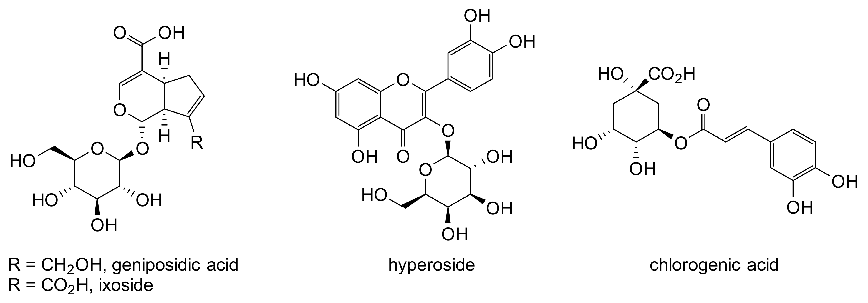
| 7 | Geniposidic acid | 10.03 | C16H22O10 | 373.1135 | 123/149/211/167/373/193 | + | + | - | - | [37] |
| 8 | Ixoside | 10.40 | C16H20O11 | 387.0927 | 181/93/343/89/59/137/163/119/71/101/205/387 | - | + | ++ | - | [38] |
| 9 | Protocatechuic acid | 11.43 | C7H6O4 | 153.0177 | 109/153/110 | - | - | - | + | [22] |
| 10 | Shanzhiside methyl ester | 12.23 | C18H28O13 | 451.1454 * | 243/101/405/451 | + | +++ | +++ | - | [39] |
| 11 | Chlorogenic acid | 12.98 | C16H18O9 | 353.0878 | - | + | - | + | Standard | |
| 12 | Rehmannioside A | 13.47 | C21H32O15 | 523.1664 | 293/89/233/71/125/477 | - | - | + | - | [37] |
| 13 | 4-Hydroxybenzoic acid | 13.80 | C7H6O3 | 137.0238 | 137/138/136/109/108/81/119/93 | - | - | - | + | [22] |
| 14 | Loganine | 14.69 | C18H28O12 | 435.1504 * | 227/101/139/435 | + | - | - | - | [40] |
| 15 | 11-Methylixoside | 15.03 | C17H22O11 | 401.1086 | 101/137/401/195/93/239/221 | - | + | ++ | - | [41] |
| 16 17 | Hyperoside Isoquercetin | 18.10 18.51 | C21H20O12 C21H20O12 | 463.0881 463.0881 | 300/463/271/255/179 300/463/271/255/179/151 | + + | - - | - - | + - | Standard Standard |
| 18 | Vanillic acid | 19.78 | C8H8O4 | 167.0338 | 167/152/111 | - | - | - | + | Standard |
| 19 | 3,4-dicaffeoylquinic acid | 24.89 | C25H24O12 | 515.1189 | - | - | + | - | Standard | |
| 20 | Azelaic acid | 26.71 | C9H16O4 | 187.0965 | 125/187/169/126/97/143 | - | - | - | + | Standard |
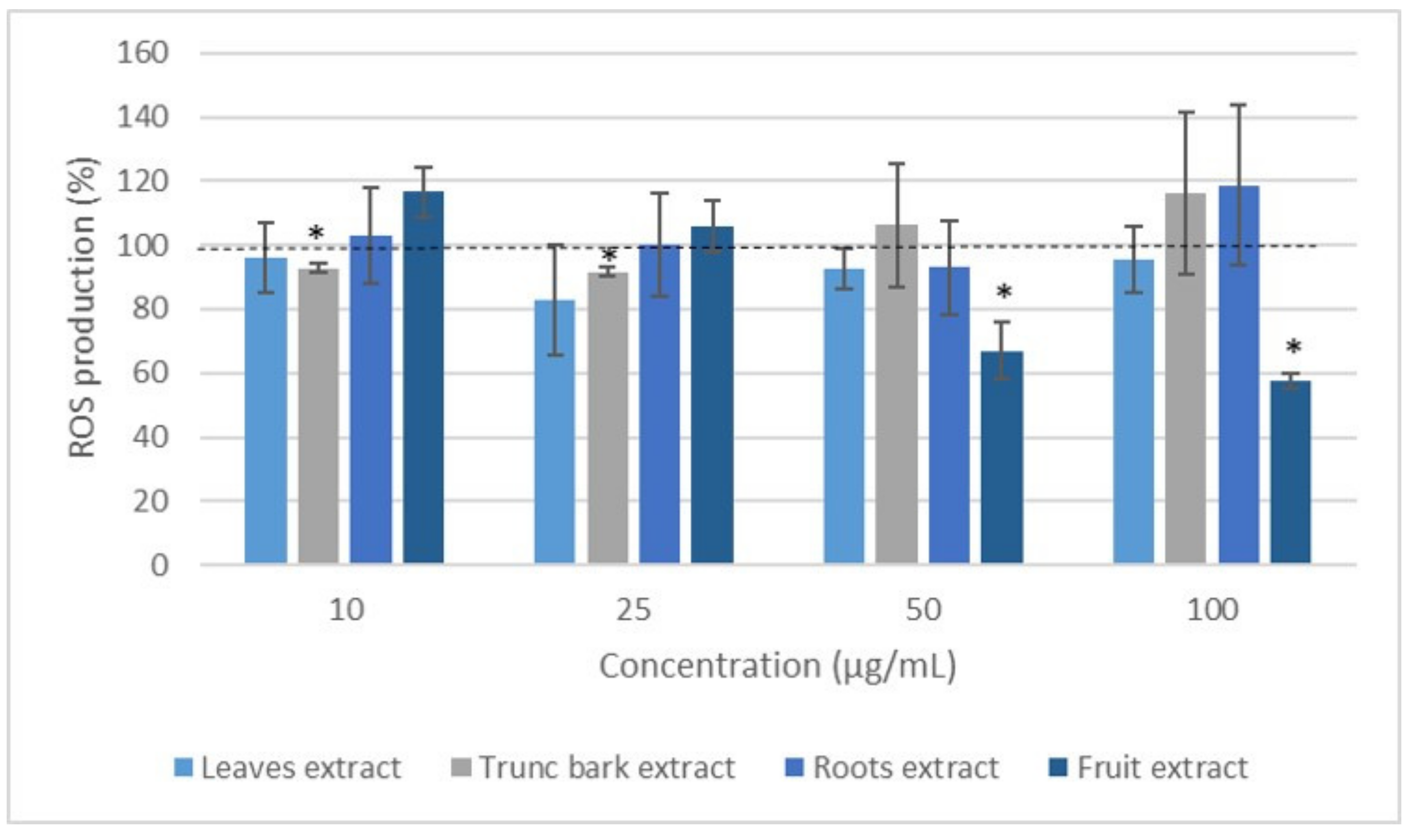
2.3. ROS Production Inhibition
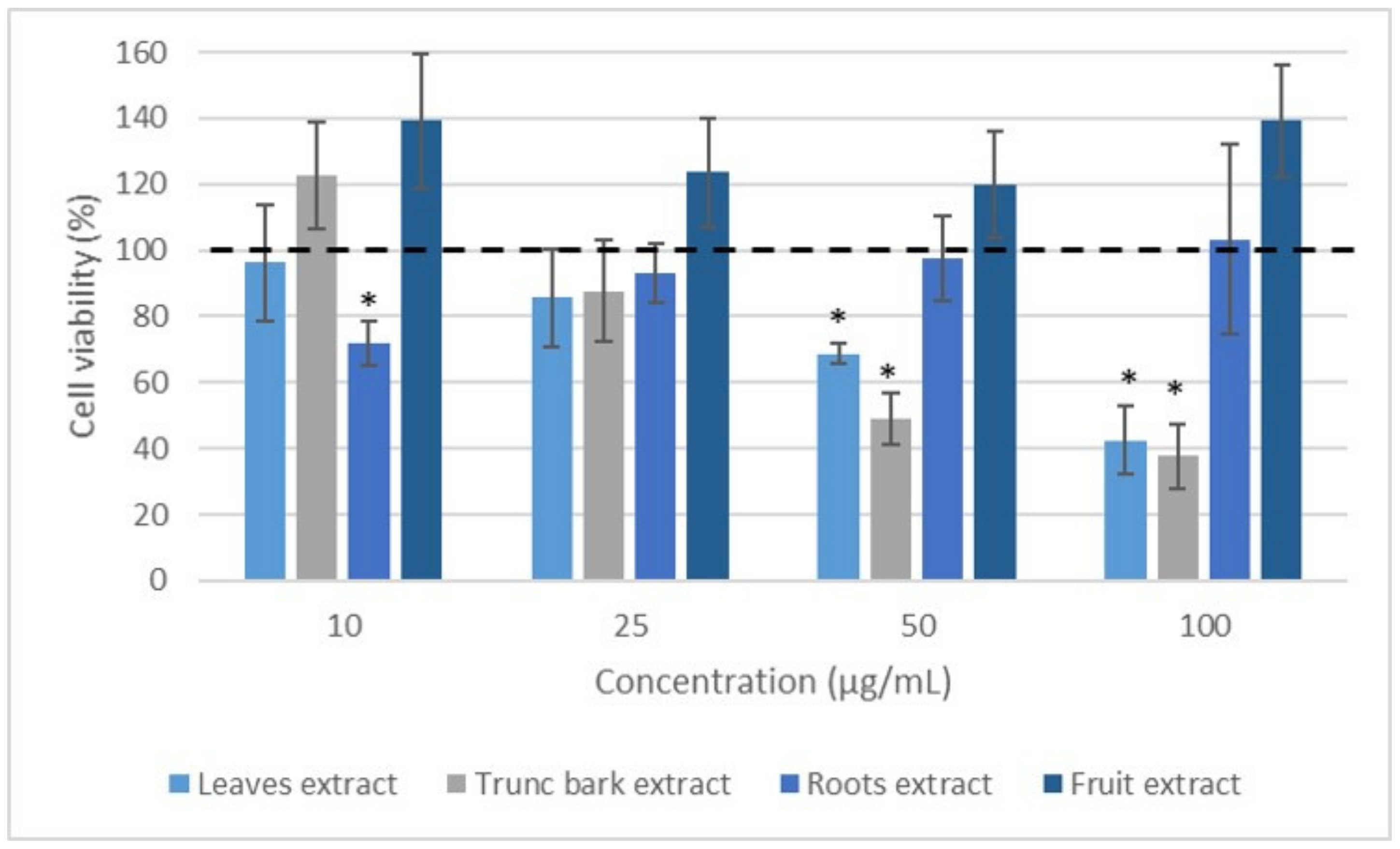
2.4. Effect of Extracts on Leukocyte Viability
3. Discussion
4. Materials and Methods
4.1. Plant Material
4.2. Cell Material
4.3. Methanolic Extracts
4.4. Determination of the Chemical Profiles
4.5. Antioxidant Properties
4.6. ROS Production Inhibition
4.7. Effect of Extracts on Leukocyte Viability
4.8. Data Analysis
5. Conclusions
Author Contributions
Funding
Institutional Review Board Statement
Informed Consent Statement
Acknowledgments
Conflicts of Interest
References
- Salawu, O.A.; Chindo, A.Y.; Tijani, A.B.; Adzu, B. Analgesic, anti-inflammatory, antipyretic and antiplasmodial effects of the methanolic extract of Crossopteryx febrifuga. J. Med. Plants Res. 2008, 2, 213–218. [Google Scholar]
- Chantaranothai, P. A revision of the genus Vitex (Lamiaceae) in Thailand. Trop. Nat. Hist. 2011, 11, 91–118. [Google Scholar]
- Adjanohoum, E.J. Contribution aux Etudes Ethnobotaniques et Floristiques en République Populaire du Congo; ACCT: Paris, France, 1988; p. 605. [Google Scholar]
- Audu, J.A. Medicinal Plants and their use in Bauchi State. Big. Field 1989, 54, 157–168. [Google Scholar]
- Bridson, D.; Verdcourt, B.; Rubiaceae, I. Flora of Tropical East Africa; aa Balkema: Rotterdam, The Netherlands, 1988; p. 2. [Google Scholar]
- Tomas-Barberan, F.A.; Hostettmann, K.A. Cytotoxic triterpenoid and flavonoids from Crossopteryx febrifuga. Planta Med. 1988, 54, 266–267. [Google Scholar] [CrossRef] [PubMed]
- Danton, O. Extraction des Substances Naturelles Antalgiques à Partir des Plantes Utilisées Dans la Pharmacopée Traditionnelle au Mali. Ph.D. Thesis, Université Clermont Auvergne, Clermont-Ferrand, France, 2017. [Google Scholar]
- Bouquet, A. Féticheurs et Médecines Traditionnelles du Congo (Brazzaville); Travaux et Documents de l’ORSTOM: Paris, France, 1972; Volume 36, p. 282. [Google Scholar]
- Elufioye, T.O.; Agbedahunsi, J.M. Antimalarial activities of Tithonia diversifolia (Asteraceae) and Crossopteryx febrifuga (Rubiaceae) on mice in vivo. J. Ethnopharmacol. 2004, 93, 167–171. [Google Scholar] [CrossRef] [PubMed]
- Gariboldi, P.; Verotta, L.; Gabetta, B. Saponins from Crossopteryx febrifuga. Phytochemistry 1990, 29, 2629–2635. [Google Scholar] [CrossRef]
- Tona, L.; Kambu, K.; Ngimbi, N.; Mesia, K.; Penge, O.; Lusakibanza, M.; Cimanga, K.; De Bruyne, T.; Apers, S.; Totte, J.; et al. Antiamoebic and spasmolytic activity of extract of some antidiarrhoeal traditional preparations used in Kinshasa Congo. Phytomedicine 2000, 7, 31–38. [Google Scholar] [CrossRef]
- Latham, P. Useful Plants of Bas-Congo Province, Democratic Republic of the Congo; DFIG: London, UK, 2004; p. 320. [Google Scholar]
- Arbonnier, M. Arbres, Arbustes et Lianes des Zones Sèches d’Afrique de L’ouest, 3rd ed.; Quae: Versailles, France, 2009. [Google Scholar]
- Kerharo, J.; Adams, J.G. La Pharmacopée Sénégalaise Traditionnelle, Plantes Médicinales et Toxiques; Vigot Frères: Paris, France, 1974. [Google Scholar]
- Ondo, J.P.; Lekana-Douki, J.P.; Bongui, J.B.; Zang Edou, E.S.; Zatra, R.; Toure-Ndoua, F.S.; Elombi, A.; Lebibi, J.; Seguin, E. In vitro antiplasmodial activity and cytotoxicity of extracts and fractions of Vitex madiensis, medicinal plant of Gabon. Trop. Med. Int. Health 2012, 17, 316–321. [Google Scholar] [CrossRef] [PubMed]
- Kubo, I.; Matsumoto, A.; Ayafor, J.F. Efficient Isolation of a Large Amount of 20-Hydroxyecdysone from Vitex madiensis (Verbenaceae) by Droplet Counter-Current Chromatography. Agr. Biol. Chem. 1984, 48, 1683–1684. [Google Scholar] [CrossRef]
- Nguyen, T.K.T.; Ninh, K.B.; Do Thi, T.; Tran, M.L.; Vu Huong, G.; Nguyen, X.N.; Phan, V.K. Ecdysteroids from leaves of Vitex trifolia. Vietnam. J. Chem. 2018, 56, 162–166. [Google Scholar]
- Meena, A.K.; Niranjan, U.S.; Rao, M.M.; Padhi, M.M.; Ramesh, B. A review of the important chemical constituents and medicinal uses of Vitex genus. Asian J. Tradit. Med. 2011, 6, 54–60. [Google Scholar]
- Lengbiye, E.M.; Koto-te- Nyiwa, N. Vitex madiensis Oliv. (Lamiaceae): Phytochemistry, pharmacology and future directions, a mini-review. J. Pharmacogn. Phytochem. 2018, 7, 244–251. [Google Scholar]
- Masateru, O.; Yasuyuki, I.; Satoko, K.; Toshihiro, N. Two news iridoids from Viticis trifoliae Fructus (Fruit of Vitex rotundifolia L.). Chem. Pharm. Bull. 1997, 45, 1094–1096. [Google Scholar]
- Chen, P.; Yi, W.; Lulin, C.; Wei, J.; Yan, N.; Qing, S.; Lu, G.; Quancheng, Z.; Licheng, Y.; Shufang, W. Comparison of the anti-inflammatory active constituents and hepatotoxic pyrrolizidine alkaloids in two Senecio plants and their preparations by LC–UV and LC–MS. J. Pharmaceut. Biomed. 2015, 115, 260–271. [Google Scholar] [CrossRef]
- Vallverdú-Queralt, A.; Boix, N.; Piqué, E.; Gómez-Catalan, J.; Medina-Remon, A.; Sasot, G.; Mercader-Martí, M.; Llobet, M.J.; Lamuela-Raventos, M.R. Identification of phenolic compounds in red wine extract samples and zebrafish embryos by HPLC-ESI-LTQ-Orbitrap-MS. Food Chem. 2015, 181, 146–151. [Google Scholar] [CrossRef]
- Elhawary, S.S.; Younis, I.Y.; El Bishbishy, M.H.; Khattab, A.R. LC–MS/MS-based chemometric analysis of phytochemical diversity in 13 Ficus spp. (Moraceae): Correlation to their in vitro antimicrobial and in silico quorum sensing inhibitory activities. Ind. Crops Prod. 2018, 126, 261–271. [Google Scholar] [CrossRef]
- Wang, Y.-H.; Avula, B.; Jadhav, B.-A.; Smillie, T.-J.; Khan, I.A. Structural characterization and identification of ecdysteroids from Sida rhombifolia L. in positive electrospray ionization by tandem mass spectrometry. Rapid Commun. Mass Spectrom. 2008, 22, 2413–2422. [Google Scholar] [CrossRef]
- Zhou, J.; Qi, Y.; Hou, Y.; Zhao, J.; Li, Y.; Xue, X.; Wu, L.; Zhang, J.; Chen, F. Quantitative determination of juvenile hormone III and 20-hydroxyecdysone in queen larvae and drone pupae of Apis mellifera by ultrasonic-assisted extraction and liquid chromatography with electrospray ionization tandem mass spectrometry. J. Chromatogr. B 2011, 879, 2533–2541. [Google Scholar] [CrossRef]
- Available online: https://mona.fiehnlab.ucdavis.edu/spectra/display/CCMSLIB00005723108 (accessed on 22 December 2022).
- Olennikova, D.N.; Kashchenkoa, N.I. Phytoecdysteroids of Serratula centauroides Herb from Cisbaikalia. Russ. J. Bioorg. Chem. 2019, 45, 913–919. [Google Scholar] [CrossRef]
- Kubo, I.; Asaka, Y.; Stout, M.J.; Nakatsu, T. Structure of a novel phytoecdysteroid, vitexirone, and efficient isolation of phytoecdysteroids from Vitex fisherii. J. Chem. Ecol. 1990, 16, 2581–2588. [Google Scholar] [CrossRef] [PubMed]
- Le Bizec, B.; Antignac, J.-P.; Monteau, F.; Andre, F. Ecdysteroids: One potential new anabolic family in breeding animals. Anal. Chim. Acta 2002, 47, 389–397. [Google Scholar] [CrossRef]
- Feng, R.; Luya, L.; Zhang, X.; Zhang, Y.; Chen, Y.; Feng, X.; Zhang, L.; Zhang, G. Assessment of a developed HPLC-MS/MS approach for determining plasma eupatorin in rats and its application in pharmacokinetics analysis. RSC Adv. 2020, 10, 32020. [Google Scholar] [CrossRef] [PubMed]
- Mittal, M.; Siddiqui, M.R.; Tran, K.; Reddy, S.P.; Malik, A.B. Reactive Oxygen Species in Inflammation and Tissue Injury. Antioxid. Redox. Signal. 2014, 20, 1126–1167. [Google Scholar] [CrossRef] [Green Version]
- Sena Filho, J.G.; Duringerc, J.; Maiaa, G.L.A.; Tavaresa, J.F.; Haroudo, X.H.S.; Sobral da Silvaa, M.; Em.dio da-Cunhaa, V.L.; Barbosa-Filhoa, J.M. Ecdysteroids from Vitex Species: Distribution and Compilation of Their 13C-NMR Spectral Data. Chem. Biodivers. 2008, 5, 707–713. [Google Scholar] [CrossRef] [PubMed]
- Sarr, S.O.; Fall, A.D.; Ngueye, R.; Diop, A.; Diatta, K.; Diop, N.; Ndiaye, B.; Diop, Y.M. Etude de l’activité antioxydante des extraits des feuilles de Vitex doniana (Verbenacea). Int. J. Biol.Chem. Sci. 2015, 9, 1263–1269. [Google Scholar] [CrossRef] [Green Version]
- Ochieng, C.O.; Nandwa, B.O. Proximate composition, phenolic content and antioxidant activities of three black plum (Vitex sp.) fruits: Preliminary results. J. Food Technol. 2010, 8, 118–125. [Google Scholar] [CrossRef]
- Said, A.A.H.; Abuotabl, E.A.; Raoof, G.F.A. Identification of Constituents from Pleiogynium timorense (Dc.) Leenh Pericarp and Seeds Using High-Performance Liquid Chromatography with Electrospray Ionization Mass Spectrometry. AASCIT J. Chem. 2017, 3, 30–36. [Google Scholar]
- Serralheiro, M.L.; Guedes, R.; Fadel, R.S.; Bendif, H. Data on identification of primary andsecondary metabolites in aqueous extract of Verbascum betonicifolium. Data Brief 2020, 32, 106146. [Google Scholar] [CrossRef]
- Maoyuan, W.; Qinglong, W.; Qing, Y.; Xiaoxia, Y.; Shixiu, F.; Zhunian, W. Comparison of Anthraquinones, Iridoid Glycosides and Triterpenoids in Morinda officinalis and Morinda citrifolia Using UPLC/Q-TOF-MS and Multivariate Statistical Analysis. Molecules 2020, 25, 160. [Google Scholar]
- Ibekwe, N.N.; Adesomoju, A.A.; Igoli, J.O.; Barry, C.E.; Okogun, J.I. An iridoid glucoside from the leaves of Pavetta crassipes. J. Chem. Soc. Niger. 2019, 44, 629–632. [Google Scholar]
- Available online: https://mona.fiehnlab.ucdavis.edu/spectra/display/Bruker_HCD_library000649 (accessed on 22 December 2022).
- Xue, G.; Zhu, M.; Meng, N.; Guan, J.; Zhang, J.; Yang, J.; Wang, Y.; Cui, Y.; Chai, X. Integrating Study on Qualitative and Quantitative Characterization of the Major Constituents in Shuanghuanglian Injection with UHPLC/Q-Orbitrap-MS and UPLC-PDA. J. Anal. Methods Chem. 2021, 22, 1–11. [Google Scholar] [CrossRef] [PubMed]
- Bealem, A.; Sakava, P.; Favier, L.; Hauchard, D.; Guegan, J.-P.; Nyemb, J.N.; Talla, E.; Henoumont, C.; Laurent, S.; Atchade De Theodore, A.; et al. Chemical Constituents of Crossopteryx febrifuga (Afzel ex G. Don) Benth (Rubiaceae), their Phytotoxicity and Antioxidant activities. Nat. Prod. Chem. Res. 2021, 9, 1000416. [Google Scholar]
- Thuy, T.T.; Porzel, A.; Ripperger, H.; Sung, T.V.; Adam, G. Chalcones and ecdysteroids from Vitex leptobotrys. Phytochemistry 1998, 49, 2603–2605. [Google Scholar] [CrossRef]
- Dos Santos, T.C.; Delle Monache, F.G.; Leita, S.G. Ecdysteroids from two Brazilian Vitex species. Fitoterapia 2001, 72, 215–220. [Google Scholar] [CrossRef]
- Suksamrarn, A.; Kumpun, S.; Yingyongnarongkul, B. Ecdysteroids of Vitex scabra Stem Bark. J. Nat. Prod. 2002, 65, 1690–1692. [Google Scholar] [CrossRef]
- Dinan, L.; Dioh, W.; Veillet, S.; Lafont, R. 20-Hydroxyecdysnoe, form Plant extracts to Clinical Use: Therapeutic Potential for the Treatment of Neuromuscular, Cardio-Metabolic and Respiratory Diseases. Biomedicines 2021, 9, 492. [Google Scholar] [CrossRef]
- Ganapaty, S.; Vidydhar, K.N. Phytoconstituents and biological activities of Vitex—A review. J. Nat. Remedies 2005, 5, 75–95. [Google Scholar]
- Guzik, T.J.; Korbut, R.; Adamek-Guzik, T. Nitric oxide and superoxide in inflammation and immune regulation. J. Physiol. Pharmacol. 2003, 54, 469–487. [Google Scholar]
- Reczek, C.R.; Chandel, N.S. The Two Faces of Reactive Oxygen Species in Cancer. Annu. Rev. Cancer Biol. 2017, 1, 79–98. [Google Scholar] [CrossRef]
- Perl, A.; Gergely, P.; Banki, K. Mitochondrial dysfunction in T cells of patients with systemic lupus erythematosus. Int. Rev. Immunol. 2004, 23, 293–313. [Google Scholar] [CrossRef]
- Herb, M.; Schramm, M. Functions of ROS in Macrophages and Antimicrobial Immunity. Antioxidants 2021, 10, 313. [Google Scholar] [CrossRef] [PubMed]
- Novo, E.; Parola, M. Redox mechanisms in hepatic chronic wound healing and fibrogenesis. Fibrogenesis Tissue Repair. 2008, 1, 5–8. [Google Scholar] [CrossRef] [PubMed] [Green Version]
- Santanam, N.; Ramachandran, S.; Parthasarathy, S. Oxygen Radicals, Antioxidants, and Lipid Peroxidation. Semin. Reprod. Med. 1998, 16, 275–280. [Google Scholar] [CrossRef] [PubMed]
- Bulger, E.M.; Maier, R.V. Antioxidants in critical illness. Arch. Surg. 2001, 136, 1201–1207. [Google Scholar] [CrossRef]
- Li, X.; Fang, P.; Mai, J.; Choi, E.T.; Wang, H.; Yang, X.-F. Targeting mitochondrial reactive oxygen species as novel therapy for inflammatory diseases and cancers. J. Hematol. Oncol. 2013, 6, 19. [Google Scholar] [CrossRef] [Green Version]
- Cai, Y.-J.; Dai, J.-Q.; Fang, J.-G.; Ma, L.-P.; Hou, L.-F.; Yang, L.; Liu, Z.-L. Antioxidative and free radical scavenging effects of ecdysteroids form Serratula strangulata. Can. J. Physiol. Pharmacol. 2002, 80, 1187–1194. [Google Scholar] [CrossRef]
- Das, N.; Mishra, S.K.; Bishatee, A.; Ali, A.S.; Bishayee, A. The Phytochemical, biological and medicinal attributes of phytoecdysteroids: An uptaded review. Acta Pharm. Sin. B 2021, 11, 1740–1766. [Google Scholar] [CrossRef]
- Akanni, O.O.; Owumi, S.E.; Adaramoye, O.A. In vitro studies to assess the antioxidative, radical scavenging and arginase inhibitory potentials of extracts from Artocarpus altilis, Ficus exasperata and Kigelia africana. Asian Pac. J. Trop. Biomed. 2014, 4, 492–499. [Google Scholar] [CrossRef] [Green Version]
- Fall, A.D.; Sy, A.N.; Fokou, J.B.H.; Fomi, J.O.N.; Dieng, M.; Dieng, S.I.M.; Bassene, E. Phytochemical screening, polyphenol content and antioxidant studies of ethanol leaf extract of Combretum aculeatum Vent. Eur. J. Med. Plants 2015, 10, 1–7. [Google Scholar] [CrossRef]
- Anyasor, G.N.; Ogunwenmo, O.; Oyelana, O.A.; Akpofunure, B.E. Phytochemical constituents and antioxidant activities of aqueous and methanol stem extracts of Costus afer Ker Gawl. (Costaceae). Afr. J. Biotechnol. 2010, 9, 4880–4884. [Google Scholar]
- Maiga, A.; Malterud, K.E.; Diallo, D.; Paulsen, B.S. Antioxidant and 15-lipoxygenase inhibitory activities of the malian medicinal plants Diospyros abyssinica (Hiern) F. white (Ebenaceae), Lannea velutina A. Rich (Anacardiaceae) and Crossopteryx febrifuga (Afzer) Benth (Rubiaceae). J. Ethnopharmacol. 2006, 104, 132–137. [Google Scholar] [CrossRef] [PubMed]
- Cholet, J.; Decombat, C.; Vareille-Delarbre, M.; Gainche, M.; Berry, A.; Senejoux, F.; Ripoche, I.; Delort, L.; Vermery, M.; Fraisse, D.; et al. In vitro anti-inflammatory and immunomodulatory activities of an extract from roots of Buplerum Rotundifolium. Medicines 2019, 6, 101. [Google Scholar] [CrossRef] [Green Version]
- Brand-Williams, W.; Cuvelier, M.E.; Berset, C. Use of free radical method to evaluate antioxidant activity. Lebensm. Wiss. Technol. 1995, 28, 25–30. [Google Scholar] [CrossRef]
- Meda, N.R.; Fraisse, D.; Gnoula, C.; Vivier, M.; Felgines, C. Characterization of antioxidants from Detarium microcarpum Guill. et Perr. leaves using HPLC-DAD coupled with pre-column DPPH assay. Eur. Food Res. Technol. 2017, 243, 1659–1666. [Google Scholar] [CrossRef]
- Sahgal, G.; Ramanathan, S.; Sasidharan, S.; Mordi, M.N.; Ismail, S.; Mansor, S.M. In vitro antioxidant and xanthine oxidase inhibitory activities of methanolic Swietenia mahagoni seed extracts. Molecules 2009, 14, 4476–4485. [Google Scholar] [CrossRef]
- Cholet, J.; Decombat, C.; Varelle-Dlarbre, M.; Gainche, M.; Berry, A.; Ogeron, C.; Ripoche, I.; Delort, L.; Vermerie, M.; Fraise, D.; et al. Comparison of The Anti-Inflammatory and Immunomodulatory Mechanisms of Two Medicinal Herbs: Meadowsweet (Filipendula ulmaria) and Harpagophytum (Harpagophytum procumbens). Int. J. Plant Anim. Environ. Sci. 2019, 9, 145–163. [Google Scholar]


| N° | Compound | Tr (min) | Formula | M-H (m/z) | M + (m/z) | MS2 (m/z) | Le | TB | RB | F | Reference |
|---|---|---|---|---|---|---|---|---|---|---|---|
| 1 | Quinic acid | 3.96 | C7H12O6 | 191.0553 | 191/85/127/93/85 | ++ | + | +++ | + | Standard | |
| 2 | Citric acid | 6.91 | C6H8O7 | 191.0189 | 191/129/11/87/85 | + | + | + | + | Standard | |
| 3 | 1-oxo-eucommiol | 7.76 | C9H14O5 | 201.0757 | 201/109/139 | ++ | + | + | + | [20] | |
| 4 | 5-Ethylidene-2-hydroxy-2-hydroxymethyl-3-methylhexanedioic acid | 9.40 | C10H16O6 | 231.0866 | 231/213/169/125/157/187/143 | + | - | - | + | [21] | |
| 5 | Protocatechuic acid | 11.49 | C7H6O4 | 153.0179 | 109/153/110 | + | - | - | - | [22] | |
| 6 | Vicenin-1 | 14.72 | C26H28O14 | 563.1413 | 563/353/383/297/473/443/297/325/503/545 | + | - | - | - | [23] | |
| 7 | Homoorientin | 15.15 | C21H20O11 | 447.0928 | 429/357/327 | + | + | + | - | Standard | |
| 8 | Orientin | 15.91 | C21H20O11 | 447.0928 | 369/357/327/299 | + | + | - | - | Standard | |
| 9 | Vitexin | 17.90 | C21H20O10 | 431.0981 | + | + | - | + | Standard | ||
| 10 | 20-Hydroxyecdysone | 19.15 | C27H44O7 | 525.3065 * | 481.3152 | 445/371/165/427/125/69/481# | +++ | + | +++ | +++ | [24,25,26] |
| 11 | Luteolin-7-O-glucuronide | 19.57 | C21H18O12 | 461.0734 | 285 | + | + | - | - | [27] | |
| 12 | 3,4-dicaffeoylquinic acid | 22.22 | C25H24O12 | 515.1201 | 353/191/179 | + | + | + | + | Standard | |
| 13 | Luteolin-4’-O-glucoside | 23.84 | C21H20O11 | 447.0939 | 285/447 | + | + | - | - | Standard | |
| 14 | Isovitexirone | 24.41 | C27H42O7 | 523.2917 * | 479.2998 | 425/443/373/123/145/219/303/407/461/479 # | + | + | - | + | [27] |
| 15 | Vitexirone | 26.77 | C27H42O7 | 523.2908 * | 479.2998 | 69/443/425/299/109/407/479/461/281/311 # | + | + | +++ | + | [28] |
| 16 | Ajugasterone C | 32.70 | C27H44O7 | 525.3061 * | 481.3152 | 427/445/81/409/299/311/481/463/189 # | + | + | ++ | - | [29] |
| 17 | Luteolin | 38.57 | C15H10O6 | 285.0405 | + | - | - | + | Standard | ||
| 18 | 3,7-Dimethylquercetin | 40.99 | C17H14O7 | 329.0664 | 314/299/271/243 | + | - | - | - | Standard | |
| 19 | Eupatorin | 41.82 | C18H16O7 | 343.0824 | 328/298/270 | + | - | - | - | [30] | |
| 20 | Phytuberin | 42.33 | C17H26O4 | 293.1759 | 236/221/293 | - | + | - | - | [31] |
| V. madiensis | C. febrifuga | Ascorbic Acid | |||||||
|---|---|---|---|---|---|---|---|---|---|
| L | TB | R | F | L | TB | R | F | ||
| IC50 (µg/mL) | 110 | 125 | 980 | 210 | 100 | 110 | 200 | 710 | 100 |
| Vitex madiensis | Crossopteryx febrifuga | |||||||
|---|---|---|---|---|---|---|---|---|
| Parts of plants | L | TB | RB | F | L | TB | RB | F |
| Mass of plants (g) | 200 | 200 | 115 | 300 | 200 | 200 | 300 | 112 |
| Mass of extract (g) | 12 | 6 | 5.8 | 12 | 38 | 20 | 42 | 3.4 |
| Yield (%) | 6 | 3 | 5 | 4 | 19 | 10 | 14 | 3 |
Disclaimer/Publisher’s Note: The statements, opinions and data contained in all publications are solely those of the individual author(s) and contributor(s) and not of MDPI and/or the editor(s). MDPI and/or the editor(s) disclaim responsibility for any injury to people or property resulting from any ideas, methods, instructions or products referred to in the content. |
© 2023 by the authors. Licensee MDPI, Basel, Switzerland. This article is an open access article distributed under the terms and conditions of the Creative Commons Attribution (CC BY) license (https://creativecommons.org/licenses/by/4.0/).
Share and Cite
Boungou-Tsona, G.; Gainche, M.; Decombat, C.; Ripoche, I.; Bikindou, K.; Delort, L.; Caldefie-Chézet, F.; Loumouamou, A.; Chalard, P. Chemical Profile, Antioxidant and Anti-Inflammatory Potency of Extracts of Vitex madiensis Oliv. and Crossopteryx febrifuga (Afzel ex G. Don). Plants 2023, 12, 386. https://doi.org/10.3390/plants12020386
Boungou-Tsona G, Gainche M, Decombat C, Ripoche I, Bikindou K, Delort L, Caldefie-Chézet F, Loumouamou A, Chalard P. Chemical Profile, Antioxidant and Anti-Inflammatory Potency of Extracts of Vitex madiensis Oliv. and Crossopteryx febrifuga (Afzel ex G. Don). Plants. 2023; 12(2):386. https://doi.org/10.3390/plants12020386
Chicago/Turabian StyleBoungou-Tsona, Ghislaine, Maël Gainche, Caroline Decombat, Isabelle Ripoche, Kevin Bikindou, Laetitia Delort, Florence Caldefie-Chézet, Aubin Loumouamou, and Pierre Chalard. 2023. "Chemical Profile, Antioxidant and Anti-Inflammatory Potency of Extracts of Vitex madiensis Oliv. and Crossopteryx febrifuga (Afzel ex G. Don)" Plants 12, no. 2: 386. https://doi.org/10.3390/plants12020386