
Four New Fungal Species in Forest Ecological System from Southwestern China

1
College of Biodiversity Conservation, Southwest Forestry University, Kunming 650224, China
2
Yingjiang Branch Bureau of Ecological Environment, Dehong 679300, China
3
Key Laboratory for Forest Resources Conservation and Utilization in the Southwest Mountains of China, Ministry of Education, Southwest Forestry University, Kunming 650224, China
*
Authors to whom correspondence should be addressed.
J. Fungi 2024, 10(3), 194; https://doi.org/10.3390/jof10030194
Submission received: 18 January 2024
/
Revised: 22 February 2024
/
Accepted: 29 February 2024
/
Published: 2 March 2024
(This article belongs to the Special Issue Polyphasic Identification of Fungi 3.0)
Abstract
:Four new wood-inhabiting fungi were found in Southwestern China within the genera Phanerochaete, Phlebiopsis, Asterostroma, and Vararia of the families Phanerochaetaceae and Peniophoraceae, belonging to the orders Polyporales and Russulales individually. Combined with their morphological characteristics and molecular biological evidence, the present study describes them as new fungal taxa. Asterostroma yunnanense is characterized by the resupinate, membranaceous to pellicular basidiomata with a cream to salmon-buff hymenial surface, hyphal system dimitic bearing simple-septa, thin- to thick-walled, yellowish brown asterosetae with acute tips, and thin-walled, echinulate, amyloid, globose basidiospores. Phanerochaete tongbiguanensis is characterized by the resupinate basidiomata with a white to cream hymenial surface, a monomitic hyphal system with simple-septa generative hyphae, the presence of subclavate cystidia covered with a lot of crystals, and oblong ellipsoid basidiospores (6–9 × 3–4.5 µm). Phlebiopsis fissurata is characterized by the membranaceous, tuberculate basidiomata with a buff to slightly brown hymenial surface, a monomitic hyphal system with simple-septa, conical cystidia, and broadly ellipsoid. Vararia yingjiangensis is characterized by a corky basidiomata with a pinkish buff to cinnamon-buff hymenial surface, cracking, yellowish dichohyphae with slightly curved tips, subulate gloeocystidia, and thick-walled, ellipsoid basidiospores (6.5–11.5 × 5–7 µm). The phylogenetic analyses of ITS + nLSU revealed that the two new species were nested into the genera Phanerochaete and Phlebiopsis within the family Phanerochaetaceae (Polyporales), in which Phanerochaete tongbiguanensis was sister to P. daliensis; Phlebiopsis fissurata was grouped with P. lamprocystidiata. Two new species were clustered into the genera Asterostroma and Vararia within the family Peniophoraceae (Russulales), in which Asterostroma yunnanense was sister to A. cervicolor; Vararia yingjiangensis formed a single branch.
1. Introduction
Over the past 30 years, wood-inhabiting basidiomycetes have been extensively studied in Chinese forests, and nearly 1600 species of wood-inhabiting basidiomycetes have been found in China [1,2,3,4,5,6,7,8,9,10]. One survey showed that 86 percent of species cause white rot, and 14 percent cause brown rot [11]. Two-order Polyporales Gäum. and Russulales Kreisel ex P.M. Kirk, P.F. Cannon, and J.C. David are diverse groups of the class Agaricomycetes Doweld (Basidiomycota R.T. Moore) [12].
The genus Asterostroma Massee belongs to the family Peniophoraceae Lotsy (Russulales, Basidiomycota), and it is typified with Corticium apalum Berk & Broome. It is characterized by the resupinate, membranaceous to pellicular basidiocarps, a dimitic (asterodimitic) hyphal system, simple-septate generative hyphae, dextrinoid asterosetae, the presence of gloeocystidia, and smooth or ornamented basidiospores with or without amyloid reactions [13,14,15,16]. Based on the MycoBank database (http://www.MycoBank.org, accessed on 22 February 2024) and the Index Fungorum (http://www.indexfungorum.org, accessed on 22 February 2024), 38 specific and infraspecific names have been registered in Asterostroma, but the actual number of the species has reached 31, and it is still poorly studied in China [17,18]. The wood-inhabiting fungal genus Phanerochaete P. Karst. belonged to the family Phanerochaetaceae Jülich (Polyporales, Basidiomycota), typified by P. alnea (Fr.) P. Karst. [19]. it is characterized by white-rot, resupinate, and membranaceous basidiocarps; a smooth or tuberculate hymenial surface; a monomitic hyphal system; generative hyphae mostly simple-septate; the presence of smooth or encrusted cystidia; and thin-walled, non-amyloid, and acyanophilous basidiospores [20,21,22,23]. Based on the MycoBank database (http://www.MycoBank.org, accessed on 22 February 2024) and the Index Fungorum (http://www.indexfungorum.org, accessed on 22 February 2024), the genus Phanerochaete has 210 specific and registered names, but the actual number of species has reached 112 [23,24,25,26,27,28]. The genus Phlebiopsis Jülich (Phanerochaetaceae, Polyporales), typified with P. gigantea (Fr.) Jülich, is characterized by a combination of resupinate to effused-reflexed basidiomata with a membranaceous to subceraceous consistency when fresh, cracked when dry, a smooth to odontoid to poroid hymenophore, a monomitic hyphal system with colorless, generative hyphae with simple-septate, hyaline cystidia that are thick-walled and encrusted, usually narrowly clavate basidia, and basidiospores that are hyaline, thin-walled, smooth, cylindrical to ellipsoid, acyanophilous, and negative in Melzer’s reagent [13,29]. So far, the MycoBank database (http://www.MycoBank.org, accessed on 22 February 2024) and Index Fungorum (http://www.indexfungorum.org, accessed on 22 February 2024) have registered 39 specific and infraspecific names for Phlebiopsis, but the actual number of the species has reached 33, and 6 species were transferred to Phaeophlebiopsis Floudas & Hibbett [26,30,31,32,33,34,35]. Recently, more than 150 specimens of the genus Phlebiopsis were collected by the mycologist from China and Southeast Asia [26,35]. The genus Vararia P. Karst. (Peniophoraceae, Russulales), typified by V. investiens (Schwein.) P. Karst., is a corticioid wood-inhabiting fungal genus with a wide distribution [13]. The genus is characterized by the resupinate basidiomata, a dimitic hyphal structure with simple-septate or clamped generative hyphae and often dextrinoid dichohyphae in Melzer’s reagent, the presence of gloeocystidia, and variously shaped smooth basidiospores with or without an amyloid reaction [13,36,37,38]. Based on the MycoBank database (http://www.MycoBank.org, accessed on 22 February 2024) and the Index Fungorum (http://www.indexfungorum.org, accessed on 22 February 2024), there are 99 specific and infraspecific names in Vararia [13,39,40,41,42,43,44]. But the actual number of species has reached 76, and they occur mainly in the tropical and subtropical areas of the world [8,42,43,44,45,46,47,48,49,50,51,52].
Pioneering research according to the family Phanerochaetaceae Jülich (Polyporales) and Peniophoraceae Lotsy (Russulales) was just the prelude to the molecular systematics of Basidiomycota [25,53,54,55]. Based on the nuclear rDNA ITS1-5.8S-ITS2 (ITS), the D1–D2 domains of 28S rDNA (28S), and the RNA polymerase II largest subunit (rpb1) genes, the phylogenetic diversity revealed that the taxa of Polyporales nested in the phlebioid clade, which included the family of Phanerochaetaceae, Irpicaceae Spirin & Zmitr., and Meruliaceae Rea, in which the result showed that 54 genera were included [12,25,54,55,56,57,58,59]. Species diversity, taxonomy, and multigene phylogeny revealed that the family Phanerochaetaceae comprises four main lineages with substantial support, including the Donkia Pilát, Phanerochaete, Phlebiopsis, and Bjerkandera P. Karst. Clades, in which Phanerochaete s.l. was defined as a polyphyletic genus based on previous phylogeny results [25]. Revisiting the taxonomy of Phanerochaete (Phanerochaetaceae, Polyporales) based on RPB1, RPB2, and the ITS and LSU revealed that Phanerochaete was further divided into four smaller clades (Phanerochaete sensu stricto, Bjerkandera, Hyphodermella J. Erikss. & Ryvarden, and Phlebiopsis); however, only Phanerochaete s.s. and Phlebiopsis clades have been previously identified [22]. The family Peniophoraceae (Russulales) was a large and rather heterogeneous family, although it appeared monophyletic in most analyses, and it was almost totally dominated by corticioid species, and the prime exception was the clavarioid genus Lachnocladium Lév. [53,60]. The phylogenetic diversity displayed by the corticioid fungal species based on 5.8S and 28S nuclear rDNA revealed that the taxa of Peniophoraceae were nested in the russuloid clade, which held a considerable share of the phylogenetic framework [14,15,16,61]. The phylogenetic research about the major clades of mushroom-forming fungi (Homobasidiomycetes) indicate that the largest resupinate forms were divided into the polyporoid clade, russuloid clade, and hymenochaetoid clade, in which Peniophora Cooke was grouped with Asterostroma and Scytinostroma Donk [54]. Re-thinking the classification of corticioid fungi to clear the phylogenetic relationships inferred from 5.8S and nLSU rDNA sequences using Bayesian analysis showed that Asterostroma, Gloiothele Bres., Peniophora, Scytinostroma, and Vararia were clustered in the family Peniophoraceae (Russulales) [41,53].
During the investigations on wood-inhabiting fungi in Yunnan province, China, four new species were found, which could not be assigned to any described species. We present the morphological and molecular phylogenetic evidence that support the recognition of these four new species in Phanerochaetaceae and Peniophoraceae based on the internal transcribed spacer (ITS) regions and the large subunit nuclear ribosomal RNA gene (nLSU) sequences.
2. Materials and Methods
2.1. Sample Collection and Herbarium Specimen Preparation
Fresh fruiting bodies of basidiomycetous macrofungi were collected from Lincang, Dehong, Yunnan province, P.R. China. Specimens were dried in an electric food dehydrator at 40 °C and then sealed and stored in an envelope bag and deposited in the herbarium of Southwest Forestry University (SWFC), Kunming, Yunnan province, P.R. China. Macromorphological descriptions are based on field notes and photos captured in the field and lab.
2.2. Molecular Phylogeny
Macromorphological descriptions and color terminology are based on field notes and photos captured in the field or lab, and they follow those of a previous study [54]. The micromorphological data were obtained from the dried specimens based on observing them under a light microscope following a previous study [55]. The following abbreviations are used: KOH = 5% potassium hydroxide water solution, CB = Cotton Blue, CB– = acyanophilous, IKI = Melzer’s reagent, IKI– = both inamyloid and indextrinoid, L = mean spore length (arithmetic average for all spores), W = mean spore width (arithmetic average for all spores), Q = variation in the L/W ratios between the specimens studied, and n = a/b (number of spores (a) measured from given number (b) of specimens).
2.3. DNA Extraction and Sequencing
According to the manufacturer’s instructions, we used the CTAB rapid plant genome extraction kit-DN14 (Aidlab Biotechnologies Co., Ltd., Kunming, China) to obtain genomic DNA from dried specimens [62]. A total of 3 µL of DNA was evenly mixed with 3 µL 5 × bromophenol blue indicator and a 3 µL DNA sample to be tested, and the samples were placed on a 1.5% agarose gel plate (containing 0.5 µg/mL EB). The DNA molecular weight was labeled DL 2000 with a molecular weight of 560–23,130 bp, and the pressure was stabilized at 90 V. Electrophoresis occurred for 30 min. The nuclear ribosomal ITS region was amplified with primers ITS5 (GGA AGT AAA AGT CGT AAC AAG G) and ITS4 (TCC TCC GCT TAT TGA TAT GC) [62]. The nuclear nLSU region was amplified with primer pair LR0R (ACC CGC TGA ACT TAA GC) and LR7 (TAC TAC CAC CAA GAT CT) [62]. The basic amplification reaction system of ITS and nLSU is shown in Table 1. And the newly generated sequences were deposited in NCBI GenBank (Table 2).
2.4. Phylogenetic Analyses
The sequences were aligned in MAFFT 7 (https://mafft.cbrc.jp/alignment/server/, 20 December 2023) using the “G-INS-i” strategy for the ITS and nLSU datasets and manually adjusted in BioEdit [63]. Sequences of Gloeoporus pannocinctus (Romell) J. Erikss. and G. dichrous (Fr.) Bres. Obtained from GenBank were selected as an outgroup for phylogenetic analysis of the ITS + nLSU phylogenetic tree (Figure 1) [64]. Sequences of Confertobasidium olivaceoalbum (Bourdot & Galzin) Jülich and Metulodontia nive (P. Karst.) Parmasto retrieved from GenBank were used as outgroups in the ITS + nLSU (Figure 2) analysis following a previous study [65]. The sequences of Phaeophlebiopsis caribbeana Floudas & Hibbett and Phlebiopsis flavidoalba (Cooke) Hjortstam were selected as an outgroup in the ITS analysis (Figure 3) following a previous study [64]. The sequences of Crystallicutis serpens (Tode) El-Gharabawy, Leal-Dutra & G.W. Griff., and Phlebia acerina Peck were selected as an outgroup for the phylogenetic analysis of ITS phylogenetic tree (Figure 4) [29]. The sequences of Confertobasidium olivaceoalbum (Bourdot & Galzin) Jülich and Scytinostroma ochroleucum Donk were selected as an outgroup for the phylogenetic analysis of ITS phylogenetic tree (Figure 5) [35]. The sequences of Peniophora incarnata (Pers.) P. Karst. and Peniophora nuda (Fr.) Bres. retrieved from GenBank were used as outgroups in the ITS (Figure 6) analysis following a previous study [65].
Maximum parsimony (MP), maximum likelihood (ML), and Bayesian inference (BI) analyses were applied to the combined three datasets [66]. BS (Branch Support) for ML (maximum likelihood) analysis was determined by 1000 bootstrap replicates, and bootstrap values were >70% [66]. MP (maximum parsimony) analysis was performed in PAUP* version 4.0b10, and parsimony bootstrap values were >50% [67]. BI (Bayesian inference) and clade robustness were assessed using bootstrap (BT) analysis with 1000 replicates, and Bayesian posterior probabilities were >0.95 [68,69]. All of the characters were equally weighted, and gaps were treated as missing data. Trees were inferred using the heuristic search option with TBR branch swapping and 1000 random sequence additions. Max trees were set to 5000, branches of zero length were collapsed, and all parsimonious trees were saved. Clade robustness was assessed using bootstrap (BT) analysis with 1000 replicates [68]. Descriptive tree statistics, tree length (TL), consistency index (CI), retention index (RI), rescaled consistency index (RC), and homoplasy index (HI) were calculated for each maximum parsimonious tree generated. The multiple sequence alignment was also analyzed using maximum likelihood (ML) in RAxML-HPC2 through the Cipres Science Gateway [69].

Table 2.
List of species, specimens, and GenBank accession numbers of sequences used in this study. The new species are in bold.
Table 2.
List of species, specimens, and GenBank accession numbers of sequences used in this study. The new species are in bold.
| Species Name | Specimen No. | GenBank Accession No. | Country | References | |
|---|---|---|---|---|---|
| ITS | nLSU | ||||
| Asterostroma bambusicola | He4132 | KY263865 | KY263871 | China | [42] |
| A. bambusicola | He4128 | KY263864 | China | [42] | |
| A. cervicolor | He4020 | KY263860 | KY263868 | China | [42] |
| A. cervicolor | He2314 | KY263859 | KY263869 | China | [42] |
| A. cervicolor | TMI:21362 | AB439560 | Japan | [17] | |
| A. cervicolor | KHL9239 | AF506408 | AF506408 | Sweden | [41] |
| A. laxum | EL33-99 | AF506410 | AF506410 | Sweden | [41] |
| A. macrosporum | TMI:25696 | AB439544 | Japan | [17] | |
| A. macrosporum | TMI:25697 | AB439545 | Japan | [17] | |
| A. medium | HFRG_EJ220212_2_FRDBI 23891920 | OQ133615 | United Kingdom | Unpublished | |
| A. medium | HFRG_EJ210127_2 FRDBI 18772203 | OL828779 | United Kingdom | Unpublished | |
| A. muscicola | He20121104-1 | KY263862 | KY263872 | China | [42] |
| A. muscicola | He4106 | KY263861 | KY263873 | China | [42] |
| A. muscicola | TMI:25860 | AB439551 | Japan | [17] | |
| A. vararioides | He4140 | KY263867 | KY263870 | China | [42] |
| A. vararioides | He4136 | KY263866 | China | [42] | |
| A. yunnanense | CLZhao 22781 * | OR048809 | OR506285 | China | Present study |
| A. yunnanense | CLZhao 22846 | OR048810 | OR506287 | China | Present study |
| A. yunnanense | CLZhao 22786 | OR048811 | OR506286 | China | Present study |
| Baltazaria galactina | CBS 752.86 | MH862034 | MH873721 | France | [70] |
| B. galactina | CBS:753.86 | MH862035 | MH873722 | France | [70] |
| B. neogalactina | CBS 755.86 | MH862037 | MH873724 | France | [70] |
| B. neogalactina | CBS:758.86 | MH862040 | MH873727 | France | [70] |
| Bjerkandera adusta | HHB-12826-Sp | KP134983 | KP135198 | USA | [22] |
| B. centroamericana | L13104sp | KY948791 | KY948855 | Costa Rica | [56] |
| Confertobasidium olivaceoalbum | FP90196 | AF511648 | AF511648 | Sweden | [41] |
| Crystallicutis serpens | HHB-15692-Sp | KP135031 | KP135200 | USA | [22] |
| Dichostereum boidinii | He4410 | MH538315 | MH538331 | China | [70] |
| D. boidinii | He5026 | MH538324 | MH538330 | China | [71] |
| D. pallescens | CBS:718.81 | MH861456 | MH873198 | USA | [70] |
| D. pallescens | CBS:719.81 | MH861457 | MH873199 | USA | [70] |
| Gloeoporus dichrous | FP-151129 | KP135058 | USA | [22] | |
| G. pannocinctus | L-15726-Sp | KP135060 | USA | [22] | |
| Lachnocladium schweinfurthianum | KM49740 | MH260033 | MH260051 | United Kingdom | [65] |
| Metulodontia nivea | NH13108 | AF506423 | AF506423 | Sweden | [41] |
| M. artocreas | GHL-2016-Oct | MH204688 | MH204692 | USA | [72] |
| Peniophora cinerea | He3725 | MK588769 | MK588809 | China | Unpublished |
| P. cinerea | CBS:261.37 | MH855905 | MH867412 | Belgium | [70] |
| P. incarnata | CBS 430.72 | MH860518 | MH872230 | Netherlands | [70] |
| P. incarnata | NH10271 | AF506425 | AF506425 | Sweden | [41] |
| P. quercina | CBS 407.50 | MH856687 | MH868204 | France | [70] |
| P. quercina | CBS:410.50 | MH856690 | MH868207 | France | [70] |
| Phaeophlebiopsis caribbeana | HHB-6990 | KP135415 | KP135243 | USA | [22] |
| P. peniophoroides | FP-150577 | KP135417 | KP135273 | USA | [22] |
| P. ravenelii | CBS:411.50 | MH856691 | MH868208 | France | [70] |
| Phanerochaete burdsallii | He 2066 | MT235690 | MT248177 | USA | Unpublished |
| P. aculeata | Wu 1809-278 | MZ422786 | MZ637178 | China | [25] |
| P. aculeata | GC 1703-117 | MZ422785 | MZ63717 | China | [25] |
| P. albida | WEI 18-365 | MZ422789 | MZ637180 | China | [25] |
| P. albida | GC 1407-14 | MZ422788 | MZ637179 | China | [25] |
| P. allantospora | KKN-111-Sp | KP135038 | KP135238 | USA | [22] |
| P. allantospora | RLG-10478 | KP135039 | USA | [22] | |
| P. alnea | K. H. Larsson 12054 | KX538924 | Norway | [73] | |
| P. alnea voucher | K. H. Larsson 12054 | KX538924 | Norway | [73] | |
| P. alpina | Wu 1308-61 | MZ422790 | MZ637182 | China | [25] |
| P. alpina | Wu 1308-77 | MZ422791 | MZ637183 | China | [25] |
| P. arizonica | RLG-10248-Sp | KP135170 | KP135239 | USA | [22] |
| P. australis | He 6013 | MT235656 | MT248136 | China | [74] |
| P. australis | HHB-7105-Sp | KP135081 | KP135240 | USA | [22] |
| P. australosanguinea | MA:Fungi:91308 | MH233925 | MH233928 | Chile | [74] |
| P. australosanguinea | MA:Fungi:91309 | MH233926 | MH233929 | Chile | [74] |
| P. bambusicola | He 3606 | MT235657 | MT248137 | China | [25] |
| P. bambusicola | Wu 0707-2 | MF399404 | MF399395 | China | [75] |
| P. brunnea | He 4192 | MT235658 | MT248138 | China | [76] |
| P. burdsallii | CFMR:RF9JR | KU668973 | USA | [27] | |
| P. burtii | HHB-4618-Sp | KP135117 | KP135241 | USA | [22] |
| P. burtii | FD-171 | KP135116 | USA | [22] | |
| P. calotricha | Vanhanen382 | KP135107 | USA | [22] | |
| P. canobrunnea | He 5726 | MT235659 | MT248139 | Sri Lanka | [75] |
| P. canobrunnea | TNM:CHWC 1506-66 | LC412095 | LC412104 | China | [76] |
| P. carnosa | He 5172 | MT235660 | MT248140 | China | [76] |
| P. carnosa | HHB-9195 | KP135129 | KP135242 | USA | [22] |
| P. chrysosporium | He 5778 | MT235661 | MT248141 | Sri Lanka | [76] |
| P. chrysosporium | HHB-6251-Sp | KP135094 | KP135246 | USA | [22] |
| P. citrinosanguinea | FP-105385-Sp | KP135100 | KP135234 | USA | [22] |
| P. concrescens | He 4657 | MT235662 | MT248142 | China | [25] |
| P. concrescens | H Spirin 7322 | KP994380 | KP994382 | Russia | [77] |
| P. conifericola | OM8110 | KP135171 | Finland | [22] | |
| P. crystallina | Chen 3823 | MZ422802 | MZ637188 | China | [25] |
| P. crystallina | Chen 3576 | MZ422801 | China | [25] | |
| P. cumulodentata | He 2995 | MT235664 | MT248144 | China | [74] |
| P. cumulodentata | LE < RUS_:298935 | KP994359 | KP994386 | Russia | [77] |
| P. cystidiata | He 4224 | MT235665 | MT248145 | China | [76] |
| P. cystidiata | TNM:Wu 1708-326 | LC412097 | LC412100 | China | [78] |
| P. daliensis | CLZhao F10107 | OP605506 | OP874696 | China | [27] |
| P. daliensis | CLZhao F10088 | OP605505 | OP874695 | China | [27] |
| P. ericina | HHB-2288 | KP135167 | KP135247 | USA | [22] |
| P. ericina | He 4285 | MT235666 | MT248146 | China | [76] |
| P. fusca | TNM:Wu 1409-163 | LC412099 | LC412106 | China | [78] |
| P. guangdongensis | Wu 1809-348 | MZ422813 | MZ637199 | China | [25] |
| P. guangdongensis | Wu 1809-319 | MZ422811 | MZ637197 | China | [25] |
| P. hainanensis | He 3562 | MT235692 | MT248179 | China | [24] |
| P. incarnata | He 20120728-1 | MT235669 | MT248149 | China | [76] |
| P. incarnata | WEI 16-075 | MF399406 | MF399397 | China | [75] |
| P. krikophora | HHB-5796 | KP135164 | KP135268 | USA | [22] |
| P. laevis | He 20120917-8 | MT235670 | MT248150 | China | [76] |
| P. laevis | HHB-15519 | KP135149 | KP135249 | USA | [22] |
| P. leptocystidiata | He 5853 | MT235685 | MT248168 | China | [76] |
| P. leptocystidiata | Dai 10468 | MT235684 | MT248167 | China | [76] |
| P. livescens | He 5010 | MT235671 | MT248151 | China | [76] |
| P. metuloidea | He 2766 | MT235682 | MT248164 | China | [76] |
| P. minor | He 3988 | MT235686 | MT248170 | China | [76] |
| P. parmastoi | He 4570 | MT235673 | MT248153 | China | [76] |
| P. porostereoides | He1902 | KX212217 | KX212221 | China | [42] |
| P. pruinosa | CLZhao 7112 | MZ435346 | MZ435350 | China | [64] |
| P. pruinosa | CLZhao 7113 | MZ435347 | MZ435351 | China | [64] |
| P. pseudomagnoliae | PP25 | KP135091 | KP135250 | South Africa | [22] |
| P. pseudosanguinea | FD-244 | KP135098 | KP135251 | USA | [22] |
| P. queletii | FP-102166-Sp | KP134995 | USA | [22] | |
| P. queletii | HHB-11463 | KP134994 | KP135235 | USA | [22] |
| P. rhizomaurantiata | CLZhao 10470 | MZ435348 | MZ435352 | China | [64] |
| P. rhizomaurantiata | CLZhao 10477 | MZ435349 | MZ435353 | China | [64] |
| P. rhizomorpha | GC 1708-335 | MZ422824 | MZ637208 | China | [24] |
| P. rhizomorpha | GC 1708-354 | MZ422825 | MZ637209 | China | [25] |
| P. rhodella | FD-18 | KP135187 | KP135258 | USA | [22] |
| P. robusta | Wu 1109-69 | MF399409 | MF399400 | China | [78] |
| P. robusta | MG265 | KP127068 | KP127069 | China | [23] |
| P. sanguineocarnosa | FD-359 | KP135122 | KP135245 | USA | [22] |
| P. sinensis | He 4660 | MT235688 | MT248175 | China | [76] |
| P. sinensis | GC 1809-56 | MT235689 | MT248176 | China | [76] |
| P. singularis | He1873 | KX212220 | KX212224 | China | [78] |
| P. spadicea | Wu 0504-15 | MZ422837 | MZ637219 | China | [25] |
| P. spadicea | Wu 0504-11 | MZ422836 | China | [25] | |
| P. stereoides | He 5824 | MT235677 | MT248158 | Sri Lanka | [76] |
| P. stereoides | He2309 | KX212219 | KX212223 | China | [42] |
| P. subcarnosa | Wu 9310-3 | MZ422841 | GQ470642 | China | [21] |
| P. subcarnosa | GC 1809-90 | MZ422840 | MZ637222 | China | [25] |
| P. subrosea | He 2421 | MT235687 | MT248174 | China | [76] |
| P. subtropica | CLZhao F8716 | OP605486 | OQ195089 | China | [27] |
| P. subtropica | CLZhao F2763 | OP605518 | OQ195090 | China | [27] |
| P. subtropica | CLZhao F8716 | OP605486 | OQ195089 | China | [27] |
| P. subtuberculata | CLZhao F5130 | OP605484 | OQ195088 | China | [27] |
| P. subtuberculata | CLZhao F6838 | OP605485 | OQ195087 | China | [27] |
| P. subtuberculata | CLZhao F6838 | OP605485 | OQ195087 | China | [27] |
| P. taiwaniana | He 5269 | MT235680 | MT248161 | Vietnam | [76] |
| P. taiwaniana | Wu 0112-13 | MF399412 | MF399403 | China | [75] |
| P. tongbiguanensis | CLZhao 30606 * | OR917875 | OR921222 | China | Present study |
| P. tongbiguanensis | CLZhao 30628 | OR917876 | China | Present study | |
| P. velutina | He 3079 | MT235681 | MT248162 | China | [76] |
| P. velutina | H:7022032 Kotiranta 25567 | KP994354 | KP994387 | Russia | [77] |
| P. yunnanensis | He 2719 | MT235683 | MT248166 | China | [76] |
| Phanerodontia magnoliae | He 3321 | MT235672 | MT248152 | China | [76] |
| Phlebiopsis albescens | He 5805 | MT452526 | China | [35] | |
| P. amethystea | CL161 | MK993644 | MK993638 | Brazil | [79] |
| P. amethystea | URM84741 | MK993645 | MK993639 | China | [66] |
| P. brunnea | He 5822 | MT452527 | MT447451 | China | [35] |
| P. brunneocystidiata | Chen 666 | MT561707 | GQ470640 | China | [21] |
| P. castanea | Viacheslav Spirin 5295 (H) | KX752610 | KX752610 | Russia | [80] |
| P. crassa | He 3349 | MT561712 | MT447407 | China | [35] |
| P. crassa | KKN-86 | KP135394 | KP135215 | USA | [22] |
| P. cylindrospora | He5932 | MT386403 | MT447444 | China | [35] |
| P. cylindrospora | He5984 | MT386404 | MT447445 | China | [35] |
| P. lamprocystidiata | He5910 | MT386383 | MT386383 | China | [35] |
| P. lamprocystidiata | He3874 | MT386382 | MT447418 | China | [35] |
| P. fissurata | CLZhao 30247 | OR917878 | OR921226 | China | Present study |
| P. fissurata | CLZhao 30147 * | OR917877 | OR921223 | China | Present study |
| P. flavidoalba | Otto Miettinen 17896 (H) | KX752607 | KX752607 | USA | [80] |
| P. flavidoalba | HHB-4617 | KP135401 | KP135401 | USA | [22] |
| P. flavidoalba | FD-263 | KP135402 | KP135271 | USA | [22] |
| P. friesii | He 5722 | MT452528 | MT447413 | Sri Lanka | [35] |
| P. friesii | He 5817 | MT452529 | MT447414 | Sri Lanka | [35] |
| P. gigantea | CBS:935.70 | MH860011 | MH871798 | Germany | [70] |
| P. gigantea | FP-70857-Sp | KP135390 | KP135272 | USA | [22] |
| P. lacerata | SWFC00003692 | MT180946 | MT180950 | China | Unpublished |
| P. lacerata | SWFC00003705 | MT180947 | MT180951 | China | Unpublished |
| P. laxa | Wu 9311_17 | MT561710 | GQ470649 | China | [21] |
| P. membranacea | He3842 | MT386400 | China | [35] | |
| P. membranacea | He3849 | MT386401 | China | [35] | |
| P. pilatii | He5114 | MT386385 | China | [35] | |
| P. pilatii | Viacheslav Spirin 5048 (H) | KX752590 | KX752590 | Russia | [80] |
| P. sinensis | He4295 | MT386395 | China | [35] | |
| P. sinensis | He4665 | MT386396 | China | [35] | |
| P. yunnanensis | CLZhao 3958 | MH744140 | MH744142 | China | [81] |
| P. yunnanensis | CLZhao 3990 | MH744141 | MH744143 | China | [81] |
| Rhizochaete belizensis | FP-150712 | KP135408 | KP135280 | Belize | [22] |
| R. flava | CFMR:PR-1141 | KY273030 | KY273033 | Puerto Rico | [82] |
| R. fouquieriae | KKN121sp | KY948786 | KY948858 | USA | [57] |
| R. radicata | FD-123 | KP135407 | KP135279 | USA | [22] |
| R. sulphurosa | URM87190 | KT003522 | KT003519 | Brazil | [83] |
| Scytinostroma alutum | CBS:766.81 | MH861486 | MH873225 | France | [70] |
| S. alutum | CBS 763.81 | MH861483 | MH873222 | France | [70] |
| S. duriusculum | CBS 757.81 | MH861477 | MH873216 | France | [70] |
| S. duriusculum | CBS:758.81 | MH861478 | MH873217 | France | [70] |
| S. ochroleucum | TAA159869 | AF506468 | AF506468 | Sweden | [41] |
| S. portentosum | EL11-99 | AF506470 | AF506470 | Sweden | [41] |
| Terana caerulea | FP-104073 | KP134980 | KP135276 | USA | [22] |
| T. caerulea | T-616 | KP135276 | USA | [22] | |
| Vararia abortiphysa | CBS:632.81 | MH861387 | MH861387 | Gabon | [70] |
| V. ambigua | CBS 634.81 | MH861388 | MH873137 | France | [70] |
| V. amphithallica | CBS:687.81 | MH861431 | MH861431 | France | [70] |
| V. aurantiaca | CBS:642.81 | MH861394 | MH861394 | Gabon | [70] |
| V. aurantiaca | CBS:641.81 | MH861393 | MH861393 | France | [70] |
| V. breviphysa | CBS:644.81 | MH861396 | MH861396 | Gabon | [70] |
| V. calami | CBS:646.81 | MH861398 | MH861398 | France | [70] |
| V. calami | CBS:648.81 | MH861399 | MH861399 | France | [70] |
| V. callichroa | CBS:744.91 | MH874000 | MH874000 | France | [70] |
| V. cinnamomea | CBS:642.84 | MH873488 | MH873488 | Madagascar | [70] |
| V. cinnamomea | CBS:641.84 | MH861794 | MH861794 | Madagascar | [70] |
| V. cremea | CBS:651.81 | MH873147 | MH873147 | France | [70] |
| V. daweishanensis | CLZhao 17911 | OP380613 | OP615103 | China | [43] |
| V. daweishanensis | CLZhao 17936 | OP380614 | OP380688 | China | [43] |
| V. dussii | CBS:655.81 | MH861405 | MH861405 | France | [70] |
| V. dussii | CBS:652.81 | MH873148 | MH873148 | France | [70] |
| V. ellipsospora | HHB-19503 | MW740328 | MW740328 | New Zealand | [43] |
| V. fragilis | CLZhao 2628 | OP380611 | China | [43] | |
| V. fragilis | CLZhao 16475 | OP380612 | OP380687 | China | [43] |
| V. fusispora | PDD:119539 | OL709443 | OL709443 | New Zealand | [43] |
| V. gallica | CBS 234.91 | MH862250 | MH873932 | Canada | [70] |
| V. gallica | CBS 656.81 | MH861406 | MH873152 | France | [70] |
| V. gillesii | CBS:660.81 | MH873153 | MH873153 | Cote d’Ivoire | [70] |
| V. gomezii | CBS:661.81 | MH873154 | MH873154 | France | [70] |
| V. gracilispora | CBS:664.81 | MH861412 | MH861412 | Gabon | [70] |
| V. gracilispora | CBS:663.81 | MH861411 | Gabon | [70] | |
| V. insolita | CBS:668.81 | MH861413 | MH861413 | France | [70] |
| V. intricata | CBS:673.81 | MH861418 | MH861418 | France | [70] |
| V. investiens | FP-151122ITS | MH971976 | MH971977 | USA | [72] |
| V. malaysiana | CBS:644.84 | MH873490 | MH873490 | Singapore | [70] |
| V. minispora | CBS:682.81 | MH861426 | MH861426 | France | [70] |
| V. ochroleuca | CBS:465.61 | MH858109 | MH858109 | France | [70] |
| V. ochroleuca | JS24400 | AF506485 | AF506485 | Norway | [41] |
| V. parmastoi | CBS:879.84 | MH861852 | MH861852 | Uzbekistan | [70] |
| V. pectinata | CBS:685.81 | MH861429 | Cote d’Ivoire | [70] | |
| V. perplexa | CBS:695.81 | MH861438 | MH861438 | France | [70] |
| V. pirispora | CBS:720.86 | MH862016 | MH862016 | France | [70] |
| V. rhombospora | CBS:743.81 | MH861470 | MH861470 | France | [70] |
| V. rosulenta | CBS:743.86 | MH862028 | France | [70] | |
| V. rugosispora | CBS:697.81 | MH861440 | MH861440 | Gabon | [70] |
| V. sigmatospora | CBS:748.91 | MH874001 | MH874001 | Netherlands | [70] |
| V. sphaericospora | CBS:700.81 | MH873185 | MH873185 | Gabon | [70] |
| V. sphaericospora | CBS:703.81 | MH861446 | MH861446 | Gabon | [70] |
| V. trinidadensis | CBS:651.84 | MH861803 | MH861803 | Madagascar | [70] |
| V. trinidadensis | CBS:650.84 | MH873495 | MH873495 | Madagascar | [70] |
| V. tropica | CBS 704.81 | MH861447 | MH873189 | France | [70] |
| V. vassilievae | UC2022892 | KP814203 | KP814203 | USA | Unpublished |
| V. verrucosa | CBS:706.81 | MH861449 | MH861449 | France | [70] |
| V. yingjiangensis | CLZhao 30284 * | OR917879 | OR921225 | China | Present study |
| V. yingjiangensis | CLZhao 30392 | OR917880 | OR921224 | China | Present study |
* Is shown in holotype.
The best-fit evolution model for each dataset for BI (Bayesian inference) was determined by using MrModeltest 2.3 [84]. BI was calculated with MrBayes3.1.2 with a general time reversible (GTR + I + G) model of DNA substitution and a gamma distribution rate variation rate variation across sites [85]. A total of four Markov chains were run for two runs from random starting trees for 2 million and 0.5 million generations for ITS + nLSU (Figure 1 and Figure 2), respectively, and based on ITS for 5 million generations (Figure 3), 0.5 million generations (Figure 4), for 0.5 million generations (Figure 5), and 0.2 million generations (Figure 6), with trees and parameters sampled every 1000 generations.
3. Results
3.1. Molecular Phylogeny
The ITS + nLSU dataset (Figure 1) included sequences from 32 fungal specimens representing 29 species. The dataset had an aligned length of 2550 characters, of which 1682 characters are constant, 424 are variable and parsimony-uninformative, and 444 are parsimony-informative. Maximum parsimony analysis yielded one equally parsimonious tree (TL = 2330, CI = 0.5408, HI = 0.4592, RI = 0.4861, RC = 0.2629). The best model for the ITS + nLSU dataset estimated and applied in the Bayesian analysis was GTR + I + G (lset nst = 6, rates = invgamma; prset statefreqpr = dirichlet (1,1,1,1). Bayesian analysis and ML analysis resulted in a similar topology to MP analysis with an average standard deviation of split frequencies = 0.008198 (BI), and the effective sample size (ESS) across the two runs is the double of the average ESS (avg ESS) = 1887. The phylogeny (Figure 1) based on the combined nLSU sequences includes six genera within the family Peniophoraceae: Bjerkandera, Phaeophlebiopsis, Phanerochaete, Phlebiopsis, Rhizochaete Gresl. and Nakasone & Rajchenb. and Terana Adans. Our current two new species were clustered into genera Phanerochaete and Phlebiopsis.
The ITS + nLSU dataset (Figure 2) included sequences from 37 fungal specimens representing 25 species. The dataset had an aligned length of 2573 characters, of which 1433 characters are constant, 383 are variable and parsimony-uninformative, and 757 are parsimony-informative. Maximum parsimony analysis yielded one equally parsimonious tree (TL = 3484, CI = 0.5347, HI = 0.4653, RI = 0.6921, RC = 0.3701). The best model for the ITS + nLSU dataset estimated and applied in the Bayesian analysis was GTR + I + G (lset nst = 6, rates = invgamma; prset statefreqpr = dirichlet (1,1,1,1). Bayesian analysis and ML analysis resulted in a similar topology to MP analysis with an average standard deviation of split frequencies = 0.005232 (BI), and the effective sample size (ESS) across the two runs is the double of the average ESS (avg ESS) = 304. The phylogeny (Figure 2) based on the combined ITS + nLSU sequences includes eight genera within the family Peniophoraceae: Asterostroma, Baltazaria Leal-Dutra, Dentinger & G.W. Griff., Dichostereum Pilát, Lachnocladium, Michenera Berk. & M.A. Curtis, Peniophora, Scytinostroma, and Vararia. Our current two new species were clustered into genera Asterostroma and Vararia.
The ITS dataset of the genus Asterostroma (Figure 3) included sequences from 18 fungal specimens representing 10 species. The dataset had an aligned length of 1560 characters, of which 983 characters are constant, 246 are variable and parsimony-uninformative, and 331 are parsimony-informative. Maximum parsimony analysis yielded one equally parsimonious tree (TL = 814, CI = 0.8710, HI = 0.1290, RI = 0.8930, RC = 0.7778). The best model for the ITS dataset estimated and applied in the Bayesian analysis was GTR + I + G (lset nst = 6, rates = invgamma; prset statefreqpr = dirichlet (1,1,1,1). Bayesian analysis and ML analysis resulted in a similar topology to MP analysis with an average standard deviation of split frequencies = 0.009408 (BI). The phylogenetic tree indicated that A. yunnanense was grouped with the close taxa A. cervicolor (Berk. & M.A. Curtis) Massee.
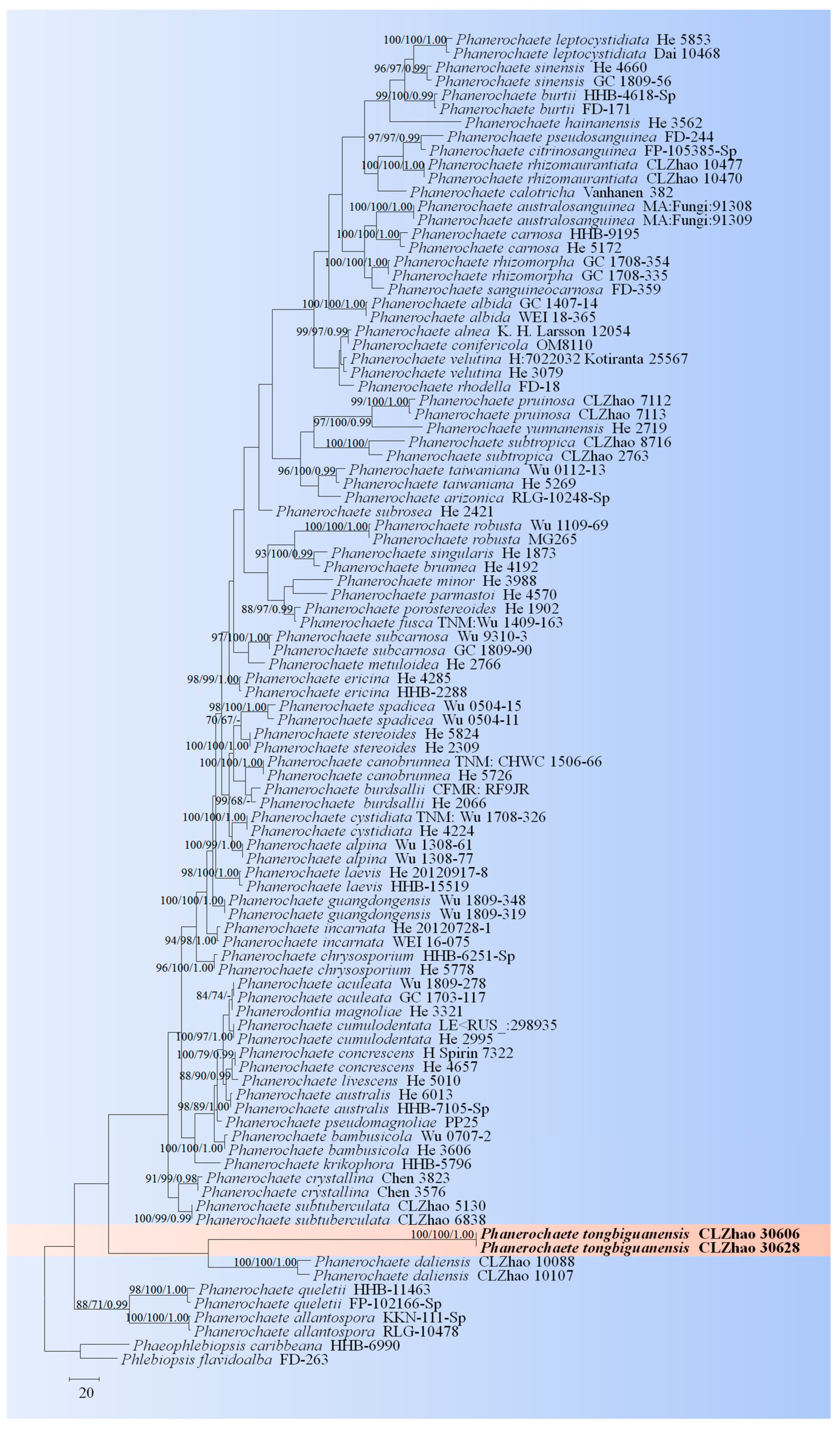
The ITS dataset of the genus Phanerochaete (Figure 4) included sequences from 96 fungal specimens representing 60 species. The dataset had an aligned length of 880 characters, of which 319 characters are constant, 77 are variable and parsimony-uninformative, and 484 are parsimony-informative. Maximum parsimony analysis yielded one equally parsimonious tree (TL = 2187, CI = 0.4015, HI = 0.5985, RI = 0.6231, RC = 0.2501). The best model for the ITS dataset estimated and applied in the Bayesian analysis was GTR + I + G (lset nst = 6, rates = invgamma; prset statefreqpr = dirichlet (1,1,1,1). Bayesian analysis and ML analysis resulted in a similar topology to MP analysis with an average standard deviation of split frequencies = 0.001737 (BI). The phylogenetic tree indicated that P. tongbiguanensis was grouped with the close taxa P. daliensis J. Yu & C.L. Zhao.
The ITS dataset of the genus Phlebiopsis (Figure 5) included sequences from 33 fungal specimens representing 20 species. The dataset had an aligned length of 665 characters, of which 392 characters are constant, 82 are variable and parsimony-uninformative, and 191 are parsimony-informative. Maximum parsimony analysis yielded six equally parsimonious trees (TL = 685, CI = 0.5650, HI = 0.4350, RI = 0.6543, RC = 0.3697). The best model for the ITS dataset estimated and applied in the Bayesian analysis was GTR + I + G (lset nst = 6, rates = invgamma; prset statefreqpr = dirichlet (1,1,1,1). Bayesian analysis and ML analysis resulted in a similar topology to MP analysis with an average standard deviation of split frequencies = 0.003384 (BI). The phylogenetic tree indicated that P. fissurata was grouped with the close taxa P. lamprocystidiata (Sheng H. Wu) Sheng H. Wu & Hallenb.
The ITS dataset of the genus Vararia (Figure 6) included sequences from 52 fungal specimens representing 40 species. The dataset had an aligned length of 796 characters, of which 148 characters were constant, 116 were variable and parsimony-uninformative, and 532 were parsimony-informative. Maximum parsimony analysis yielded one equally parsimonious tree (TL = 4063, CI = 0.3104, HI = 0.6896, RI = 0.4313, and RC = 0.1339). The best model for the ITS dataset estimated and applied in the Bayesian analysis was GTR + I + G. The Bayesian and ML analyses resulted in a similar topology to that of the MP analysis with split frequencies = 0.000442 (BI). The phylogram inferred from ITS sequences (Figure 6) revealed that V. yingjiangensis was grouped with six close taxa, namely V. ambigua Boidin, Lanq. & Gilles, V. ellipsospora G. Cunn., V. fragilis L. Zou & C.L. Zhao, V. gallica (Bourdot & Galzin) Boidin, V. ochroleuca (Bourdot & Galzin) Donk and V. tropica A.L. Welden.
3.2. Taxonomy
MycoBank no.: 851416
Holotype—China, Yunnan province, Lincang, Fengqing County, Yaojie Town, Xingyuan Village, 24°58′ N, 99°92′ E, altitude 1660 m asl., on the fallen branch of angiosperm, leg. C.L. Zhao, 20 July 2022, CLZhao 22781 (SWFC).
Etymology—Yunnanense (Lat.): referring to the locality (Yunnan province) of the type specimen.
Fruiting body—Basidiomata annual, resupinate, membranaceous to pellicular, soft, without odor and taste when fresh, up to 110 mm long, 60 mm wide, and 280 µm thick. Hymenial surface smooth, cream when fresh, cream to salmon-buff, sometimes cracked when dried. Sterile margin thinning out, becoming indistinct and concolorous with hymenophore surface, up to 1 mm.
Hyphal system—Dimitic, generative hyphae bearing simple-septa, scattered, thick-walled, colorless, 2–4 µm in diameter, IKI-, CB-, tissues unchanged in KOH. Asterosetae in subiculum are abundant, predominant, yellowish brown, thick-walled, 2–4 µm in diameter, regularly star-shaped, weakly dextrinoid, rays up to 60 µm long, with acute tips, CB-, tissues unchanged in KOH.
Hymenium—Gloeocystidia subulate, thick-walled, with a basal simple septum, 34.5–54 × 7–10 µm. Basidia cylindrical, colorless, with four sterigmata and a basal simple-septum, 31–38 × 4–5 µm.
Basidiospores—Globose, colorless, thin-walled, echinulate, amyloid, 4.5–6 × 4–5 µm, L = 5.11 µm, W = 4.33 µm, Q = 1.07–1.18 (n = 60/2).
Additional specimens examined (paratypes)—China, Yunnan province, Lincang, Fengqing County, Yaojie Town, Xingyuan Village, 24°58′ N, 99°92′ E, altitude 1660 m asl., on the trunk of angiosperm, leg. C.L. Zhao, 20 July 2022, CLZhao 22786 (SWFC); on the fallen branch of angiosperm, leg. C.L. Zhao, 20 July 2022, CLZhao 22846 (SWFC).
MycoBank no.: 851417
Holotype—China, Yunnan province, Dehong, Yingjiang County, Tongbiguan Provincial Nature Reserve, 24°71′ N, 97°94′ E, altitude 2000 m asl., on the fallen branch of angiosperm, 20 July 2023, CLZhao 30606 (SWFC).
Etymology—Tongbiguanensis (Lat.): referring to the locality (Tongbiguan) of the type specimen.
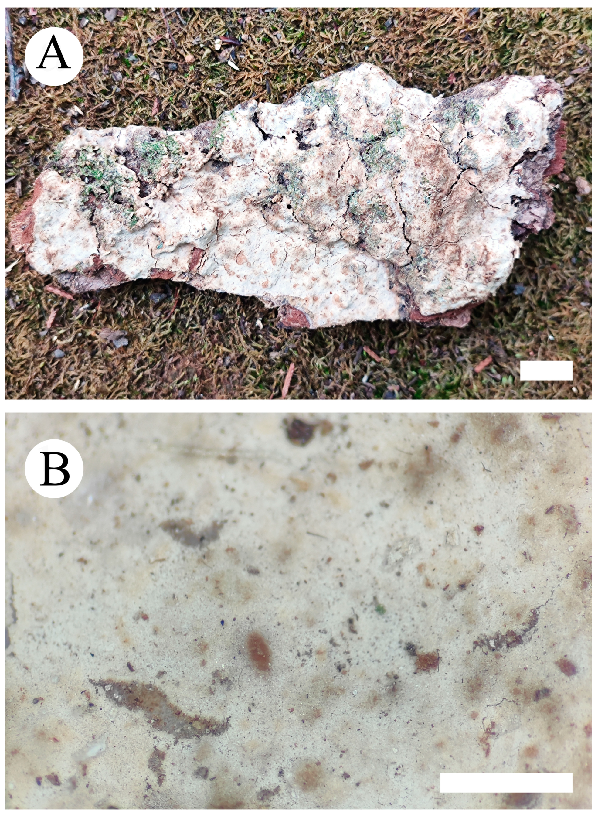
Fruiting body—Basidiomata annual, resupinate, thin, adnate, leather, without odor and taste when fresh, up to 70 mm long, 10 mm wide, 70–130 µm thick. Hymenial surfaces are smooth, white to cream when fresh, to cream to slightly buff upon drying. Sterile margins are distinct, whitish, and up to 1 mm.
Hyphal system—Monomitic, generative hyphae bearing simple-septa, thick-walled, 3–4 µm in diameter, branched, colorless, IKI-, CB-; tissues unchanged in KOH; subhymenial hyphae densely covered by crystals.
Hymenium—Cystidia subclavate, colorless, covered with a lot of crystals, thick-walled, 32–41 × 6.5–11 µm. Basidia subclavate to cylindrical, with four sterigmata and a basal simple septum, 17–26 × 6–7 µm.
Basidiospores—Oblong ellipsoid, colorless, thin-walled, smooth, IKI-, CB-, 6–9 × 3–4.5 µm, L = 7.48 µm, W = 4.02 µm, Q = 1.84–1.88 (n = 60/2).
Additional specimen examined (paratype)—China, Yunnan province, Dehong, Yingjiang County, Tongbiguan Provincial Nature Reserve, 24°71′ N, 97°94′ E, altitude 2000 m asl., on the fallen branch of angiosperm, 20 July 2023, CLZhao 30628 (SWFC).
MycoBank: 851421
Holotype—China, Yunnan province, Dehong, Yingjiang County, Tongbiguan Provincial Nature Reserve, 24°71′ N, 97°94′ E, altitude 2000 m asl., on the fallen branch of angiosperm, 19 July 2023, CLZhao 30147 (SWFC).
Etymology—Referring to the cracking hymenial surface.
Fruiting body—Basidiomata annual, resupinate, adnate, membranaceous, without odor and taste when fresh, up to 100 mm long, 70 mm wide, 100–210 µm thick. Hymenial surface tuberculate, white when fresh, white to buff to slightly brown upon drying, sometimes sparsely and deeply cracked with age. Sterile margins are distinct, white, and up to 2 mm.
Hyphal system—Monomitic, generative hyphae bearing simple-septa, colorless, thick-walled, branched, interwoven, 4–5 µm in diameter, IKI-, CB-; tissues unchanged in KOH.
Hymenium—Cystidia conical, colorless, covered with a lot of crystals, thick-walled, 27–48 × 6–11 µm. Basidia clavate, with four sterigmata and a basal simple septum, 16–26 × 5–7 µm.
Basidiospores—Broadly ellipsoid, thin-walled, colorless, smooth, IKI-, CB-, 4–6.5 × 3–4 µm, L = 5.03 µm, W = 3.59 µm, Q = 1.33–1.47 (n = 60/2).
Additional specimen examined (paratype)—China, Yunnan province, Dehong, Yingjiang County, Tongbiguan Provincial Nature Reserve, 24°71′ N, 97°94′ E, altitude 2000 m asl., on the fallen branch of angiosperm, 19 July 2023, CLZhao 30247 (SWFC).
MycoBank no.: 851424
Holotype—China, Yunnan province, Dehong, Yingjiang County, Tongbiguan Provincial Nature Reserve, 24°71′ N, 94°52′ E, altitude 1500 m asl., on fallen branch of angiosperm, 19 July 2023, CLZhao 30284 (SWFC).
Etymology—Yingjiangensis (Lat.): referring to the locality (Yingjiang) of the type specimen.
Fruiting body—Basidiomata annual, adnate, corky, without odor and taste when fresh, up to 80 mm long, 40 mm wide, 80–120 µm thick. Hymenial surface smooth, cream to pinkish buff when fresh, pinkish buff to cinnamon-buff when dry, cracking with age. Sterile margin thin, indistinct, slightly cream to pinkish buff, up to 2 mm.
Hyphal system—Dimitic, generative hyphae bearing simple-septa, colorless, thin- to thick-walled, occasionally branched, interwoven, 3–4 µm in diameter, IKI-, CB-, tissues unchanged in KOH. Dichohyphae yellowish, capillary, distinctly thick-walled, up to 1.4 μm in diameter and with acute tips, moderately dextrinoid in Melzer’s reagent; more frequently branched.
Hymenium—Gloeocystidia two types, (i) Gloeocystidia subulate, usually with a constriction at the tip, colorless, obviously thick-walled, smooth, 25–42.5 × 5–11 µm; (ii) Gloeocystidia subulate, usually with two constrictions at the tip, colorless, obviously thick-walled, smooth, 28–35 × 6–10 µm. Basidia rare; basidioles cylindrical, dominant, thin-walled, 13–26 × 4.5–10 µm.
Basidiospores—Ellipsoid, slightly thick-walled, colorless, smooth, amyloid, CB-, 6.5–11.5 × 5–7 µm, L = 9.34 µm, W = 6.08 µm, Q = 1.5–1.6 (n = 60/2).
Additional specimen examined (paratype)—China, Yunnan province, Dehong, Yingjiang County, Tongbiguan Provincial Nature Reserve, 24°71′ N, 97°52′ E, altitude 1500 m asl., on fallen branch of angiosperm, 19 July 2023, CLZhao 30392 (SWFC).
4. Discussion
The family-level classification for the order Polyporales (Basidiomycota) revealed that the two taxa of Phanerochaete daliensis and Phlebiopsis lamprocystidiata nested into the family Phanerochaetaceae within the residual polyporoid clade based on the molecular systematics study amplifying the ITS, nLSU, RPB1, and RPB2 genes [21,27]. Seven genera, Asterostroma, Dichostereum, Gloiothele, Peniophora, Scytinostroma, Vararia, and Vesiculomyces E. Hagstr., were grouped together and clustered within the family Peniophoraceae [18]. In the present study, four new species were nested into the families Phanerochaetaceae and Peniophoraceae; from the phylogram of the ITS + nLSU data, the new species Phanerochaete tongbiguanensis were grouped into Phanerochaete, and the taxon Phlebiopsis fissurata was grouped into genus Phlebiopsis (Figure 1); the new species Asterostroma yunnanense was grouped into Asterostroma, and Vararia yingjiangensis was clustered into Vararia (Figure 2).
Based on ITS topology (Figure 3), the present study revealed that the new species Asterostroma yunnanense was grouped with two close taxa, A. cervicolor and A. vararioides S.L. Liu & S.H. He. However, morphologically, A. cervicolor is distinct from A. yunnanense by the thin-walled marginal hyphae (2–5 µm diameter) and thin-walled aerial hyphae 1–5 µm diameter, and smaller gloeocystidia (20–30 × 7–15 µm) [86]. The species A. vararioides can be distinguished by its grayish brown to dark brown hymenial surface, thin-walled generative hyphae, presence of the dichohyphidia and thin-walled, longer gloeocystidia (30–60 × 5–11 µm), larger subcylindrical to fusoid basidia (30–65 × 7–11 µm), and larger, smooth basidiospores measuring 5.5–7.5 × 5–7 µm [18].
The phylogenetic tree (Figure 4) based on the ITS data showed that the new taxon P. tongbiguanensis was grouped with the species P. daliensis and P. subtropica J. Yu & C.L. Zhao. However, morphologically, P. daliensis is distinct from P. tongbiguanensis by its grandinioid hymenophore, ellipsoid e to cylindrical, thick-walled, smaller basidiospores (3–6 × 1.8–3 µm) [27]. The species P. subtropica is distinguished from P. tongbiguanensis by its fusiform cystidia and smaller basidia (12–21 × 3–5 µm) and ellipsoid basidiospores measuring as 3.0–4.8 × 2.4–3.4 µm [27]. Phylogenetic tree analysis (Figure 5) revealed that the new species P. fissurata was grouped with the species P. lamprocystidiata and then closely clustered with P. yunnanensis C.L. Zhao and P. gigantea (Fr.) Jülich. However, morphologically, P. lamprocystidiata is distinct from P. fissurata by its grayish yellow hymenial surface and distinct lamprocystidia [31]. The taxon P. yunnanensis is distinct from P. fissurata by having the smaller, narrowly clavate to subcylindrical basidia (10–21 × 3.5–4.5 µm) and smaller basidiospores measuring as 3.5–4.5 × 2.5–3.5 µm. Another species P. gigantea can be distinguished by its greyish-white to buff basidimata, larger cystidia (50–80 × 10–15 µm), and narrowly ellipsoid, smaller basidiospores (6.5–8 × 3–3.5 µm) [13].
Based on the ITS phylogenetic analysis (Figure 6), the new species Vararia yingjiangensis is closely grouped with six taxa, namely V. ambigua, V. ellipsospora, V. fragilis, V. gallica, V. ochroleuca, and V. tropica. However, morphologically, V. ochroleuca is distinct from V. yingjiangensis by having the slightly thick-walled gloeocystidia, thin-walled generative hyphae, and both smaller gloeocystidia (16–34 × 4.5–7.5 µm) and basidiospores measuring as 2.6–3.8 × 2–3.2 µm [87]. The taxon V. gallica is distinct from V. yingjiangensis by having the longer basidiospores measuring as 9–12 × 3.5–5 µm [24]. The species V. ellipsospora is distinct from V. yingjiangensis by having the fimbriate basidiomata, generative hyphae with clamped connection, and flexuous to cylindrical gloeocystidia [45]. Vararia fragilis differs from V. yingjiangensis by having smaller, elliptical to ovoid gloeocystidia measuring as 5.8–16 × 3.5–7 µm. The taxon V. ambigua differs from V. yingjiangensis by its thin-walled and smaller spores measuring as 3–8 × 3–5 µm. The species V. tropica can be distinguished by its wider, oblong basidiospores (10–12 × 7–8 µm) [88].
Based on the phylogenetic and morphological research results, more and more new wood-inhabiting fungi are being found and reported [1,43,54,55,89,90,91,92]. In the present study, four new taxa from the subtropics are described based on morphological and molecular phylogenetic analyses, which can enrich the wood-inhabiting fungal diversity in China and the world.
Author Contributions
Conceptualization, C.Z. and J.Z.; methodology, C.Z. and Y.D.; software, C.Z. and Y.D.; validation, C.Z. and Y.D.; formal analysis, C.Z. and Y.D.; investigation, J.L., C.Z. and Y.D.; resources, J.L., C.Z. and J. Z.; writing—original draft preparation, C.Z. and Y.D.; writing—review and editing, C.Z. and Y.D.; visualization, C.Z.; supervision, C.Z.; project administration, C.Z.; funding acquisition, C.Z. and J.Z. All authors have read and agreed to the published version of the manuscript.
Funding
The research was supported by the National Natural Science Foundation of China (Project No. 32170004, U2102220), Forestry Innovation Programs of Southwest Forestry University (Grant No: LXXK-2023Z07), and the High-level Talents Program of Yunnan province (YNQR-QNRC-2018-111).
Institutional Review Board Statement
Not applicable for studies involving humans or animals.
Informed Consent Statement
Not applicable for studies involving humans.
Data Availability Statement
Publicly available datasets were analyzed in this study.
Conflicts of Interest
The authors declare no conflicts of interest.
References
- Dai, Y.C. A revised checklist of corticioid and hydnoid fungi in China for 2010. Mycoscience 2011, 52, 69–79. [Google Scholar] [CrossRef]
- Dai, Y.C. Pathogenic wood-decaying fungi on woody plants in China. Mycosystema 2012, 31, 493–509. [Google Scholar]
- Sun, Y.F.; Costa-Rezende, D.H.; Xing, J.H.; Zhou, J.L.; Zhang, B.; Gibertoni, T.B.; Gates, G.; Glen, M.; Dai, Y.C.; Cui, B.K. Multigene phylogeny and taxonomy of Amauroderma s. lat. (Ganodermataceae). Persoonia 2019, 44, 206–239. [Google Scholar] [CrossRef]
- Sun, Y.F.; Xing, J.H.; He, X.L.; Wu, D.M.; Song, C.G.; Liu, S.; Vlasák, J.; Gates, G.; Gibertoni, T.B.; Cui, B.K. Species diversity, sys-tematic revision and molecular phylogeny of Ganodermataceae (Polyporales, Basidiomycota) with an emphasis on Chinese collections. Stud. Mycol. 2022, 101, 287–415. [Google Scholar] [CrossRef]
- Li, Q.S.; Wei, Y.L. Species composition and distribution characteristics of wood-decaying fungi in tropical areas of China. Mycosystema 2021, 40, 2653–2672. [Google Scholar] [CrossRef]
- Dai, Y.C.; Fan, L.F.; Chen, J.J.; Wu, C.P.; Wu, Y.D.; Yuan, Y. Diversity of Heterobasidion causing root and butt rotting of conifers and proposal for entry wood quarantine aiming at Heterobasidion species. Mycosystema 2021, 40, 1958–1964. [Google Scholar] [CrossRef]
- Dai, Y.C.; Liu, H.G.; Wu, F.; Si, J.; Yuan, Y.; He, S.H.; Cui, B.K.; Zhou, M.; Zhao, Q. Resources and Diversity of Wood-Decaying Fungi in Yunnan; Science Press: Beijing, China, 2022; p. 574. [Google Scholar]
- Dai, Y.C.; Yang, Z.L.; Cui, B.K.; Wu, G.; Yuan, H.S.; Zhou, L.W.; He, S.H.; Ge, Z.W.; Wu, F.; Wei, Y.L.; et al. Diversity and systematics of the important macrofungi in Chinese forests. Mycosystema 2021, 40, 770–805. [Google Scholar] [CrossRef]
- Liu, S.; Chen, Y.Y.; Sun, Y.F.; He, X.L.; Song, C.G.; Si, J.; Liu, D.M.; Gates, G.; Cui, B.K. Systematic classification and phylogenetic relationships of the brown rot fungi within the Polyporales. Fungal Divers. 2023, 118, 1–94. [Google Scholar] [CrossRef]
- Liu, S.; Han, M.L.; Xu, T.M.; Wang, Y.; Wu, D.M.; Cui, B.K. Taxonomy and phylogeny of the Fomitopsis pinicola complex with de-scriptions of six new species from East Asia. Front. Microbiol. 2021, 12, 644979. [Google Scholar] [CrossRef]
- Yuan, Y.; Bian, L.S.; Wu, Y.D.; Chen, J.J.; Wu, F.; Liu, H.G.; Zeng, G.Y.; Dai, Y.C. Species diversity of pathogenic wood-rotting fungi (Agaricomycetes, Basidiomycota) in China. Mycology 2023, 14, 204–226. [Google Scholar] [CrossRef] [PubMed]
- He, M.Q.; Zhao, R.L.; Hyde, K.D.; Begerow, D.; Kemler, M.; Yurkov, A.; McKenzie, E.H.C.; Raspé, O.; Kakishima, M.; Sánchez-Ramírez, S.; et al. Notes, outline and divergence times of Basidiomycota. Fungal Diver. 2019, 99, 105–367. [Google Scholar]
- Bernicchia, A.; Gorjón, S.P. Fungi Europaei 12: Corticiaceae s.l.; Edizioni Candusso: Alassio, Italy, 2011. [Google Scholar]
- Larsson, K.H.; Larsson, E.; Kõljalg, U. High phylogenetic diversity among corticioid homobasidiomycetes. Mycol. Res. 2004, 108, 983–1002. [Google Scholar] [CrossRef]
- Parmasto, E. The Lachnocladiaceae of the Soviet Union. With a Key to the Boreal Species; Institute of Zoology and Botany, Academy of Sciences of the Estonian S.S.R.: Tartu, Estonia, 1971; p. 167. [Google Scholar]
- Hallenberg, N.; Eriksson, J. The Lachnocladiaceae and Coniophoraceae of North Europoe; Fungiflora: Oslo, Norway, 1985; p. 96. [Google Scholar]
- Suhara, H.; Maekawa, N.; Ushijima, S.; Kinjo, K.; Hoshi, Y. Asterostroma species (Basidiomycota) from mangrove forests in Japan. Mycoscience 2010, 51, 75–80. [Google Scholar] [CrossRef]
- Liu, S.L.; Tian, Y.; Nie, T.; Thawthong, A.; Hyde, K.D.; Xu, L.L.; He, S.H. Updates on East Asian Asterostroma (Russulales, Basidiomycota): New species and new records from Thailand and China. Mycol. Prog. 2017, 16, 667–676. [Google Scholar] [CrossRef]
- Karsten, P. Kritisk öfversigt af Finlan’s Basidsvamper (Basidiomycetes, Gastero- & Hxmenomycetes). Bidr. Till Kännedom Finl. Nat. Och Folk. 1889, 48, 1–470. [Google Scholar]
- Wu, S.H. Six new species of Phanerochaete from Taiwan. Bot. Bull. Acad. Sin. 2000, 41, 165–174. [Google Scholar]
- Wu, S.H.; Nilsson, H.R.; Chen, C.T.; Yu, S.Y.; Hallenberg, N. The white-rotting genus Phanerochaete is polyphyletic and distributed throughout the phleboid clade of the Polyporales (Basidiomycota). Fungal Diver. 2010, 42, 107–118. [Google Scholar] [CrossRef]
- Floudas, D.; Hibbett, D.S. Revisiting the taxonomy of Phanerochaete (Polyporales, Basidiomycota) using a four gene dataset and extensive ITS sampling. Fungal Biol. 2015, 119, 679–719. [Google Scholar] [CrossRef] [PubMed]
- Ghobad-Nejhad, M.; Liu, S.L.; Langer, E.; Dai, Y.C. Molecular and morphological evidence reveal a new non-cystidiate species belonging to the core Phanerochaete (Polyporales). Mycol. Prog. 2015, 14, 68. [Google Scholar] [CrossRef]
- Boonmee, S.; Wanasinghe, D.N.; Calabon, M.S.; Huanraluek, N.; Chandrasiri, S.K.U.; Jones, G.E.B.; Rossi, W.; Leonardi, M.; Singh, S.K.; Rana, S.; et al. Fungal Diver. notes 1387–1511: Taxonomic and phylogenetic contributions on genera and species of fungal taxa. Fungal Diver. 2021, 111, 1–335. [Google Scholar] [CrossRef] [PubMed]
- Chen, C.C.; Chen, C.Y.; Wu, S.H. Species diversity, taxonomy and multi-gene phylogeny of phlebioid clade (Phanerochaetaceae, Irpicaceae, Meruliaceae) of Polyporales. Fungal Diver. 2021, 111, 337–442. [Google Scholar] [CrossRef]
- Li, Y.; Chen, C.C.; He, S.H. New corticioid taxa in Phanerochaetaceae (Polyporales, Basidiomycota) from East Asia. Front. Microbiol. 2023, 14, 1093096. [Google Scholar] [CrossRef]
- Yu, J.; Cai, J.; Liu, H.G.; Zhao, C.L. Molecular systematics and taxonomy reveal three new taxa in Phanerochaete (Phanerochaetaceae, Polyporales) from the Yunnan–Guizhou plateau, East Asia. Mycol. Prog. 2023, 22, 59. [Google Scholar] [CrossRef]
- Zhang, Q.Y.; Liu, Z.B.; Liu, H.G.; Si, J. Two new corticioid species of Phanerochaetaceae (Polyporales, Basidiomycota) from Southwest China. Front. Cell. Infect. Microbiol. 2023, 13, 1105918. [Google Scholar] [CrossRef]
- Jülich, W. Studies in resupinate Basidiomycetes V. Some new genera and species. Persoonia 1978, 10, 137–140. [Google Scholar]
- Dhingra, G.S. The genus Phlebiopsis in the Eastern Himalayas. Nova Hedwig. 1987, 44, 221–227. [Google Scholar]
- Wu, S.H. Two new species of Phanerochaete from Taiwan. Mycotaxon 2004, 90, 423–429. [Google Scholar]
- Priyanka; Dhingra, G.S.; Kaur, N. Phlebiopsis mussooriensis (Agaricomycetes), a new corticioid species from India. Mycotaxon 2011, 115, 255–258. [Google Scholar] [CrossRef]
- Kaur, G.; Singh, A.P.; Dhingra, G.S. Phlebiopsis punjabensis, a new species from India. Mycotaxon 2015, 130, 907–909. [Google Scholar] [CrossRef]
- Xu, T.M.; Zeng, Y.F.; Cheng, Y.H.; Zhao, C.L. Phlebiopsis lacerata sp. nov (Polyporales, Basidiomycota) from southern China. Phytotaxa 2020, 440, 268–280. [Google Scholar] [CrossRef]
- Zhao, Y.N.; He, S.H.; Nakasone, K.K.; Wasantha, K.L.; Chen, C.C.; Liu, S.L.; Ma, H.X.; Huang, M.R. Global phylogeny and taxonomy of the wood-decaying fungal genus Phlebiopsis (Polyporales, Basidiomycota). Front. Microbiol. 2021, 12, 622460. [Google Scholar] [CrossRef]
- Karsten, P.A. Kritisk Ofversigt af Finlands Basidsvampar. Biology 1898, 3, 1–36. [Google Scholar]
- Boidin, J.; Lanquetin, P. Vararia subgenus Vararia (Basidiomycetes, Lachnocladiaceae): Étude spèciale des espèces d’ Afrique intertropicale. Bull. Soc. Mycol. Fr. 1975, 91, 457–513. [Google Scholar]
- Boidin, J. Application du concept biologique del’espèce aux Basidiomycètes. Le genre Vararia section Vararia au Gabon. Cryptogam. Mycol. 1980, 1, 265–384. [Google Scholar]
- Boidin, J.; Lanquetin, P. Le genre Scytinostroma Donk (Basidiomycetes, Lachnocladiaceae). Bibl. Mycol. 1987, 114, 1–130. [Google Scholar]
- Stalpers, J.A. The aphyllophoraceous fungi II. Keys to the species of the Hericiales. Stud. Mycol. 1996, 40, 1–183. [Google Scholar]
- Larsson, E.; Larsson, K.H. Phylogenetic relationships of russuloid basidiomycetes with emphasis on aphyllophoralean taxa. Mycologia 2003, 95, 1037–1065. [Google Scholar] [CrossRef]
- Liu, S.L.; He, S.H. The genus Vararia (Russulales, Basidiomycota) in China. Two new species and two new Chinese records. Nord. J. Bot. 2016, 34, 553–558. [Google Scholar] [CrossRef]
- Zou, L.; Zhang, X.L.; Deng, Y.L.; Zhao, C.L. Four new wood-inhabiting fungal species of Peniophoraceae (Russulales, Basidiomycota) from the Yunnan-Guizhou Plateau, China. J. Fungi 2022, 8, 1227. [Google Scholar] [CrossRef] [PubMed]
- Deng, Y.L.; Zhao, C.L. The molecular phylogeny and morphology revealed a new wood-rotting fungus Vararia yunnanensis (Peniophoraceae, Russulales) in Yunnan Province, China. Phytotaxa 2023, 583, 39–49. [Google Scholar] [CrossRef]
- Cunningham, G.H. Thelephoraceae of New Zealand. Part IV. The genus Vararia. Trans. Proc. R. Soc. N. Z. 1955, 82, 973–985. [Google Scholar]
- Gilbertson, R.L. Some species of Vararia from temperate North America. Pap. Mich. Acad. Sci. 1965, 50, 161–184. [Google Scholar]
- Boidin, J. Basidiomycètes Lachnocladiaceae résupinés de la Republique Centrafricaine. Cah. Maboké 1967, 5, 23–35. [Google Scholar]
- Pouzar, Z. Taxonomic studies in resupinate fungi I. Česká Mykol. 1982, 36, 141–145. [Google Scholar]
- Boidin, J.; Gilles, G. Contribution à la connaissance du genre Vararia (Basidiomycotina). Bull. Soc. Mycol. 1999, 115, 115–139. [Google Scholar]
- Duhem, B.; Buyck, B. On two new tropical Vararia (Russulales, Basidiomycota) with extremely small, racemose dichohyphidia. Cryptogam. Mycology 2012, 33, 427–437. [Google Scholar]
- Sanyal, S.K.; Dhingra, G.S.; Singh, A.P. Vararia longicystidiata sp. nov. (Agaricomycetes) from India. Mycotaxon 2012, 120, 357–360. [Google Scholar] [CrossRef]
- Nakasone, K.K. Taxonomic studies in Chrysoderma, Corneromyces, Dendrophysellum, Hyphoradulum, and Mycobonia. Mycotaxon 2015, 130, 369–397. [Google Scholar] [CrossRef]
- Larsson, K.H. Re-thinking the classification of corticioid fungi. Mycol. Res. 2007, 111, 1040–1063. [Google Scholar] [CrossRef] [PubMed]
- Luo, K.Y.; Zhao, C.L. A molecular systematics and taxonomy research on Trechispora (Hydnodontaceae, Trechisporales): Concentrating on three new Trechispora species from East Asia. J. Fungi 2022, 8, 1020. [Google Scholar] [CrossRef] [PubMed]
- Zhao, C.L.; Qu, M.H.; Huang, R.X.; Karunarathna, S.C. Multi-gene phylogeny and taxonomy of the wood-rotting fungal genus Phlebia sensu lato (Polyporales, Basidiomycota). J. Fungi 2023, 9, 320. [Google Scholar] [CrossRef]
- Binder, M.; Hibbett, D.S.; Larsson, K.H.; Larsson, E.; Langer, E.; Langer, G. The phylogenetic distribution of resupinate forms across the major clades of mushroom-forming fungi (Homobasidiomycetes). Syst. Biodivers. 2005, 3, 113–157. [Google Scholar] [CrossRef]
- Binder, M.; Justo, A.; Riley, R.; Salamov, A.; Lopez-Giraldez, F.; Sjökvist, E.; Copeland, A.; Foster, B.; Sun, H.; Larsson, E.; et al. Phylogenetic and phylogenomic overview of the Polyporales. Mycologia 2013, 105, 350–1373. [Google Scholar] [CrossRef]
- Justo, A.; Miettinen, O.; Floudas, D.; Ortiz-Santana, B.; Sjökvist, E.; Lindner, D.; Nakasone, K.; Niemelä, T.; Larsson, K.H.; Ryvarden, L.; et al. A revised family-level classifcation of the Polyporales (Basidiomycota). Fungal Biol. 2017, 121, 798–824. [Google Scholar] [CrossRef]
- Haelewaters, D.; Wijayawardene, N.N.; Hyde, K.D.; Dai, D.Q.; Sánchez-García, M. Outline of Fungi and fungus-like taxa-2021. Mycosphere 2022, 13, 53–453. [Google Scholar] [CrossRef]
- Reid, D.A. A Monograph of the Stipitate Stereoid Fungi; J. Cramer: Weinheim, Germany, 1965. [Google Scholar]
- Miller, S.L.; Larsson, E.; Larsson, K.H.; Verbeken, A.; Nuytinck, J. Perspectives in the new Russulales. Mycologia 2006, 98, 960–970. [Google Scholar] [CrossRef] [PubMed]
- White, T.J.; Bruns, T.; Lee, S.; Taylor, J. Amplification and direct sequencing of fungal ribosomal RNA genes for phylogenetics. PCR Protoc. A Guide Methods Appl. 1990, 18, 315–322. [Google Scholar] [CrossRef]
- Hall, T.A. BioEdit: A user-friendly biological sequence alignment editor and analysis program for Windows 95/98/NT. Nucleic Acids Symp. Ser. 1999, 41, 95–98. [Google Scholar]
- Wang, D.Q.; Zhao, C.L. Morphological and phylogenetic evidence for recognition of two new species of Phanerochaete from East Asia. J. Fungi 2021, 7, 1063. [Google Scholar] [CrossRef] [PubMed]
- Leal-Dutra, C.A.; Neves, M.A.; Griffith, G.W.; Reck, M.A.; Clasen, L.A.; Dentinger, B.T.M. Reclassification of Parapterulicium Corner (Pterulaceae, Agaricales), contributions to Lachnocladiaceae and Peniophoraceae (Russulales) and introduction of Baltazaria gen. nov. MycoKeys 2018, 37, 39–56. [Google Scholar] [CrossRef] [PubMed]
- Zhao, C.L.; Wu, Z.Q. Ceriporiopsis kunmingensis sp. nov. (Polyporales, Basidiomycota) evidenced by morphological characters and phylogenetic analysis. Mycol. Prog. 2017, 16, 93–100. [Google Scholar] [CrossRef]
- Swofford, D.L. PAUP*: Phylogenetic Analysis Using Parsimony (* and Other Methods), Version 4.0b10; Sinauer Associates: Sunderland, MA, USA, 2002.
- Felsenstein, J. Confidence intervals on phylogenetics: An approach using bootstrap. Evolution 1985, 39, 783–791. [Google Scholar] [CrossRef]
- Miller, M.A.; Pfeiffer, W.; Schwartz, T. The CIPRES Science Gateway: Enabling high-impact science for phylogenetics researchers with limited resources. Assoc. Comput. Mach. 2012, 39, 1–8. [Google Scholar] [CrossRef]
- Vu, D.; Groenewald, M.; Vries, M.; Gehrmann, T.; Stielow, B.; Eberhardt, U.; Al-Hatmi, A.; Groenewald, J.Z.; Cardinali, G.; Houbraken, J.; et al. Large-scale generation and analysis of filamentous fungal DNA barcodes boosts coverage for kingdom fungi and reveals thresholds for fungal species and higher taxon delimitation. Stud. Mycol. 2019, 92, 135–154. [Google Scholar] [CrossRef]
- Liu, S.L.; Nakasone, K.K.; He, S.H. Michenera incrustata sp. nov. (Peniophoraceae, Russulales) from southern China. Nova Hedwig. 2019, 108, 197–206. [Google Scholar] [CrossRef]
- Liu, S.L.; He, S.H. Taxonomy and phylogeny of Dichostereum (Russulales), with descriptions of three new species from southern China. MycoKeys 2018, 40, 111–126. [Google Scholar] [CrossRef]
- Spirin, V.; Malysheva, V.; Miettinen, O.; Vlasák, J.; Alvarenga, R.L.M.; Gibertoni, T.B.; Ryvarden, L.; Larsson, K.K. On Protomerulius and Heterochaetella (Auriculariales, Basidiomycota). Mycol. Prog. 2019, 18, 1079–1099. [Google Scholar] [CrossRef]
- Phookamsak, R.; Hyde, K.D.; Jeewon, R.; Bhat, D.J.; Jones, E.B.G.; Maharachchikumbura, S.; Raspé, O.; Karunarathna, S.C.; Wanasinghe, D.; Hongsanan, S.; et al. Fungal diversity notes 929–1035: Taxonomic and phylogenetic contributions on genera and species of fungi. Fungal Divers. 2019, 95, 1–273. [Google Scholar] [CrossRef]
- Wu, S.H.; Chen, Y.P.; Wei, C.L.; Floudas, D.; Dai, Y.C. Two new species of Phanerochaete (Basidiomycota) and redescription of P. robusta. Mycol. Prog. 2017, 17, 425–435. [Google Scholar] [CrossRef]
- Xu, Y.L.; Cao, Y.F.; Nakasone, K.K.; Chen, C.C.; He, S.H. Taxonomy and phylogeny of Phanerochaete sensu stricto (Polyporales, Basidiomycota) with emphasis on Chinese collections and descriptions of nine new species. Mycosphere 2020, 11, 1527–1552. [Google Scholar] [CrossRef]
- Volobuev, S.; Okun, M.; Ordynets, A.; Spirin, V. The Phanerochaete sordida group (Polyporales, Basidiomycota) in temperate Eurasia, with a note on Phanerochaete pallida. Mycol. Prog. 2015, 14, 80. [Google Scholar] [CrossRef]
- Wu, S.H.; Chen, C.C.; Wei, C.L. Three new species of Phanerochaete (Polyporales, Basidiomycota). MycoKeys 2018, 41, 91–106. [Google Scholar] [CrossRef] [PubMed]
- Lima, V.X.; Lira, C.S.; Chikowski, R.S.; Santos, C.; Lima, N.; Gibertoni, T.B. Additions to neotropical stereoid fungi (Polyporales, Basidiomycota): One new species of Lopharia and one new combination in Phlebiopsis. Mycol. Prog. 2020, 19, 31–40. [Google Scholar] [CrossRef]
- Miettinen, O.; Spirin, V.; Vlasák, J.; Rivoire, B.; Stenroos, S.; Hibbett, D. Polypores and genus concepts in Phanerochaetaceae (Polyporales, Basidiomycota). MycoKeys 2016, 17, 1–46. [Google Scholar] [CrossRef]
- Zhao, C.L.; Liu, X.F.; Ma, X. Phlebiopsis yunnanensis sp. nov. (Polyporales, Basidiomycota) evidenced by morphological characters and phylogenetic analysis. Nova Hedwig. 2019, 108, 265–279. [Google Scholar] [CrossRef]
- Nakasone, K.K.; Draeger, K.R.; Ortiz-Santana, B. A contribution to the taxonomy of Rhizochaete (Polyporales, Basidiomycota). Cryptogam. Mycol. 2017, 38, 81–99. [Google Scholar] [CrossRef]
- Chikowski, R.S.; Larsson, K.H.; Gibertoni, T.B. Three new combinations in Rhizochaete (Agaricomycetes, Fungi) and a new record to the Brazilian Amazonia. Nova Hedwig. 2016, 102, 185–196. [Google Scholar] [CrossRef]
- Nylander, J.A.A. MrModeltest v2; Program Distributed by the Author; Evolutionary Biology Centre, Uppsala University: Upp-sala, Sweden, 2004. [Google Scholar]
- Ronquist, F.; Teslenko, M.; van der Mark, P.; Ayres, D.L.; Darling, A.; Hohna, S.; Larget, B.; Liu, L.; Suchard, M.A.; Huelsenbeck, J.P. Mrbayes 3.2: Efficient bayesian phylogenetic inference and model choice across a large model space. Syst. Biol. 2012, 61, 539–542. [Google Scholar] [CrossRef]
- Massee, G.E. A monograph of the Thelephoraceae. Part I Bot. J. Linn. Soc. 1889, 25, 107–155. [Google Scholar] [CrossRef]
- Karasiński, D. Polish resupinate Russulales: The genus Vararia. Acta Mycol. 2010, 45, 45–56. [Google Scholar] [CrossRef]
- Welden, A.L. West Indian species of Vararia with notes on extralimital species. Mycologia 1965, 57, 502–520. [Google Scholar] [CrossRef]
- Yang, Y.Y.; Jiang, Q.Q.; Li, Q.; Yang, J.W.; Cha, L.J.; Cheng, L.; Yang, S.Q.; Zhao, C.L.; Zhou, H.M. Molecular systematics and taxonomic analyses of three new wood-inhabiting fungi of Hyphoderma (Hyphodermataceae, Basidiomycota). J. Fungi 2023, 9, 1044. [Google Scholar] [CrossRef] [PubMed]
- Bian, L.S.; Wu, F.; Dai, Y.C. Two new species of Coltricia (Hymenochaetaceae, Basidiomycota) from Southern China based onevidence from morphology and DNA sequence data. Mycol. Prog. 2016, 15, 27. [Google Scholar] [CrossRef]
- Zhao, C.L.; Wu, F.; Dai, Y.C. Leifiporia rhizomorpha gen. et sp. nov. and L. eucalypti comb. nov. in Polyporaceae (Basidiomycota). Mycol. Prog. 2016, 15, 799–809. [Google Scholar] [CrossRef]
- Guan, Q.X.; Zhao, T.J.; Zhao, C.L. Morphological characters and phylogenetic analyses reveal two new species of Peniophorella from Southern China. Mycol. Prog. 2020, 19, 397–404. [Google Scholar] [CrossRef]
Figure 1.
Maximum parsimony is a strict consensus tree illustrating the phylogeny of two new species and related genera in the family Phanerochaetaceae based on ITS + nLSU sequences. The new species are marked with asterisks.
Figure 1.
Maximum parsimony is a strict consensus tree illustrating the phylogeny of two new species and related genera in the family Phanerochaetaceae based on ITS + nLSU sequences. The new species are marked with asterisks.

Figure 2.
Maximum parsimony is a strict consensus tree illustrating the phylogeny of two new species and related genera in the family Peniophoraceae based on ITS + nLSU sequences. The new species are marked with asterisks.
Figure 2.
Maximum parsimony is a strict consensus tree illustrating the phylogeny of two new species and related genera in the family Peniophoraceae based on ITS + nLSU sequences. The new species are marked with asterisks.

Figure 3.
Maximum parsimony is a strict consensus tree illustrating the phylogeny of the new species and related species in the genus Asterostroma based on ITS sequences.
Figure 3.
Maximum parsimony is a strict consensus tree illustrating the phylogeny of the new species and related species in the genus Asterostroma based on ITS sequences.

Figure 4.
Maximum parsimony is a strict consensus tree illustrating the phylogeny of the new species and related species in the genus Phanerochaete based on ITS sequences.
Figure 4.
Maximum parsimony is a strict consensus tree illustrating the phylogeny of the new species and related species in the genus Phanerochaete based on ITS sequences.

Figure 5.
Maximum parsimony is a strict consensus tree illustrating the phylogeny of the new species and related species in the genus Phlebiopsis based on ITS sequences.
Figure 5.
Maximum parsimony is a strict consensus tree illustrating the phylogeny of the new species and related species in the genus Phlebiopsis based on ITS sequences.

Figure 6.
Maximum parsimony is a strict consensus tree illustrating the phylogeny of the new species and related species in the genus Vararia based on ITS sequences.
Figure 6.
Maximum parsimony is a strict consensus tree illustrating the phylogeny of the new species and related species in the genus Vararia based on ITS sequences.

Figure 7.
Basidiomata of Asterostroma yunnanense (holotype): the front of the basidiomata (A); characteristic hymenophore (B). Bars: (A) = 1 cm and (B) = 1 mm.
Figure 7.
Basidiomata of Asterostroma yunnanense (holotype): the front of the basidiomata (A); characteristic hymenophore (B). Bars: (A) = 1 cm and (B) = 1 mm.

Figure 8.
Microscopic structures of Asterostroma yunnanense (holotype): basidiospores (A), basidioles (B), basidia (C), gloeocystidia (D), asterosetae from subiculum (E), and a section of hymenium (F). Bars: (A–F) = 10 µm.
Figure 8.
Microscopic structures of Asterostroma yunnanense (holotype): basidiospores (A), basidioles (B), basidia (C), gloeocystidia (D), asterosetae from subiculum (E), and a section of hymenium (F). Bars: (A–F) = 10 µm.

Figure 9.
Basidiomata of Phanerochaete tongbiguanensis (holotype): the front of the basidiomata (A); characteristic hymenophore (B). Bars: (A) = 1 cm and (B) = 1 mm.
Figure 9.
Basidiomata of Phanerochaete tongbiguanensis (holotype): the front of the basidiomata (A); characteristic hymenophore (B). Bars: (A) = 1 cm and (B) = 1 mm.

Figure 10.
Microscopic structures of Phanerochaete tongbiguanensis (holotype): basidiospores (A), basidioles (B), basidia (C), cystidia (D), and A section of hymenium (E). Bars: (A–E) = 10 µm.
Figure 10.
Microscopic structures of Phanerochaete tongbiguanensis (holotype): basidiospores (A), basidioles (B), basidia (C), cystidia (D), and A section of hymenium (E). Bars: (A–E) = 10 µm.

Figure 11.
Basidiomata of Phlebiopsis fissurata (holotype): the front of the basidiomata (A); characteristic hymenophore (B). Bars: (A) = 1 cm and (B) = 1 mm.
Figure 11.
Basidiomata of Phlebiopsis fissurata (holotype): the front of the basidiomata (A); characteristic hymenophore (B). Bars: (A) = 1 cm and (B) = 1 mm.

Figure 12.
Microscopic structures of Phlebiopsis fissurata (holotype): a section of hymenium (A), basidiospores (B), basidioles (C), basidia (D), and cystidia (E). Bars: (A–E) = 10 µm.
Figure 12.
Microscopic structures of Phlebiopsis fissurata (holotype): a section of hymenium (A), basidiospores (B), basidioles (C), basidia (D), and cystidia (E). Bars: (A–E) = 10 µm.

Figure 13.
Basidiomata of Vararia yingjiangensis (holotype): the front of the basidiomata (A); characteristic hymenophore (B). Bars: (A) = 1 cm and (B) = 1 mm.
Figure 13.
Basidiomata of Vararia yingjiangensis (holotype): the front of the basidiomata (A); characteristic hymenophore (B). Bars: (A) = 1 cm and (B) = 1 mm.

Figure 14.
Microscopic structures of Vararia yingjiangensis (holotype): basidiospores (A), basidioles (B), gloeocystidia (C,D), dichohyphae (E), and a section of hymenium (F). Bars: (A–F) = 10 µm.
Figure 14.
Microscopic structures of Vararia yingjiangensis (holotype): basidiospores (A), basidioles (B), gloeocystidia (C,D), dichohyphae (E), and a section of hymenium (F). Bars: (A–F) = 10 µm.

Table 1.
PCR reaction system and reaction conditions.
| Genes | Primers | Temperature | Time | |
|---|---|---|---|---|
| ITS | Primer (10 µmol/L) (ITS 5) Primer (10 µmol/L) (ITS 4) | Predegeneration 94 °C | 1.5 min | 35 cycles |
| Denaturation 94 °C | 30 s | |||
| Renaturation 55 °C | 45 s | |||
| Extend 72 °C | 1 min | |||
| Extend 72 °C | 10 min | |||
| Save 4 °C | — | |||
| nLSU | Primer (10 µmol/L) (LROR) Primer (10 µmol/L) (LR 7) | Predegeneration 94 °C | 1.5 min | |
| Denaturation 94 °C | 20 s | |||
| Renaturation 48 °C | 1.5 min | |||
| Extend 72 °C | 1.5 min | |||
| Extend 72 °C | 5 min | |||
| Save 4 °C | — |
Disclaimer/Publisher’s Note: The statements, opinions and data contained in all publications are solely those of the individual author(s) and contributor(s) and not of MDPI and/or the editor(s). MDPI and/or the editor(s) disclaim responsibility for any injury to people or property resulting from any ideas, methods, instructions or products referred to in the content. |
© 2024 by the authors. Licensee MDPI, Basel, Switzerland. This article is an open access article distributed under the terms and conditions of the Creative Commons Attribution (CC BY) license (https://creativecommons.org/licenses/by/4.0/).
Share and Cite
MDPI and ACS Style
Deng, Y.; Li, J.; Zhao, C.; Zhao, J. Four New Fungal Species in Forest Ecological System from Southwestern China. J. Fungi 2024, 10, 194. https://doi.org/10.3390/jof10030194
AMA Style
Deng Y, Li J, Zhao C, Zhao J. Four New Fungal Species in Forest Ecological System from Southwestern China. Journal of Fungi. 2024; 10(3):194. https://doi.org/10.3390/jof10030194
Chicago/Turabian StyleDeng, Yinglian, Jinfa Li, Changlin Zhao, and Jian Zhao. 2024. "Four New Fungal Species in Forest Ecological System from Southwestern China" Journal of Fungi 10, no. 3: 194. https://doi.org/10.3390/jof10030194
Note that from the first issue of 2016, this journal uses article numbers instead of page numbers. See further details here.