Proceeding Paper

Evaluation of Antioxidant Properties of Choloroform Extract of Chasmanthera dependens Roots †

Department of Biochemistry, University of Nigeria, Nsukka 41001, Enugu State, Nigeria
*
Author to whom correspondence should be addressed.
Presented at the 1st International E-Conference on Antioxidants in Health and Disease, 1–15 December 2020; Available online: https://cahd2020.sciforum.net/.
Med. Sci. Forum 2021, 2(1), 21; https://doi.org/10.3390/CAHD2020-08606
Published: 30 November 2020

Abstract

:
Chasmanthera dependens is a medicinal plant with wide application in African traditional medicine for the management of several pathologies. We report the antioxidant properties of the chloroform extract of C. Dependens root (CECDR) as to auspiciously provide scientific information that could explain some of the reported roles of the plant in human diseases. In vitro studies assayed for 1,1-diphenyl2-picrylhydrazyl (DPPH) scavenging, hydrogen peroxide scavenging, ferric reducing antioxidant power (FRAP), and total antioxidant capacity (TAC). DPPH radical scavenging activity was concentration dependent with an EC50 of 647.67 μg/mL. CECDR showed a positive hydrogen peroxide scavenging with EC50 of 57.78 ± 2.93 μg/mL relative to ascorbic acid standard (EC50: 90.06 μg/mL). The ferric reducing antioxidant power of CECDR at 15.6 μg/mL was 21.18 ± 0.15 µg gallic acid equivalents (GAE) while that of 1000 μg/mL was 0.03 ± 0.00 µg GAE. CECDR at a concentration 15.6 μg/mL showed a TAC of 15.22 ± 7.81 µg ascorbic acid equivalents (AAE) while that of 1000 μg/mL was 0.03 ± 0.00 µg AAE. In vivo analysis of CCl4-induced Wistar rats showed a significant increase (p < 0.05) in the concentrations of superoxide dismutase, catalase, and glutathione peroxidase in the treated groups relative to the untreated control. In conclusion the observed antioxidant properties of CECDR could be attributed to its rich phytochemical repertoire.

1. Introduction

In Africa, plants are used widely for the treatment of various ailments, such as malaria, diabetes, venereal diseases, hypertension, and different infectious diseases, even though little is known about their mode of action [1,2,3]. The rich phytochemical constituents of the extracts of these plants are believed to be largely responsible for their observed therapeutic effects some of which are anti-inflammatory, antioxidant, and anti-plasmodial.
Many diseases are not unconnected to oxidative stress caused by the activities of reactive oxygen species, such as hydroxyl free radicals, superoxide anion, hydrogen peroxide, and lipid peroxyl [4]. Oxidative stress leads to lipid peroxidation, protein and DNA damage, and occurs when the body’s defence mechanism responsible for antioxidant activities is overwhelmed by free radicals (both endogenous and exogenous) [5]. This has inspired diverse studies searching for medicinal plants with antioxidant activities, some of which have been identified like Carissa edulis, Acacia ataxacantha, Cucurbita maxima, Peganum harmala, and Curcuma caesia [6,7,8,9,10]. Lateef et al. [11] demonstrated the antioxidant activity of silver nanoparticles synthesized using bio resources of which Cola nitida, a medicinal plant was a part.
Chasmanthera dependens known locally as “ato” by the Yoruba indigenous group of Nigeria is a shrub found mostly in the savannah and forest margin of many African countries like Sierra Leone, Zimbabwe, and Nigeria [12,13]. In Nigeria, the stem of C dependens, together with other plants, is used in the treatment of convulsions and epilepsy, while in Congo, its root sap is applied on wounds to stop bleeding [14,15]. C. dependens has been shown from previous studies, to possess analgesic and anti-inflammatory [16,17], anti-ulcerogenic [18], fertility-enhancing [19], hepatocurative [20], and nephroprotective [21] effects.
However, this study seeks to evaluate the in vivo and in vitro free radical scavenging of the chloroform extract of Chasmanthera dependens root.

2. Materials and Methods

2.1. Plant Materials

Fresh roots of Chasmanthera dependens (6 kg) were obtained from Orba Local Government Area of Enugu state Nigeria. Mr Ozioko Alfred a taxonomist at the Bioresources Development and Conservation Program Research Centre, Nsukka identified the plant roots. Voucher specimen was deposited in the herbarium unit of the Department of Botany, University of Nigeria, Nsukka for reference purposes.

2.2. Study Animals

In total, 18 albino Wistar mice were used for the acute toxicity studies, 24 adult albino Wistar female rats, weighing between 143 and 210 g for in vivo antioxidant study. The rats where obtained from the animal unit of the Faculty of Zoology and Environmental Biology, University of Nigeria, Nsukka. They were acclimatized for a week under standard laboratory conditions at the Animal Farm of the Department of Biochemistry.

2.3. Chemicals and Reagents

Analytical grade chemicals were used for the study.

2.4. Extraction Procedure

The roots of Chasmanthera dependens plant were harvested and dried for 2 weeks under room temperature (25 °C to 45 °C) and was ground into powder.
The dried roots of Chasmanthera dependens were pulverized into a coarse form using a mechanical grinder. Powdered sample was soaked in 50% chloroform in a 3000 mL conical flask and the top sealed with foil and taped to prevent evaporation. The system was shook vigorously and then allowed to stand for 48 h with occasional stirring. The mixture was thereafter filtered using a mesh and then a Whatman No. 1 filter paper. The process was repeated twice and the resulting chloroform filtrate concentrated using a rotary evaporator at 45 °C to obtain the crude extract. A known weight of the dry extract was determined, then stored in a refrigerator at 4 °C under antiseptic conditions until needed.

2.5. Acute Toxicity Study of CECDR

Using the method described by Lorke [22], the acute toxicity and lethality (LD50) of the chloroform extract of C. dependens root in mice (n = 18) was estimated. In stage one of the test, animals received oral administration of 10, 100, and 1000 mg/kg b.w of extract (n = 3) and observed for 24 h for mortality. The test proceeded to the second stage where the mice (n = 3) received 1600, 2900, and 5000 mg/kg doses of CECDR. They were also observed for 24 h for signs of behavioural changes or mortality.
LD 50 Minimal   dose   at   which   death   occurred   ×   Maximal   dose   at   which   a   number   of   death   was   recorded

2.5.1. Qualitative and Quantitative Phytochemical Analysis of CECDR

Phytochemical analyses were carried out to establish the presence of plant secondary metabolites in CECDR using the methods outlined by [23,24,25,26,27,28].

2.5.2. Quantitative Diphenylpicryl Hydrazyl (DPPH) Radical Scavenging Assay

This was determined using a slight modification of the method of Gyamfi [29]. CECDR (1 mL) at different concentration was diluted 2-fold (1.25–160 µg/mL). Methanol (80%) was mixed with 0.5 mL of 0.082 mm DPPH in methanol, shaken vigorously and then allowed to stand in the dark for 25 min at room temperature. Then, 1 mole of 0.076 mm of DPPH in methanol was used as the negative control and L-ascorbic acid as the positive control. The absorbance of the assay was measured at 517 nm and DPPH radical scavenging activity calculated using the equation:
A 0 A s A 0 × 100 = % Scavenging   activity
  • A0 = absorbance of control
  • As = absorbance of CECDR

2.5.3. Ferric Reducing Antioxidant Power (FRAP) Assay

The ferric reducing power of CECDR was determined as described by Sahreen et al. [30]. This assay is based on the reduction of Fe3+ to Fe2+ by CECDR in acidic medium [31].

2.5.4. Hydrogen Peroxide (H2O2) Assay

The hydrogen peroxide scavenging ability of CECDR was determined according to the method of Ruch et al. [32]

2.5.5. Total Antioxidant Capacity (TAC) Assay

The total antioxidant capacity is carried out by the phosphomolybdate assay system [33] and is based on the reduction of Mo (VI) to Mo (V) by CECDR. The antioxidant capacity (TAC) is expressed as equivalents of ascorbic acid and calculated using the equation:
TAC ( % ) = A 0 A s A 0 × 100
  • A0 = absorbance of control
  • As = absorbance of CECDR

2.5.6. Evaluation of Endogenous Antioxidant

Antioxidant enzymes; superoxide dismutase (SOD), catalase (CAT), and glutathione peroxidase (GPx), and lipid peroxidation marker; malondialdehyde (MDA) were determined using the methods of [34,35], from the serum.

2.6. Experimental Animals

A total of twenty-four (24) Wistar albino female rats were acclimatized for 7 days and then housed in 6 separate cages consisting of 4 rats each. A modification of the method of Karthikeyan and Deepa (2010) was used. The route of administration of CCl4 was by intraperitoneal injection which was given every seventy-two (72) hours for ten (10) days. Group 1 = Normal control (administered with normal saline); Group 2 = Experimental control (CCl4 induced and untreated); Group 3 = Low dose (CCl4 administered and treated with 200 mg/kg body weight of CECDR); Group 4 = Mid-dose (CCl4 administered and treated with 400 mg/kg body weight of CECDR); Group 5 = High dose (CCl4 administered and treated with 600 mg/kg body weight of CECDR); Group 6 = Standard control (CCl4 administered and treated with 200 mg/kg body weight of Silymarin).

2.7. Statistical Analysis

Data obtained from this study was analysed using IBM Statistical Product and Service Solution (version 20) and the result expressed as mean ± standard deviation.

3. Results

3.1. Acute Toxicity and Lethality Study of CECDR

There was no toxicity or lethality observed in the extract up to 5000 mg/kg body weight. This suggests that the LD50 of CECDR is above 5000 mg/kg body weight.

3.2. Qualitative Phytochemical Analysis of Chloroform Extract of Chasmanthera Dependens

The qualitative phytochemical analysis showed an abundance of alkaloids, flavonoids, and steroids in followed by moderate quantities of tannins, phenolics, carbohydrates, and terpenes. Reducing sugar and saponins was detected in low concentration while glycoside was undetected (Table 1).

3.3. Quantitative Phytochemical Analysis of CECDR

In the quantitative analysis of CECDR, phenolics (4907.25 ± 75.33) were found to have the highest concentration. It also gave a high yield of alkaloids (1975 ± 68.84), carbohydrates (449.64 ± 59.30), flavonoids (319.79 ± 98.93), and terpenes (674.51 ± 46.18) but reducing sugar (80.8 ± 36.1911.44), tannins (22.47 ± 11.44), and steroids (9.25 ± 0.14) were found in lower concentration (Table 2).

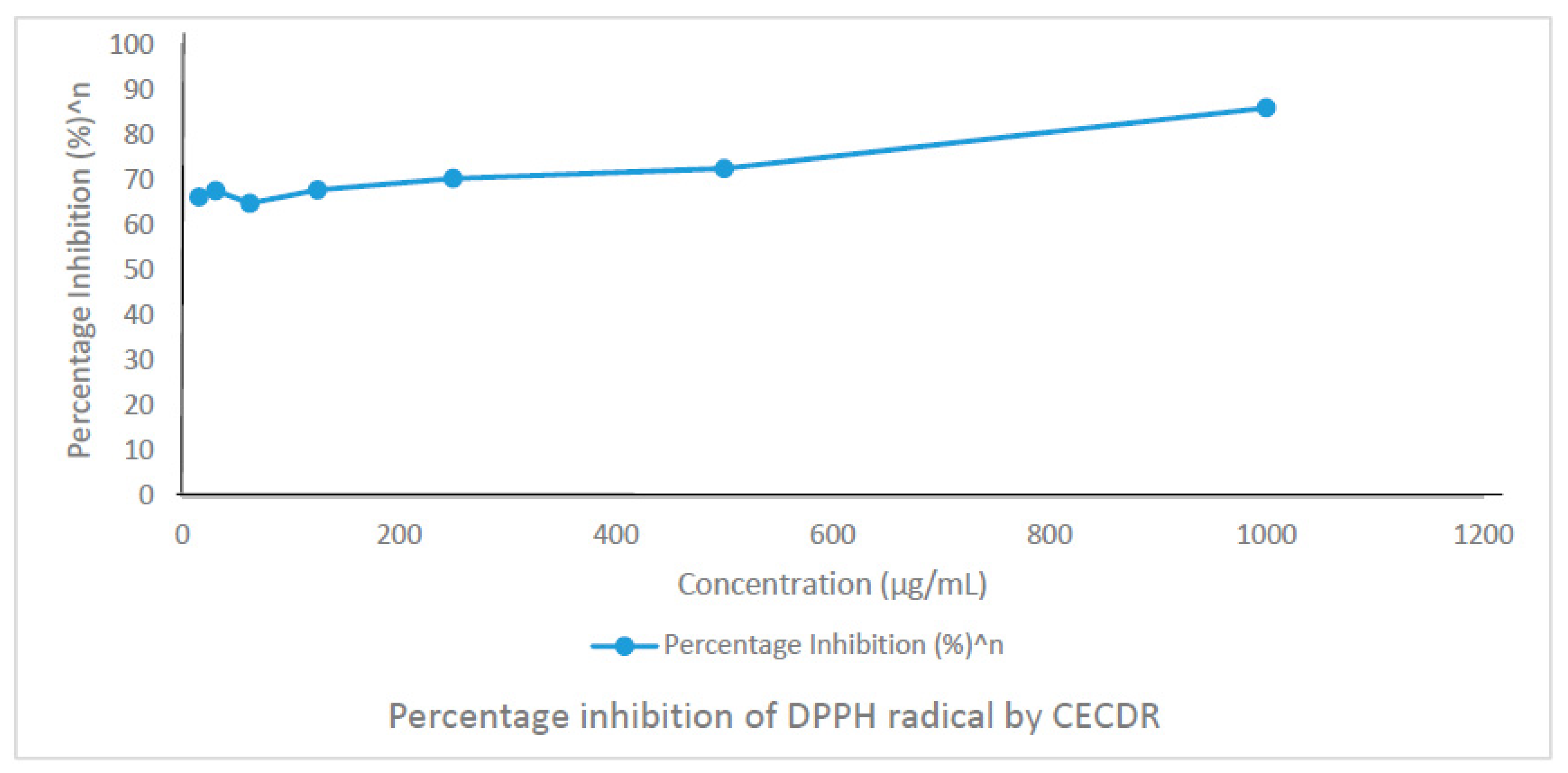
3.4. Effect of CECDR on DPPH Radical Scavenging Activity

Figure 1 shows the quantitative DPPH analysis and revealed DPPH radical scavenging activity of CECDR. The percentage inhibition was concentration dependent with an effective concentration (EC50) of −646.67 µg/mL compared to the ascorbic acid standard with EC50 of −10.58 µg/mL. The percentage inhibition against concentration showed a statistically positive correlation (p < 0.05) (R2) correlation of 0.9548. n = 3.

3.5. Effect of CECDR on Hydrogen Peroxide (H2O2)

The H2O2 scavenging activity of the chloroform extract of Chasmanthera dependens is shown in Figure 2 below. The EC50 was found to be 58.78 ± 2.93 µg/mL.

3.6. Effect of Chloroform Extract of C. dependens on Ferric Reducing Antioxidant Power (FRAP)

The Ferric reducing antioxidant power (FRAP) of CECDR was concentration dependent. A 15.6 µg/mL of CECDR showed Ferric reducing antioxidant power of 21.18 ± 0.15 µg GAE while that of 1000 µg/mL showed FRAP of 0.03 ± 0.00 µg GAE (Table 3).

3.7. Effect of Chloroform Extract of Chasmanthera dependens on Total Antioxidant Capacity (TAC)

The total antioxidant capacity (TAC) of the chloroform extract of C. dependens showed a concentration dependent antioxidant activity. The concentration 15.6 µg/mL showed a TAC of 15.22 ± 7.81 µg AAE while that of 1000 µg/mL showed TAC of 0.03 ± 0.00 µg AAE (Table 4).

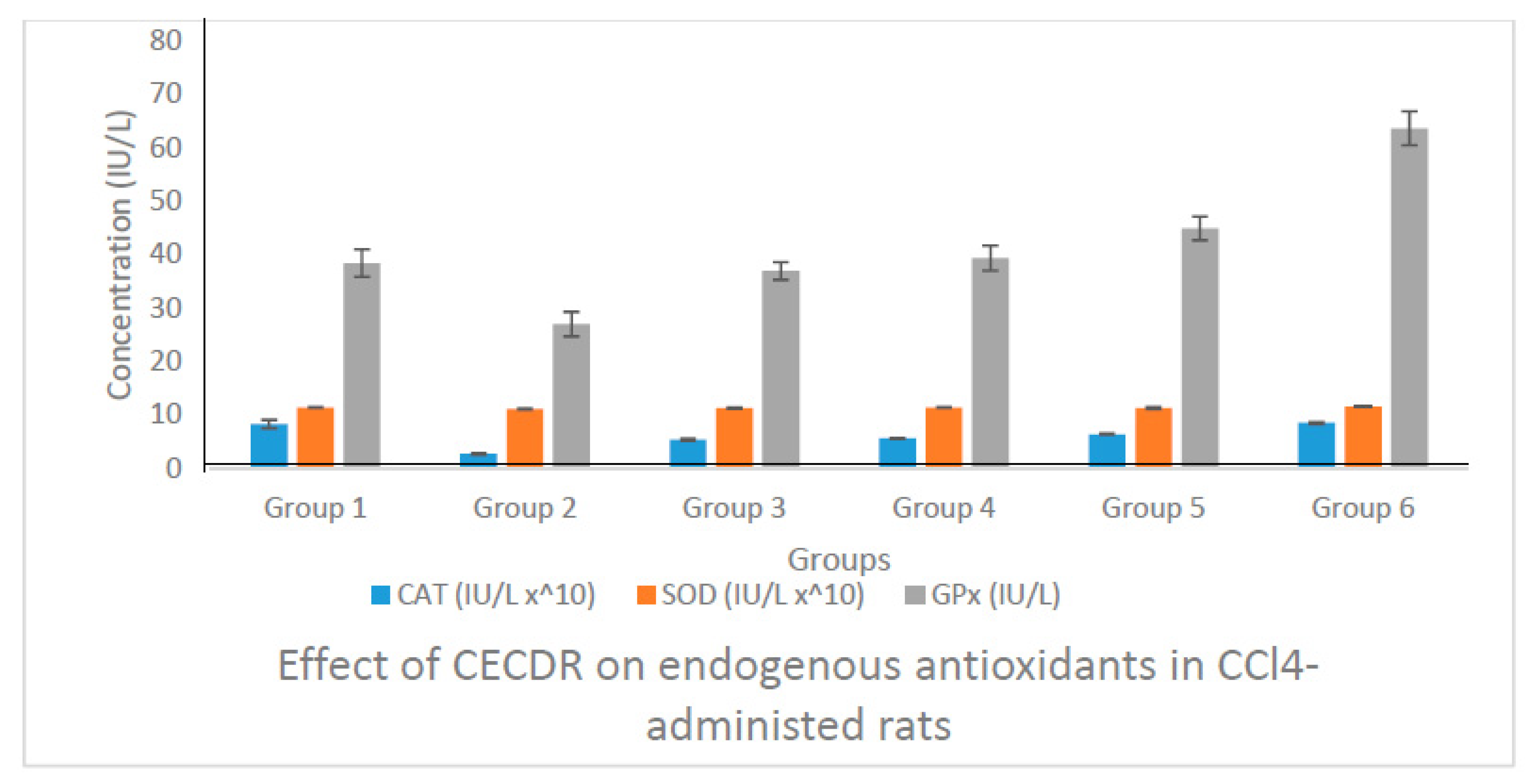
3.8. Result of the Biochemical Analysis

The endogenous antioxidants were evaluated as shown below.

3.8.1. Catalase (CAT) Activity of Rats Administered with CECDR

There is a significant (p < 0.05) decrease in the mean concentration of catalase in the experimental control (Group 2) compared with that of normal control (Group 1), whereas treatment with C. dependens showed significant (p < 0.05) increase in the catalase activity test groups (Groups 3, 4, and 5), as well as treatment with the standard drug, silymarin in the standard control.

3.8.2. Superoxide Dismutase (SOD) Activity of Rats Administered with CECDR

SOD level was non-significantly (p > 0.05) reduced in the untreated group (Group 2), and non-significantly (p > 0.05) increased in the test groups (Groups 3, 4, and 5), as well as the standard control (group 6) when compared with the normal control.

3.8.3. Glutathione Peroxidase (GPx) Activity of Rats Administered with CECDR

GPx activity in the untreated group showed significant (p < 0.05) decrease compared to the normal control, while treatment with CECDR markedly increased GPx concentration in all the test groups. Treatment with the standard drug, silymarin in Group 6 also showed significant (p < 0.05) increase in the activity of GPx compared to the untreated group.

3.8.4. Concentration of Malondialdehyde (MDA)

The concentration of MDA was significantly (p < 0.05) increased in the untreated group compared to the normal control, while the treated groups (Group 3 and 4) administered 200 mg/kg and 400 mg/kg body weight of CECDR showed significant (p < 0.05) decrease compared to the untreated group, as well as with the group treated with standard drugs, silymarin (Group 6) (Figure 3 and Figure 4).
Results are expressed in mean ± SD (n = 4).

4. Discusion

In many regions of the world, traditional medicines are a part of everyday life [36]. In this study, we investigate the antioxidant properties of the chloroform extract of the root of a medicinal plant, Chasmanthera dependens. CECDR showed positive test for the presence of phenols, alkaloids, carbohydrates, flavonoids, terpenes, reducing sugar, tannins, and steroids which is in agreement with the findings of Ogbozor and Anosike [20]. Flavonoids and phenols are known to have free radicals scavenging ability and antioxidant property [37]. Therefore, these metabolites are believed to be responsible for the observed therapeutic effects of CECDR. The Ferric reducing antioxidant power (FRAP) of CECDR was concentration dependent (Table 3). Also, the total antioxidant capacity (TAC) of the chloroform extract of C. dependens showed a concentration dependent antioxidant activity (Table 4). The results showed that the DPPH radical scavenging activities of CECDR increase gradually (p < 0.05) as the concentration increases. CECDR (EC50 of −646.67 µg/mL) in higher concentration possess higher antioxidant potential compared to the standard ascorbic acid (EC50 of −10.58 µg/mL). The strong antioxidant activities of CECDR might be correlated with the high level of phenolics and flavonoids. CECDR having an EC50 of −58.78 ± 2.93 µg/mL was found to have higher H2O2 radical scavenging activity than ascorbic acid (EC50 of 90.06 µg/mL). At 15.6 µg/mL, CECDR had a FRAP of 21.18 ± 0.15 µg GAE while at 1000 µg/mL, it demonstrated a FRAP of 0.03 ± 0.00 µg GAE. This showed that the Ferric reducing antioxidant power of CECDR increases with decreasing concentration of the extract. The CECDR at the concentrations 15.6 µg/mL and 1000 µg/mL exhibited a total antioxidant capacity (TAC) of 15.22 ± 7.81 µg AAE and 0.03 ± 0.00 µg AAE, respectively. This also indicates a significant increase (p < 0.05) in TAC with decreasing concentration of the extract.
A significant (p < 0.05) reduction in the concentration of endogenous antioxidant enzymes (catalase, superoxide dismutase, and glutathione peroxidase) was observed in Group 2 due to CCl4 administration without treatment (Figure 3), suggesting that the free radicals generated by CCl4 overwhelmed the endogenous antioxidant enzymes activities. However, treatment with CECDR resulted in significant elevation (p < 0.05) elevation across the test groups. This could be attributed to the fact that CECDR is rich in antioxidants which revived and enhanced the activities of the endogenous antioxidant enzymes. Flavonoids, terpenoids, and phenolics found in CECDR are important antioxidants that can aid in combating lipid peroxidation and oxidative stress induced by CCl4. Additionally, administration of CCl4 significantly (p < 0.05) elevated the concentration of malondialdehyde (Figure 4) in Group 2 (CCl4—induced and untreated) rats compared to those in Group 1 (Normal control). This could be due to lipid peroxidation by the free radical generated by CCl4. The test groups showed a significant (p < 0.05) reduction in their malondialdehyde concentration, suggesting that C. dependens abated lipid peroxidation.

5. Conclusions

In conclusion, the results obtained from this present study shows that the chloroform extract of Chasmanthera dependens have potent antioxidant effect and this can be credited to the antioxidant activities of its constituent phytochemicals, such as flavonoids, phenolics, terpenes, which can enhance the activities of endogenous antioxidants.

Institutional Review Board Statement

The study was conducted according to the guidelines of the National Institute of Health on the care and use of laboratory animals and approved by the Ethical Clearance Committee for Biological Sciences, University of Nigeria Nsukka with the Approval Number: UNN/FBS/EC/1050.

Informed Consent Statement

Not applicable.

References

  1. Ogunlesi, M.; Okiei, W.; Ademoye, M.; Osibote, A.E. Analysis of essential oil from the stem of Chasmanthera dependens. J. Nat. Prod. 2009, 3, 47–53. [Google Scholar]
  2. Shrikant, S.S.; Gupta, B.L. Antimicrobial activity of medicinal plants on urinary tract pathogens. Int. J. Pharm. Pharm. 2012, 4, 1. [Google Scholar]
  3. Monon, K.; Karamoko, O.; Goueh, G.; Abuo, O.; Adama, C. Study on ethnopharmacological and phytochemical screening of some plants involved in the treatment of abdominal infection in the department of Kouto (COTE D’ IVOIRE). Sch. J. Appl. Med. Sci. 2013, 1, 56–61. [Google Scholar]
  4. Vijaya, L.; Phatak, A.; Chandra, N. Antioxidant and free radical scavenging activity of Hygrophila schulli (Buch.-Ham.) Almeida and Almeida seeds. Adv. Bio Res. 2010, 1, 7. [Google Scholar]
  5. Chun-Weng, P.; Sri, N.M.; Halijah, I.; Norhanom, A.W. Antioxidant properties of crude and fractionated extracts of Alpinia mutica rhizomes and their total phenolic content. Afr. J. Pharm. Pharmacol. 2011, 5, 842–852. [Google Scholar]
  6. Amoussa, A.M.; Lagnika, L.; Tchatchedre, M.; Laleye, A.; Sanni, A. Acute toxicity and antifungal effects of Acacia ataxacantha (Bark). Int. J. Pharmacogn. Phytochem. Res. 2015, 7, 661–668. [Google Scholar]
  7. Reenu, J.; Azeez, S.; Bhageerathy, C. In vitro antioxidant potential in sequential extracts of curcuma caesia roxb. rhizomes. Indian J. Pharm. Sci. 2015, 77, 41. [Google Scholar] [CrossRef] [PubMed] [Green Version]
  8. Kanwal, R.; Arshad, M.; Bibi, Y.; Asif, S.; Khalil, S. Evaluation of Ethnopharmacological and Antioxidant Potential of Zanthoxylum armatum DC. J. Chem. 2015, 2015, 925654. [Google Scholar] [CrossRef]
  9. Fanta, Y.; Taiwe, S.G.; Ngatcha, Z.S.; Khan, M.A.; Agbor, G.A.; Ur-Rahman, N.; Ngo, B.E. Quantification of Bioactive compounds and evaluation of the antioxidant activity of Carissa edulis Valh (Apocynaceae) leaves. Sci. World J. 2019, 2019, 7549620. [Google Scholar]
  10. Ozioko, S.C.; Madueke, A.C.; Anosike, A.C. Phytochemical composition and free radical scavenging activity of Cucurbita maxima fruit juice. Res. J. Adv. Sci. 2020, 1, 17–26. [Google Scholar]
  11. Lateef, A.; Ojo, S.A.; Elegbede, J.A.; Azeez, M.A.; Yekeen, T.A.; Akinboro, A. Evaluation of some biosynthesized silver nanoparticles for biomedical applications: Hydrogen peroxide scavenging, anticoagulant and thrombolytic activities. J. Clust. Sci. 2016, 28, 1379–1392. [Google Scholar] [CrossRef]
  12. Borokini, T.I.; Lawal, I.O. Traditional medicine practices among the Yoruba people of Nigeria: A historical perspective. J. Med. Plants Stud. 2014, 2, 20–33. [Google Scholar]
  13. Use of Tropical Plant Database, UTPD. Chasmanthera dependens. Available online: http://:tropical.theferns.info (accessed on 28 April 2016).
  14. Mosango, D.M. Chasmanthera dependens Hochst. Flora 2008, 27, 1844. [Google Scholar]
  15. Wahab, O.M. Ethnomedicinal Antiepileptic plants used in parts of Oyo and Osun States, Nigeria. BRI 2015, 8, 77–81. [Google Scholar]
  16. Onabanjo, A.O.; John, T.A.; Sokale, A.A.; Samuel, O.T. Analgesic and Antiinflammatory Effects of Chasmanthera dependens. Int. J. Pharmacogn. 1991, 29, 24–28. [Google Scholar] [CrossRef]
  17. Kola-Mustapha, A.T.; Yohanna, K.A.; Ghazali, Y.O.; Ayotunde, H.T. Design, formulation and evaluation of Chasmanthera dependens Hochst and Chenopodium ambrosioides Linn based gel for its analgesic and anti- inflammatory activities. Heliyon 2020, 6, e04894. [Google Scholar] [CrossRef]
  18. Augustine, C.; Anosike, C. Anti-Ulcerogenic Effect of the Chloroform Extract of Chasmanthera dependens on Indomethacin and Aspirin-Induced Ulcer in Rats. J. Nutr. Ecol. Food Res. 2017, 4, 131–137. [Google Scholar] [CrossRef]
  19. Quadri, A.L.; Yakubu, M.T. Fertility enhancing activity and toxicity profile of aqueous extract of Chasmanthera dependens roots in male rats. Andrologia 2017, 49, e12775. [Google Scholar] [CrossRef]
  20. Ogbozor, C.S.; Anosike, C.A. The Effect of Chloroform Extract of Chasmenthera dependens on Carbon Tetrachloride (Ccl4) Induced Hepatotoxicity. Glob. Sci. J. 2020, 8. [Google Scholar]
  21. Abiola, T.S.; Susan, A.O.; Olusegun, B.O. Tannin-Rich Extract of Chasmanthera dependens Stem Potential in Piroxicam-Induced Nephrotoxicity in Adult Male Wistar Rats. Am. J. Mol. Biol. 2020, 10, 29–43. [Google Scholar] [CrossRef] [Green Version]
  22. Lorke, D. A new practical approach to practical acute toxicity testing. Acute Toxicol. 1983, 53, 275–289. [Google Scholar] [CrossRef]
  23. Wallis, T.E. Textbook of Pharmacognosy, 5th ed.; J and A Churchil Ltd.: London, UK, 1967; pp. 81–82. [Google Scholar]
  24. Harborne, J.B. Phytochemical Methods, 11th ed.; Chapman and Hall: New York, NY, USA, 1973. [Google Scholar]
  25. Swain, T. Tannins and Lignins. In Herbivores: Their Interaction with Secondary Plant Metabolites; Rosenthal, G.A., Janzen, D.H., Eds.; Academic Press: New York, NY, USA, 1979; pp. 657–682. [Google Scholar]
  26. Trease, G.E.; Evans, W.C. Pharmacognosy, 12th ed.; Bailliere Tridal: London, UK, 1983. [Google Scholar]
  27. Brunner, J.H. Direct spectrophotometric determination of saponins. Anal. Chem. 1984, 34, 1314–1326. [Google Scholar]
  28. Sofowora, E.A. Medicinal Plants and Traditional Medicine in Africa, 3rd ed.; Spectrum Books Ltd.: Ibadan, Nigeria, 2008; p. 439. [Google Scholar]
  29. Gyamfi, M.A.; Yonamine, M.; Aniya, Y. Free radical scavenging activity of medicinal herbs of Ghana: Thonningia sanguinea on experimentally induced liver injuries. Gen. Pharmacol. 1999, 32, 661–667. [Google Scholar] [CrossRef]
  30. Sahreen, S.; Khan, M.R.; Khan, R.A. Evaluation of antioxidant activities of various solvent extracts of Carissa Opaca fruits. Food Chem. 2010, 122, 1205–1211. [Google Scholar] [CrossRef]
  31. Benzie, I.F.; Strain, J.J. The ferric reducing ability of plasma (FRAP) as ameasure of “antioxidant power”: The FRAP assay. Anal. Biochem. 1996, 239, 70–76. [Google Scholar] [CrossRef] [PubMed] [Green Version]
  32. Ruch, R.J.; Cheng, S.J.; Klaunig, J.E. Prevention of cytotoxicity and inhibition of intracellular communication by antioxidant catechins isolated from Chinese green tea. Carcinogenesis 1989, 10, 1003. [Google Scholar] [CrossRef] [PubMed]
  33. Garrat, D.C. The Quantitative Analysis of Drugs. J. Pharm. Pharmacol. 1964, 16, 772. [Google Scholar]
  34. Paglia, D.E.; Valentine, W.N. Studies on the qualitative and quantitative characterization of erythrocyte glutathione peroxidase. J. Lab. Clin. Med. 1967, 70, 158–169. [Google Scholar] [PubMed]
  35. Satoh, K. Serum lipid peroxide in cerebrovascular disorders determined by a new colorimetric method. Clin. Chim. Acta 1978, 90, 37–43. [Google Scholar] [PubMed]
  36. Bussmann, R.W.; Sharon, D. Traditional medicinal plant use in Northern Peru: Tracking two thousand years of healing culture. J. Ethnobiol. Ethnomed. 2006, 2, 47. [Google Scholar] [CrossRef] [PubMed] [Green Version]
  37. Sarian, M.N.; Ahmed, Q.U.; So’ad, S.Z.M.; Alhassan, A.M.; Murugesu, S.; Perumal, V.; Mohamad, S.N.A.S.; Khatib, A.; Latip, J. Antioxidant and antidiabetic effects of flavonoids: A structure-activity relationship based study. BioMed Res. Int. 2017, 2017, 8386065. [Google Scholar] [CrossRef] [PubMed]
Figure 1. Percentage inhibition of DPPH radical by CECDR.
Msf 02 00021 g001
Figure 2. Percentage inhibition of (H2O2) by CECDR.
Msf 02 00021 g002
Figure 3. Effect of CECDR on endogenous antioxidants in CCl4-administed rats.Results are expressed in mean ± SD (n = 4).
Msf 02 00021 g003
Figure 4. Effect of CECDR on malondialdehyde concentration in CCl4-administed rats.
Msf 02 00021 g004
Table 1. Qualitative phytochemicals of CECDR.
ConstituentsRelative Abundance
Alkaloids+++
Phenolics++
SaponinsND
Tanins++
Carbohydrate++
Flavonoids+++
Steroids+++
GlycosidesND
Terpenoids++
Reducing sugar+
Key: +++ = phytochemical present in high concentration; ++ = Present in moderate concentration; + = Present in small concentration, ND = Not detected.
Table 2. The quantitative phytochemical analysis of CECDR.
Phytochemical ConstituentsBioavailability (mg)
Phenolics4907.25 ± 75.33
Alkaloids1975 ± 68.84
Carbohydrates449.64 ± 59.30
Flavonoids319.79 ± 98.93
Terpenes674.51 ± 46.18
Reducing sugar80.8 ± 36.19
Saponins-
Tannins22.47 ± 11.44
Steroids4.55 ± 0.32
Results from the quantitative analysis are expressed in means ± SD of triplicate
determinations (n = 10)
Table 3. Ferric reducing antioxidant power (FRAP) of CECDR.
Concentration (µg/mL)Ferric Reducing Antioxidant Power (FRAP) (µg GAE)
15.621.18 ± 0.15
31.10.72 ± 0.06
62.50.37 ± 0.03
1250.19 ± 0.01
2500.11 ± 0.00
5000.05 ± 0.00
10000.03 ± 0.00
Table 4. Total antioxidant capacity (TAC) of CECDR.
Concentration (µg/mL)Total Antioxidant Capacity (TAC) (µg AAE)
15.615.22 ± 7.81
31.117.71 ± 7.35
62.57.83 ± 3.70
1253.82 ± 0.74
2503.06 ± 0.33
5002.40 ± 0.13
10001.68 ± 0.02
Publisher’s Note: MDPI stays neutral with regard to jurisdictional claims in published maps and institutional affiliations.

