Morphology of Marsilea Systematic position Kingdom : Plantae - CEC
Morphology of Marsilea Systematic position Kingdom : Plantae - CEC
Morphology of Marsilea Systematic position Kingdom : Plantae - CEC

Create successful ePaper yourself
Turn your PDF publications into a flip-book with our unique Google optimized e-Paper software.
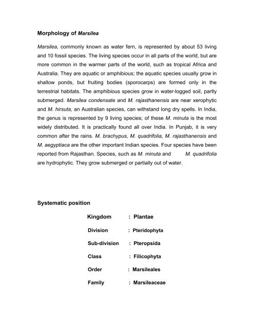
<strong>Morphology</strong> <strong>of</strong> <strong>Marsilea</strong><br />
<strong>Marsilea</strong>, commonly known as water fern, is represented by about 53 living<br />
and 10 fossil species. The living species occur in all parts <strong>of</strong> the world, but are<br />
more common in the warmer parts <strong>of</strong> the world, such as tropical Africa and<br />
Australia. They are aquatic or amphibious; the aquatic species usually grow in<br />
shallow ponds, but fruiting bodies (sporocarps) are formed only in the<br />
terrestrial habitats. The amphibious species grow in water-logged soil, partly<br />
submerged. <strong>Marsilea</strong> condensate and M. rajasthanensis are near xerophytic<br />
and M. hirsuta, an Australian species, can withstand long dry spells. In India,<br />
the genus is represented by 9 living species; <strong>of</strong> these M. minuta is the most<br />
widely distributed. It is practically found all over India. In Punjab, it is very<br />
common after the rains. M. brachypus, M. quadrifolia, M. rajasthanensis and<br />
M. aegyptiaca are the other important Indian species. Four species have been<br />
reported from Rajasthan. Species, such as M. minuta and M. quadrifolia<br />
are hydrophytic. They grow submerged or partially out <strong>of</strong> water.<br />
<strong>Systematic</strong> <strong>position</strong><br />
<strong>Kingdom</strong> : <strong>Plantae</strong><br />
Division : Pteridophyta<br />
Sub-division : Pteropsida<br />
Class : Filicophyta<br />
Order : <strong>Marsilea</strong>les<br />
Family : <strong>Marsilea</strong>ceae
Genus : <strong>Marsilea</strong> Linn.<br />
Common Name : Water Fern<br />
The plant body <strong>of</strong> <strong>Marsilea</strong> is sporophytic having diploid chromosome<br />
number in its somatic cells. During sexual reproduction, it produces male and<br />
female gametophytes, which are haploid in nature and form the sperm and<br />
egg, respectively. The sporophyte is differentiated into rhizome, roots, and<br />
leaves.<br />
Rhizome (Stem): The stem is a long and slender rhizome (Fig. 1). It creeps<br />
either on the surface like stolon, or slightly below the surface <strong>of</strong> the soil like<br />
rhizome. It grows extensively and is branched. The branches arise at the<br />
bases <strong>of</strong> petioles and are extra-axillary in <strong>position</strong>, arising in the lateral or<br />
oblique <strong>position</strong>. They run in all directions and may get rooted at the nodes. In<br />
this way, a single plant may cover an extensive area <strong>of</strong> about 20 metre’s<br />
diameter or even more. The stem is divisible into distinct nodes and<br />
internodes. The internodes are long in the hydrophytic species, and short in<br />
the sub-terrestrial or xerophytic species.<br />
Fig. 1 Creeping and slender rhizome <strong>of</strong> <strong>Marsilea</strong> sp.
Root: The primary root formed on the stem is short-lived and is soon replaced<br />
by adventitious roots, which arise gradually at the nodes on the underside <strong>of</strong><br />
the stem (Fig. 1). Sometimes, roots may also arise at internodes (<strong>Marsilea</strong><br />
aegyptiaca), or laterally (M. minuta). The roots are thin and may be branched<br />
or unbranched. They develop in an acropetal sequence, i.e. the youngest root<br />
is towards the apex <strong>of</strong> the rhizome. The number <strong>of</strong> roots at a node and their<br />
size vary considerably.<br />
Leaves: The leaves arise from the upper side <strong>of</strong> the stem and are arranged in<br />
two alternate rows (Fig. 1). They are long-petioled and compound. The<br />
petioles <strong>of</strong> the submerged plants are long, thin and flexible, with the lamina<br />
floating on the surface <strong>of</strong> water, while the leaves <strong>of</strong> plants growing in mud or<br />
on land have upright, short petioles, with lamina held in a spreading <strong>position</strong>.<br />
The lamina is usually divided into four leaflets <strong>of</strong> the same size. They spring<br />
from the tip <strong>of</strong> the petiole, so that the leaf apparently looks quadrifoliate.<br />
Occasionally, the number <strong>of</strong> leaflets may be 5 or 6, or even 8, instead <strong>of</strong> the<br />
usual 4. In outline the leaflets are obovate, elliptical, or wedge-shaped, with<br />
entire or dentate margins.<br />
According to Puri and Garg (1953), the leaf <strong>of</strong> <strong>Marsilea</strong> is pinnately<br />
compound, with four pinnules borne on the slender rachis; two pinnules are<br />
noticeably higher than the other two, and are inserted on the rachis in<br />
alternate fashion. A leaflet has many dichotomously branched veins, which<br />
are joined with each other by transverse bands and their ends unite to form<br />
marginal loops (Fig. 2).
Fig. 2 Dichotomously branched and petiolate leaves <strong>of</strong> <strong>Marsilea</strong> sp.<br />
At the base <strong>of</strong> the petiole many bean-shaped or oval and stalked<br />
sporocarps develop (Fig. 3).<br />
Fig. 3 Bean-shaped or oval and stalked sporocarps at the base <strong>of</strong> petioles in <strong>Marsilea</strong><br />
sp.
Anatomy <strong>of</strong> <strong>Marsilea</strong><br />
Rhizome (Stem)<br />
Epidermis is single-layered, made up <strong>of</strong> compactly arranged thick-<br />
walled cells.<br />
Cortex is differentiated into three regions, viz. outer cortex, the middle<br />
cortex and the inner cortex:<br />
a. outer cortex is aerenchymatous, one to several cells in<br />
thickness, sometimes also consists <strong>of</strong> tannin cells,<br />
b. middle cortex consists <strong>of</strong> sclerenchymatous tissue filled with air<br />
cavities arranged in the form <strong>of</strong> ring, and<br />
c. inner cortex is solid tissue <strong>of</strong> several cells in thickness. The<br />
inner layer <strong>of</strong> inner cortex is parenchymatous, filled with starch,<br />
while outer region <strong>of</strong> inner cortex is sclerenchymatous in nature.<br />
The vascular cylinder is siphonostele; limited externally and internally<br />
by endodermis, hence called outer endodermis and inner endodermis,<br />
respectively.<br />
The siphonostele is medullated. The xylem is in the form <strong>of</strong> ring.<br />
Phloem is present on both sides <strong>of</strong> xylem. Such a stele is called<br />
amphiphloic siphonostele.<br />
The de<strong>position</strong> <strong>of</strong> the several tissues from inwards is outer endodermis,<br />
outer pericycle, outer phloem, xylem, inner phloem, inner pericycle and<br />
inner endodermis (Fig. 4).
Petiole<br />
Fig. 4 <strong>Marsilea</strong>: Transverse section <strong>of</strong> rhizome; a part cellular<br />
Single layered epidermis made up <strong>of</strong> rectangular cells.<br />
The outer cortex consists <strong>of</strong> few layers <strong>of</strong> thin walled cells; middle<br />
cortex is aeranchymatous consisting <strong>of</strong> ring <strong>of</strong> air-chambers; while the<br />
inner cortex is a solid, compact tissue several cells deep. The inner<br />
layers <strong>of</strong> inner cortex are paranchymatous and filled with starch. Here<br />
and there tannin-filled cells also occur.<br />
The stele is covered externally by a single-layered distinct endodermis.<br />
It is triangular in outline, lies in the centre, and has a single vascular<br />
bundle. The xylem is ‘V’ shaped, with opening towards the axis.<br />
The order <strong>of</strong> the tissues from inner to outer side <strong>of</strong> the stele is xylem,<br />
xylem parenchyma, phloem, pericycle and endodermis (Fig. 5).
Leaflet<br />
Fig. 5 <strong>Marsilea</strong>:Transverse section <strong>of</strong> petiole; a part cellular<br />
It consists <strong>of</strong> upper and lower epidermal layers, each made up <strong>of</strong> a<br />
single layer <strong>of</strong> paranchymatous cells. The continuity <strong>of</strong> epidermis is<br />
interrupted by slightly sunken stomata. Stomata are restricted to the<br />
upper epidermis in floating leaves.<br />
The ground tissue (mesophyll) lies between the upper and lower<br />
epidermis and is differentiated into palisade and spongy parenchyma:<br />
The palisade part lies just beneath the upper epidermis and consists<br />
<strong>of</strong> columnar cells rich in chloroplasts, while the spongy part faces<br />
the lower epidermis and consists <strong>of</strong> rounded cells<br />
In case <strong>of</strong> submerged species, there is no distinction between<br />
palisade and spongy tissues.
Root<br />
Vascular bundles are embedded in the mesophyll tissue. They are<br />
concentric in nature. Each bundle has a central core <strong>of</strong> xylem,<br />
surrounded by phloem.<br />
The arrangement <strong>of</strong> tissues in the stele is xylem, phloem and bundle<br />
sheath (Fig. 6).<br />
Fig. 6 <strong>Marsilea</strong>: Transverse section <strong>of</strong> lamina.<br />
It consists <strong>of</strong> piliferous layer instead <strong>of</strong> epidermis, made up <strong>of</strong><br />
compactly arranged biconvex cells.<br />
Beneath piliferous layer, the cortex is made up <strong>of</strong> an outer cortex and<br />
inner cortex:<br />
Outer cortex is aerenchymatous, consisting <strong>of</strong> large air chambers<br />
arranged in the form <strong>of</strong> ring.<br />
Inner cortex is compact, made up <strong>of</strong> rounded cells containing starch.<br />
Inner cortex is delimited by a single layer <strong>of</strong> distinct endodermis.
Vascular bundle lies within the endodermis, and is usually diarch and<br />
exarch.<br />
The xylem is plate-like and occupies the centre <strong>of</strong> stele. The<br />
protoxylem, consisting <strong>of</strong> two small mass <strong>of</strong> cells, is towards the<br />
periphery (pericycle), while metaxylem is large, plate-like and occupies<br />
the centre.<br />
There is no medullary tissue (pith) in the stele and arrangement <strong>of</strong><br />
tissue is metaxylem, protoxylem, phloem, pericycle and endodermis<br />
(Fig. 7).<br />
Fig. 7 <strong>Marsilea</strong>: Transverse section <strong>of</strong> root.<br />
Reproduction in <strong>Marsilea</strong><br />
<strong>Marsilea</strong> is a sporophyte and reproduces asexually both by vegetative means<br />
as well as by means <strong>of</strong> spores.<br />
Vegetative reproduction: In <strong>Marsilea</strong>, vegetative reproduction occurs by the<br />
formation <strong>of</strong> tubers, which are small, bud-like structures containing reserve<br />
food material, arising from some subterranean branches <strong>of</strong> the rhizome.
These tubers serve as perennating organs, and are capable <strong>of</strong> tiding over the<br />
unfavourable conditions. On the return <strong>of</strong> favourable conditions, these tubers<br />
germinate to form new plants. Tuber formation has been reported in a few<br />
<strong>Marsilea</strong> species, such as M. minuta and M. hirsuta.<br />
Reproduction by spores (spore formation): <strong>Marsilea</strong> is a heterosporous<br />
fern, which produces two types <strong>of</strong> spores – microspores and megaspores in<br />
separate sporangia, borne in special bean-shaped bodies called the<br />
sporocarps.<br />
Sporocarps: Sporocarp is a bean-shaped to ovoid, nutlike structure, attached<br />
to the basal part <strong>of</strong> the petiole with the help <strong>of</strong> a stalk. It is green and s<strong>of</strong>t<br />
when young, but turns dark-brown at maturity. Usually one sporocarp is<br />
present at the base <strong>of</strong> each petiole, but in some species, the number varies<br />
from 2 – 20. Sometimes, the attachment <strong>of</strong> sporocarps with the petiole shows<br />
so much variation that different species can be distinguished on this particular<br />
character (Fig. 8); for example in M. polycarpa many sporocarps are attached<br />
on one side <strong>of</strong> the petiole in a single vertical row. In M. quadrifolia, pedicels<br />
(stalks) are united with one another, and then jointly inserted on the petiole. In<br />
M. minuta, the stalks <strong>of</strong> all the sporocarps though free, are attached to the<br />
petiole at a single point.
Fig. 8 <strong>Marsilea</strong>: different modes <strong>of</strong> attachment <strong>of</strong> sporocarps to the petiole – A,<br />
M. polycarpa; B, M. quadrifolia; C, M. minuta; and D, M. vestita.<br />
Structure <strong>of</strong> sporocarp: The sporocarp is differentiated into a stalk (pedicel)<br />
and a body. The stalk is fused laterally to the back <strong>of</strong> the body <strong>of</strong> the<br />
sporocarp, generally forming a distinct ridge called ‘raphe’. In some species,<br />
the distal (upper) end <strong>of</strong> the raphe is marked by one or two teeth-like<br />
projections, known as tubercles.<br />
Internal structure <strong>of</strong> the sporocarp: The internal structure <strong>of</strong> the sporocarp<br />
can be studied under the following headings:<br />
Sporocarp wall: The wall <strong>of</strong> the sporocarp is very hard, thick and highly<br />
resistant to mechanical injury. It is differentiated into an outer epidermis, a<br />
middle hypodermis, and an inner paranchymatous zone. The epidermis is<br />
made up <strong>of</strong> cuboidal cells, covered with a thick layer <strong>of</strong> cuticle. A large<br />
number <strong>of</strong> sunken stomata are present in the epidermis. The hypodermis<br />
consists <strong>of</strong> two layers <strong>of</strong> radially-elongated palisade-like cells, which are<br />
compactly arranged without any intercellular spaces between them. The inner<br />
paranchymatous cells <strong>of</strong> this zone form a gelatinous ring inside the sporocarp<br />
wall (Fig. 9).<br />
Fig. 9 Transverse section <strong>of</strong> the wall <strong>of</strong> <strong>Marsilea</strong> sporocarp: A, young stage; B, old<br />
stage.<br />
Sori: When we cut the horizontal section <strong>of</strong> sporocarp, a ring appears in the<br />
form <strong>of</strong> a dorsal and a ventral mass. In this plane, both micro- and
megasporangia are visible as sori. The sori are the reproductive structures<br />
arranged in the two alternating rows in the cavity <strong>of</strong> the sporocarp (Fig. 10).<br />
Each sorus has a receptacle which contains one terminal megasporangium<br />
and two microsporangia on the lateral sides. It is surrounded by an indusium.<br />
The sori overlap each other and the indusia <strong>of</strong> adjacent sori are partially<br />
fused.<br />
Fig. 10 <strong>Marsilea</strong> sp.: A, Vertical transverse section <strong>of</strong> sporocarp; B, horizontal<br />
longitudinal section <strong>of</strong> sporocarp.<br />
The number <strong>of</strong> sori in a sporocarp varies from two in M. aegyptica to twenty in<br />
M. quadrifolia and M. vestita. There are 11 – 12 sori in M. minuta. Each sorus<br />
bears both mega and microsporangia. The former are short-stalked and are<br />
arranged in a row at the tip <strong>of</strong> the receptacle, whereas the latter are long-<br />
stalked and arise on the sides. The number <strong>of</strong> micro- and megasporangia<br />
varies with species. In M. minuta, a sorus has 4-8 megasporangia and 8-13<br />
microsporangia.<br />
Development <strong>of</strong> sporocarp: Sporocarp originates from one <strong>of</strong> the marginal<br />
cells <strong>of</strong> a very young leaf, when it is 6-8 cells high. One <strong>of</strong> the marginal cell<br />
cuts <strong>of</strong>f segments on either side and one <strong>of</strong> the derivatives acts as the apical
cell for the second sporocarp. This process is repeated several times<br />
depending on the number <strong>of</strong> sporocarps formed at the base <strong>of</strong> a leaf. The<br />
sporocarp, initially cuts <strong>of</strong>f segments on either side and forms a mass <strong>of</strong><br />
undifferentiated cells, which later curves markedly so that its distal end is<br />
directed in a horizontal plane. Subsequently, two rows <strong>of</strong> soral mother cells<br />
are differentiated on the ventral side <strong>of</strong> the young sporocarp. The differential<br />
growth <strong>of</strong> soral mother cells and their derivatives, results in the formation <strong>of</strong><br />
involutions on the upper surface <strong>of</strong> the sorus. These involutions grow into arc-<br />
shaped depressions called soral canals. The soral mother cells divide to form<br />
receptacular surface in the alternating rows. The sporangial initial develops on<br />
the receptacular surface in acropetal succession, i.e. first sporangial initial is<br />
formed at the apex <strong>of</strong> receptacle and it forms megasporangia. Subsequently,<br />
two initials are formed on the lateral sides <strong>of</strong> the receptacle. They functions as<br />
microsporangial initial, each forming a microsporangium (Fig. 11).
Fig. 11 Diagrams showing developmental stages <strong>of</strong> micro- and megasporangia <strong>of</strong><br />
<strong>Marsilea</strong> sp.<br />
Development <strong>of</strong> sporangia: Since <strong>Marsilea</strong> is a heterosporous fern,<br />
megasporangia (macrosporangia) produce megaspores and the<br />
microsporangia produce microspores. The sporangia develop in a<br />
basipetalous manner on the receptacle. The megasporangia develop first at<br />
the top <strong>of</strong> the receptacle and are, therefore, older, while the microsporangia<br />
develop later along the sides <strong>of</strong> the receptacle. The development <strong>of</strong><br />
microsporangium, as well as <strong>of</strong> megasporangium is almost similar. The<br />
sporangial initial divides transversely, forming an outer and an inner cell. The<br />
entire sporangium develops from the outer cell, the inner cell taking no part in
its development. The outer cell <strong>of</strong> sporangium, which is destined to form<br />
spore mother cell, undergoes repeated divisions. Each spore mother cell<br />
undergoes reduction division and forms four haploid spores. In<br />
megasporangia, all but one spore degenerates to form a nutritive liquid, which<br />
grows in size at the expense <strong>of</strong> others, forming a single large functional<br />
megaspore (Fig. 12). In microsporangia, almost all the spores in a<br />
microsporangium are functional. They are smaller in size.<br />
Fig. 12 <strong>Marsilea</strong> sp.: mature megaspore within the megasporangium, showing very<br />
thick and well-differentiated wall.<br />
Dehiscence <strong>of</strong> sporocarp: There is no special mechanism for the liberation<br />
<strong>of</strong> sporangia from the sporocarp. They are set free only when the wall <strong>of</strong> the<br />
sporocarp splits open. As the sporocarp breaks at one point due to absorption<br />
<strong>of</strong> water, the sori come out along the gelatinous ring <strong>of</strong> sporocarp (Fig. 13).<br />
These sori function as mother cells <strong>of</strong> the gematophytic generation.
Fig. 13 Diagrams showing stages in the dehiscence <strong>of</strong> <strong>Marsilea</strong> sporocarps.<br />
Gematophytic generation<br />
The microspores and the megaspores germinate separately to produce<br />
male (micro-) and the female (mega-) gametophytes. The microspores<br />
germinate immediately after shedding, forming small biconvex prothallial cell<br />
and a large apical cell. The apical cell by repeated divisions ultimately forms<br />
androcytes or antherizoid- mother cells. Each androcyte metamorphoses into<br />
a cork screw-shaped multiflagellate antherizoid, characterized by the<br />
presence <strong>of</strong> a prominent terminal vesicle (Fig. 14).<br />
Fig. 14 <strong>Marsilea</strong>: screwshaped<br />
multiflagellate antherizoid, characterized by<br />
the presence <strong>of</strong> a prominent terminal vesicle.<br />
Similarly, megaspore undergoes division, forming small nipple-shaped<br />
apical cell and a large basal prothallial cell. The apical cell forms the female
gametophyte, while prothallial cell provides nutrition to the developing<br />
gematophytes (Fig. 15).<br />
Fig. 15 <strong>Marsilea</strong>: germination and development <strong>of</strong> the female<br />
gametophyte.<br />
Fertilization: The neck canal cells and the ventral canal cell degenerates to<br />
form a mucilaginous mass, which attracts sperms chemotactically. One <strong>of</strong> the<br />
sperms fuses with the egg to accomplish fertilization and form the diploid<br />
zygote. The zygote is the mother cell <strong>of</strong> the next sporophytic generation and<br />
develops into a young sporophyte within 2-4 days after fertilization.<br />
Embryogeny: The fertilized egg (zygote) enlarges in its size and secretes a<br />
thin cellulose wall around it. It enters into rapid divisions, to give rise to a<br />
young sporophyte within 2 - 4 days. Zygote divides vertically and produces<br />
two unequal cells. The larger cell is known as the epibasal cell, and the<br />
smaller one as the hypobasal cell. These two cells undergo transverse<br />
divisions to form a quadrant embryo. The cells <strong>of</strong> the quadrant embryo show<br />
definite relation to the primary growth <strong>of</strong> the embryo. Epibasal half produces
shoot and leaf. Hypobasal half produces root and foot. Hence the<br />
development is described as lateral. In quadrant embryo, the next divisions<br />
are irregular. At the time <strong>of</strong> differentiation <strong>of</strong> different primordia in the embryo,<br />
the adjoining cells <strong>of</strong> megagametophyte divide periclinally, forming a thick<br />
calyptras, which encloses and protects the developing embryo. The calyptra<br />
is greenish in colour. Large number <strong>of</strong> rhizoids may develop from the base <strong>of</strong><br />
calyptra. The embryo gives rise to the young seedling that penetrates through<br />
the calyptra and grows into a sporophyte (Fig. 16).<br />
Fig. 16 <strong>Marsilea</strong>: stages in the development <strong>of</strong> embryo - A-E, early developmental<br />
stages; F, mature female gametophyte bearing young embryo surrounded by the<br />
calyptra; G, median-longitudinal section <strong>of</strong> the mature female gametophyte.<br />
Life cycle: In the life cycle <strong>of</strong> <strong>Marsilea</strong>, we find alternation <strong>of</strong> generations.<br />
<strong>Marsilea</strong> plant is a sporophyte, which is the dominant phase in the entire life<br />
cycle. The sporophyte comprises rhizome, roots and leaves. It reproduces<br />
vegetatively, as well as by means <strong>of</strong> spores. The spores are formed in<br />
sporangia on specialized structures called sporocarps. The sporocarps bear<br />
two kinds <strong>of</strong> asexual spores which are formed after meiosis and are, thus<br />
haploid (meiospores). The smaller spores are known as microspores. They<br />
are produced within microsporangia. The larger spores are known as<br />
megaspores, which are formed in the megasporangia. Both the sporangia are
produced within the fruit body, the sporocarp. The whole life cycle <strong>of</strong> <strong>Marsilea</strong><br />
is depicted in Fig. 17.<br />
Fig. 17 <strong>Marsilea</strong>: diagrammatic representation <strong>of</strong> life cycle.